. 2001 Jul 2;20(13):3459-72.
doi: 10.1093/emboj/20.13.3459.

Pin1 is overexpressed in breast cancer and cooperates with Ras signaling in increasing the transcriptional activity of c-Jun towards cyclin D1

Affiliations

Pin1 is overexpressed in breast cancer and cooperates with Ras signaling in increasing the transcriptional activity of c-Jun towards cyclin D1

G M Wulf et al. EMBO J. .

Abstract

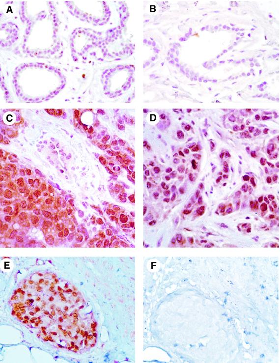
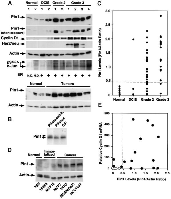
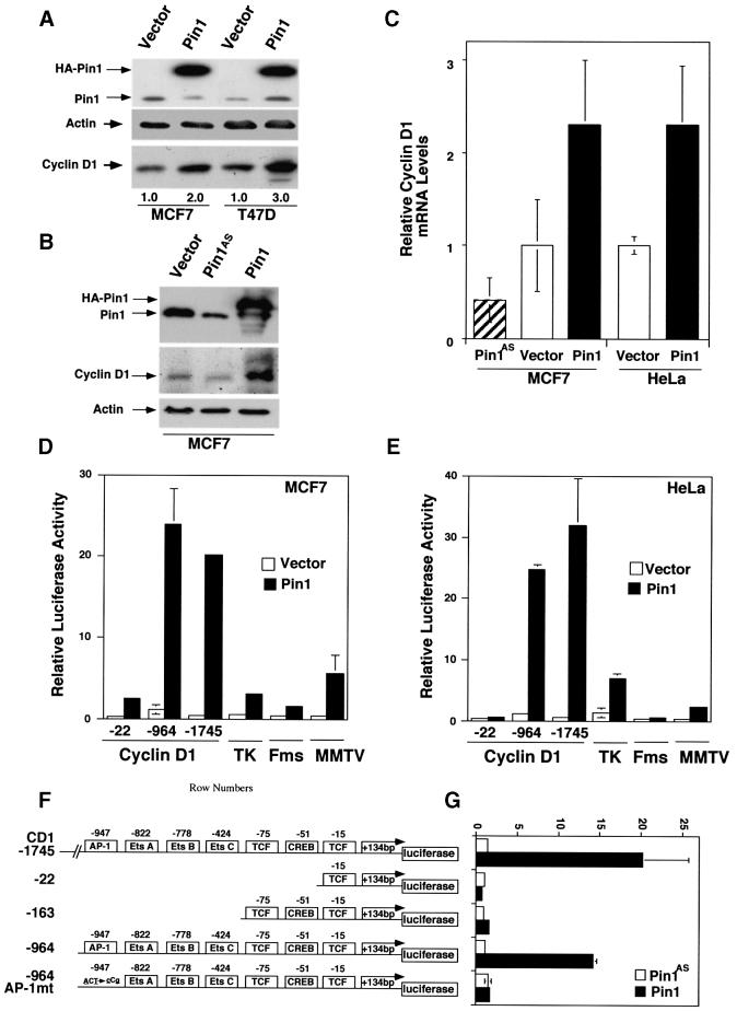
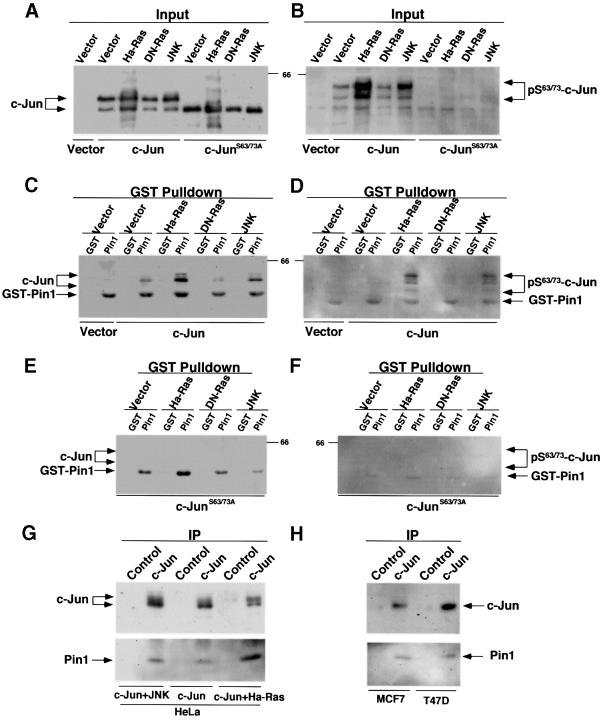
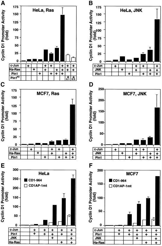
Phosphorylation on serines or threonines preceding proline (Ser/Thr-Pro) is a major signaling mechanism. The conformation of a subset of phosphorylated Ser/Thr-Pro motifs is regulated by the prolyl isomerase Pin1. Inhibition of Pin1 induces apoptosis and may also contribute to neuronal death in Alzheimer's disease. However, little is known about the role of Pin1 in cancer or in modulating transcription factor activity. Here we report that Pin1 is strikingly overexpressed in human breast cancers, and that its levels correlate with cyclin D1 levels in tumors. Overexpression of Pin1 increases cellular cyclin D1 protein and activates its promoter. Furthermore, Pin1 binds c-Jun that is phosphorylated on Ser63/73-Pro motifs by activated JNK or oncogenic Ras. Moreover, Pin1 cooperates with either activated Ras or JNK to increase transcriptional activity of c-Jun towards the cyclin D1 promoter. Thus, Pin1 is up-regulated in human tumors and cooperates with Ras signaling in increasing c-Jun transcriptional activity towards cyclin D1. Given the crucial roles of Ras signaling and cyclin D1 overexpression in oncogenesis, our results suggest that overexpression of Pin1 may promote tumor growth.


Figures

None
Fig. 1. Immunostaining of Pin1 in human breast cancer. Sections from paraffin-embedded tissues were subjected to an antigen retrieval treatment, followed by immunostaining with anti-Pin1 antibodies. Non-cancerous tissues (A and B; normal breast with mild fibrocystic changes) show weak, but detectable, Pin1 staining, while invasive ductal carcinomas (C and D) or ductal carcinoma in situ (E) show intense Pin1 staining. To show the specificity of Pin1 antibodies, Pin1-specific antibodies were first depleted using GST–Pin1 beads and then used to stain the breast sections (F).
None
Fig. 2. Pin1 overexpression in human breast cancer cell lines and patient tissues, and its correlation with the Bloom and Richardson grade of tumors. (A) Comparison of Pin1 levels and known breast tumor markers in normal and cancerous human breast tissues. Normal breast and cancer tissues were pulverized in liquid nitrogen, and equal amounts of total protein were separated on SDS-containing gels and transferred to membranes. The membranes were cut into five pieces and subjected to immunoblotting analysis using antibodies to Pin1, cyclin D1, HER2/neu, phosphorylated Ser63/73-c-Jun and actin, respectively. The estrogen receptor status was determined by radioimmunoassay and defined as positive when its levels were >10 fmol/l. The estrogen receptor status in normal controls was not determined (N.D.). Note that Pin1 was detected in immunoblots as a doublet due to phosphorylation. (B) Phosphatase treatment abolishes the double-band pattern of Pin1 in immunoblots. Tumor cell lysates were treated either with a mixture of PP1 and PP2A (PPases) in the presence (lane 1) or absence (lane 2) of the phosphatase inhibitor okadaic acid (Inh.), or CIP (lane 3). (C) Pin1 levels in 10 normal breast tissues and different stages of 51 human breast cancer samples. Pin1 levels were determined by immunoblotting analysis, as in (A), and semi-quantified using Imagequant. Actin was used as an internal control, and the Pin1 level in each sample was expressed as the Pin1:actin ratio. (D) Comparison of Pin1 levels in mammary epithelial cell lines. The same amounts of total lysates prepared from normal human mammary epithelial cell lines (Normal), spontaneously immortalized normal human mammary epithelial cell lines (Immortalized) and human breast carcinoma-derived cell lines (Cancer) were subjected to immunoblotting analysis with Pin1 or actin antibodies. (E) Correlation of Pin1 protein levels with cyclin D1 mRNA. RNA was isolated from six normal and 16 cancerous tissues, cDNA synthesized and subjected to real-time PCR for the quantitative analysis of cyclin D1 mRNA expression. The Pearson correlation coefficient was 0.47 (p <0.05).
None
Fig. 3. Pin1 elevates cyclin D1 protein and activates the cyclin D1 promoter via the AP-1 site. (A) Increase in cellular cyclin D1 protein by Pin1. MCF7 or T47D cells were transfected with Pin1 or control vector, followed by immunoblotting analysis of the cell lysates with antibodies against Pin1 and cyclin D1, with actin as a control. Cyclin D1 levels were semi-quantified using Imagequant and are presented below the image; the level in the vector control was defined as 1. (B) Manipulation of Pin1 levels in cells causes changes in cyclin D1 levels. MCF7 cells were transiently transfected with the control vector or a construct expressing HA-Pin1 or antisense Pin1 (Pin1AS), followed by immunoblotting analysis with anti-cyclin D1, -Pin1 or -actin antibodies. (C) Overexpression and depletion of Pin1 increase and decrease levels of cyclin D1 mRNA. MCF7 or HeLa cells were transfected with constructs encoding for Pin1 sense, antisense or vector control as indicated in the figure. After 24 h, mRNA was isolated, cDNA synthesized and subjected to real-time PCR to obtain relative cyclin D1 mRNA levels. (D and E) Activation of cyclin D1, but not TK, c-fms or MMTV promoter by Pin1. MCF7 (D) or HeLa (E) cells were transiently transfected with Pin1 or the vector and various reporter constructs, followed by assaying the luciferase activity. pRL-TK Renilla luciferase reporter construct was co-transfected in each sample to normalize for transfection efficiency. The activity of the reporter luciferase was expressed relative to that in control vector-transfected cells, which is defined as 1. All results are expressed as ± SD of independent duplicate cultures. Note that to detect the maximal effect of Pin1 on various promoters, we used 0.5 µg of Pin1 cDNA per transfection in this experiment, which was higher than in other experiments described here. (F) Schematic representation of cyclin D1 (CD1) pA3LUC basic reporter constructs and its mutants. Possible transcription factor-binding sites are indicated. –964CD1AP-1mt was same as the wild-type –964CD1construct except for two base-pair substitutions at the consensus AP-1 site. (G) Activation of the cyclin D1 promoter by Pin1 via the AP-1 site. HeLa cells were co-transfected with various cyclin D1 reporter constructs as indicated in (F) and Pin1 sense or antisense (Pin1AS) construct, followed by assaying the luciferase activity. Note that for this experiment 200 ng Pin1 sense or antisense cDNA were used, while in subsequent co-transfection experiments only 50 ng/assay were used.
None
Fig. 4. Pin1 binds to c-Jun phosphorylated on Ser63/73-Pro. (A and B) Modulation of c-Jun phosphorylation by Ras or JNK. HeLa cells were co-transfected with c-Jun or c-JunS63/73A and Ha-Ras, DN-Ras, activated JNK or control vector. Cells were harvested and cellular proteins were subjected to immunoblotting analysis with antibodies against c-Jun (A) or phosphorylated Ser63/73-c-Jun (B). (C and D) Interaction between Pin1 and c-Jun phosphorylated on Ser63/73-Pro. The same cellular proteins as those described in (A) were incubated with GST–agarose beads that had been pre-incubated with either GST alone or GST–Pin1. Proteins associated with the beads were subjected to immunoblotting analysis with antibodies against c-Jun (C) or phosphorylated Ser63/73-c-Jun (D). Note that GST–Pin1 was non-specifically recognized by monoclonal antibodies, as shown previously (Yaffe et al., 1997; Lu et al., 1999b). (E and F) No interaction between Pin1 and c-JunS63/73A. The same cellular proteins as those described in the (A) were incubated with GST–agarose beads containing GST or GST–Pin1, and bound proteins were subjected to immunoblotting analysis with antibodies against c-Jun (E) or phosphorylated Ser63/73-c-Jun (F). (G and H) Co-immunoprecipitation of transfected (G) or endogenous (H) c-Jun with endogenous Pin1. HeLa cells were co-transfected with c-Jun and Ha-Ras or JNK. c-Jun was immunoprecipitated from transfected HeLa cells (G) or non-transfected breast cancer cell lines (H) with polyclonal c-Jun antibodies or non-related antibodies (Control), and then subjected to immunoblotting using monoclonal anti-c-Jun antibodies (upper panel) or anti-Pin1 antibodies (lower panel).
None
Fig. 5. Pin1 cooperates either with Ha-Ras or activated JNK in enhancing the activity of c-Jun to activate the cyclin D1 promoter. HeLa cells (A, B and E) or MCF7 (C, D and F) were co-transfected with vector, c-Jun, or c-Jun with or without Ha-Ras (A and C) or activated JNK (B and D) and then subjected to the luciferase assay with the –964 cyclin D1 Luc promoter construct as reporter gene. In the same system, a reporter gene construct with an AP-1 site mutant fails to respond to Pin1 in combination with c-Jun, JNK or Ha-Ras (E and F).
None
Fig. 6. The effects on the transcriptional activity of c-Jun depend on the phosphoprotein-binding and PPIase activity of Pin1 as well as phosphorylation of c-Jun on Ser63/73. (A) Abolishing the Pin1 effect by inactivating its PPIase activity. HeLa cells were co-transfected with vectors, c-Jun, or c-Jun + Ha-Ras, and Pin1 or its PPIase-negative mutant Pin1R68,69A, and then subjected to luciferase assay. Pin1R68,69A fails to isomerize phosphorylated Ser/Thr-Pro bonds (Yaffe et al., 1997). (B) Abolishing the Pin1 effect by inactivating its phosphoprotein-binding activity. HeLa cells were co-transfected with vectors, c-Jun, or c-Jun + Ha-Ras and green fluorescent protein (GFP)–Pin1 or its WW domain point mutants, and then subjected to luciferase assay. GFP–Pin1W34A and GFP–Pin1S16E did not bind phosphoproteins, as shown (Lu et al., 1999b). Note that GFP fusion proteins were used because the WW domain Pin1 mutants were not stable in cells, but they were stable as GFP fusion proteins, although expressed at reduced levels (data not shown). Although the absolute maximal luciferase activity was not as high as in other experiments, which is likely to be due to lower levels of GFP fusion proteins being expressed, the overall trends were the same. (C) Inhibiting the ability of Pin1 to increase the c-Jun activity by DN-Ras. Cells were co-transfected with c-Jun or c-Jun + Pin1, and increasing amounts of DN-Ras, and then subjected to the luciferase assay. (D and E) Abolishing the cooperative effect between Pin1 and Ha-Ras or activated JNK by mutating c-Jun phosphorylation sites Ser63/73. HeLa cells were co-transfected with various amounts of Pin1, c-Jun or c-JunS63/73A construct, and Ha-Ras (D) or activated JNK (E) and then subjected to the luciferase assay.
None
Fig. 7. Role of Pin1 in regulating the transcriptional activity of phosphorylated c-Jun towards the cyclin D1 promoter. Oncogenic Ha-Ras activates JNKs, which phosphorylate c-Jun on two critical amino terminal Ser-Pro motifs, enhancing its transcriptional activity. Pin1 is up-regulated in breast cancer and functions as a potent regulator of phosphorylated c-Jun to induce cyclin D1 expression, presumably by altering the conformation of the phosphorylated Ser-Pro motifs (insert). Double arrows, up-regulation; the asterisk indicated the activated form of proteins.

Similar articles

Cited by

References

    1. Abate C., Luk,D. and Curran,T. (1991) Transcriptional regulation by Fos and Jun in vitro: interaction among multiple activator and regulatory domains. Mol. Cell. Biol., 11, 3624–3632. - PMC - PubMed
    1. Albanese C. et al. (1999) Activation of the cyclin D1 gene by the E1A-associated protein p300 through AP-1 inhibits cellular apoptosis. J. Biol. Chem., 274, 34186–34195. - PubMed
    1. Albanese C., Johnson,J., Watanabe,G., Eklund,N., Vu,D., Arnold,A. and Pestell,R.G. (1995) Transforming p21ras mutants and c-Ets-2 activate the cyclin D1 promoter through distinguishable regions. J. Biol. Chem., 270, 23589–23597. - PubMed
    1. Angel P., Smeal,T., Meek,J. and Karin,M. (1989) Jun and v-jun contain multiple regions that participate in transcriptional activation in an interdependent manner. New Biol., 1, 35–43. - PubMed
    1. Arber N. et al. (1997) Antisense to cyclin D1 inhibits the growth and tumorigenicity of human colon cancer cells. Cancer Res., 57, 1569–1574. - PubMed

Publication types

MeSH terms