. 2001 Jul 2;20(13):3506-17.
doi: 10.1093/emboj/20.13.3506.

Spt16-Pob3 and the HMG protein Nhp6 combine to form the nucleosome-binding factor SPN

Affiliations

Spt16-Pob3 and the HMG protein Nhp6 combine to form the nucleosome-binding factor SPN

T Formosa et al. EMBO J. .

Abstract

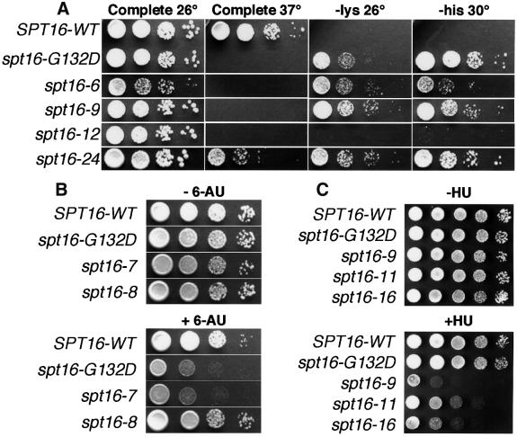
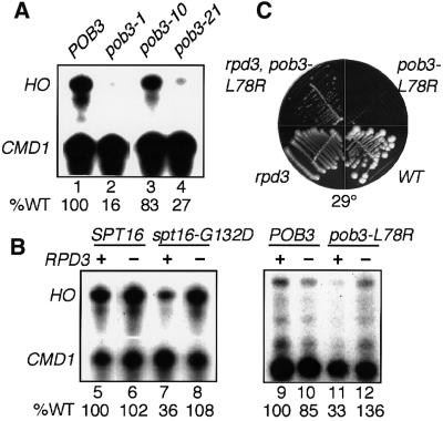
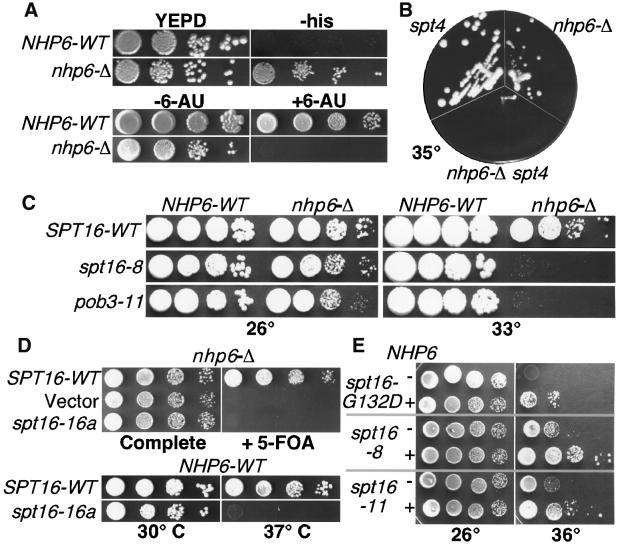
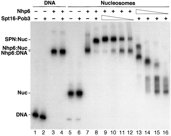
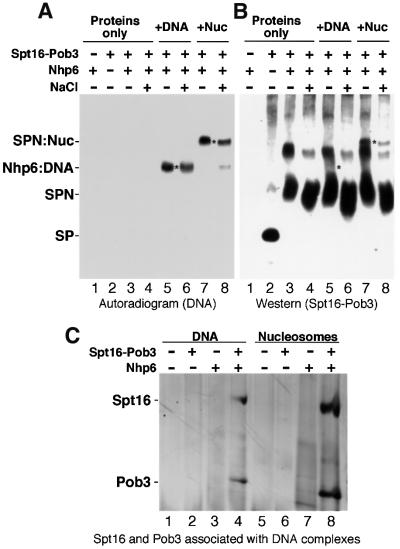
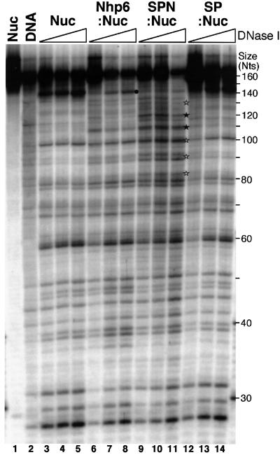
Yeast Spt16/Cdc68 and Pob3 form a heterodimer that acts in both DNA replication and transcription. This is supported by studies of new alleles of SPT16 described here. We show that Spt16-Pob3 enhances HO transcription through a mechanism that is affected by chromatin modification, since some of the defects caused by mutations can be suppressed by deleting the histone deacetylase Rpd3. While otherwise conserved among many eukaryotes, Pob3 lacks the HMG1 DNA-binding motif found in similar proteins such as the SSRP1 subunit of human FACT. SPT16 and POB3 display strong genetic interactions with NHP6A/B, which encodes an HMG1 motif, suggesting that these gene products function coordinately in vivo. While Spt16-Pob3 and Nhp6 do not appear to form stable heterotrimers, Nhp6 binds to nucleosomes and these Nhp6-nucleosomes can recruit Spt16-Pob3 to form SPN-nucleosomes. These complexes have altered electrophoretic mobility and a distinct pattern of enhanced sensitivity to DNase I. These results suggest that Spt16-Pob3 and Nhp6 cooperate to function as a novel nucleosome reorganizing factor.


Figures

None
Fig. 1. Alleles of SPT16 cause variable effects. (A) Strain 7784-1-1 (his4-912δ lys2-128δ spt16-Δ) carrying pTF128 or its derivatives with the alleles shown were grown and aliquots of 10-fold dilutions were placed on complete synthetic medium or medium lacking lysine or histidine and incubated at the temperature indicated. (B) Strains 4053-5-2 URA+ (SPT16-WT) and the isogenic 7782 URA+ set with the spt16 alleles indicated were diluted as in (A) and incubated at 30°C on synthetic medium lacking uracil and either without (– 6-AU) or with (+ 6-AU) 75 µg/ml 6-AU. (C) As in (A), except that cells were incubated at 26°C on rich medium without (–HU) or with (+HU) 90 mM HU.
None
Fig. 2. Mutations in SPT16 and POB3 decrease transcription of HO and are suppressed by loss of RPD3. (A) and (BHO and CMD1 transcripts were measured as described in Materials and methods. The amount of HO transcript was normalized to wild type (% WT shown) assuming constant CMD1 expression (the relative efficiency of labeling of each probe causes variable ratios of HO to CMD1 in each experiment). RNA was isolated from 7697 (pob3-Δ) with pTF139 (YCp LEU2 POB3, Schlesinger and Formosa, 2000) carrying the POB3 alleles indicated (lanes 1–4), and from the congenic strains DY150, DY1539, DY5391, DY5394, DY5699, DY7380, DY7379 and DY7375 (lanes 5–12). (C) Congenic W303 strains with a deletion of RPD3, the pob3-L78R mutation, neither mutation (WT) or both mutations were placed on rich medium at 29°C.
None
Fig. 3. Genetic effects of deleting or overexpressing NHP6. (A) DY2623 (his4-912δ) and DY6863 (his4-912δ nhp6a/b-Δ) were grown to saturation, diluted and placed on either rich medium (YEPD) or medium lacking histidine (–his). DY150 URA+ (NHP6-WT) and DY2382 (nhp6-Δ) were tested on 6-AU as in Figure 1B. (B) Congenic W303 strains lacking SPT4, both copies of NHP6 or all three genes were placed on rich medium at 35°C. (C) 7737-3-2 (spt16-Δ NHP6-WT) and 7847-2-4 (spt16-Δ, nhp6a-Δ, nhp6b-Δ) carrying pTF128 (YCp LEU2 SPT16) with the SPT16 alleles indicated, or 7697 (pob3-Δ) and 7746–5-4 (pob3-Δ, nhp6a-Δ, nhp6b-Δ) carrying pTF139-11 (YCp LEU2 pob3-11), were diluted and placed on rich medium at 26 or 33°C. (D) 7847-2-4 (spt16-Δ) with pCDC68 (YEp URA3 SPT16) was transformed with pTF128, YCplac111 (Gietz and Sugino, 1988; vector), or pTF128-16a, grown in rich medium, washed and dilutions were placed on complete synthetic medium or medium containing 5-FOA. In the bottom panel, dilutions of 7737-3-2 (spt16-Δ) with pTF128 or pTF128-16a were placed at the temperatures shown. (E) 7737-3-2 carrying pTF128 with the SPT16 alleles shown was transformed with YEplac195 (Gietz and Sugino, 1988, –NHP6) or pTF146 (+NHP6), and dilutions were placed on medium lacking uracil at the temperatures indicated.
None
Fig. 4. Nhp6 interacts with nucleosomes to form a binding site for Spt16–Pob3. Purified His10–Nhp6 (+ = 30 pmol, lanes 13–16 are 15, 7.5, 3.8 and 1.9 pmol) and Spt16–Pob3 (+ = 1.5 pmol, lanes 9–12 are 0.75, 0.38, 0.19 and 0.09 pmol) were mixed with 0.1 pmol of a 200 bp DNA fragment (lanes 1–4) or the same fragment incorporated into nucleosomes (lanes 5–16). After native PAGE, the DNA was detected by autoradiography. The positions of the free DNA (DNA), nucleosomes (Nuc) and complexes described in the text are indicated.
None
Fig. 5. Spt16–Pob3 binds specifically to Nhp6–nucleosome complexes. (A) and (B) Nhp6 (lane 1), Spt16–Pob3 (lane 2) or both (lanes 3–8) were incubated alone (lanes 1–4) or with either DNA (lanes 5 and 6) or nucleosomes (lanes 7 and 8) at 30 (–) or 350 mM (+) NaCl, and electrophoresed as in Figure 4. DNA (A) or Spt16–Pob3 (B) were then detected in duplicate gels by autoradiography or with antisera. Asterisks mark equivalent positions in the two panels, and complexes are labeled as described in the text. (C) Samples in 30 mM NaCl were prepared as in Figure 4 except that 0.6 pmol of DNA or nucleosomes, 2 pmol of Spt16–Pob3 and 40 pmol of Nhp6 were mixed in 11 µl. After electrophoresis, regions containing DNA were excised and subjected to SDS–PAGE, then stained with silver (Ausubel et al., 1994). Lanes 1–8 are as in Figure 4, and the forms excised were those labeled in that figure as DNA (lanes 1 and 2), Nhp6:DNA (lanes 3 and 4), Nuc (lanes 5 and 6), Nhp6:Nuc (lane 7) and SPN:Nuc (lane 8).
None
Fig. 6. Sensitivity of nucleosome complexes to DNase I. Nucleosomes (Nuc) were mixed with Nhp6 (Nhp6:Nuc), SPN (SPN:Nuc) or Spt16–Pob3 (SP:Nuc) as in Figure 4, except the DNA was a restriction fragment with 167- (labeled) and 163-nucleotide strands (see Materials and methods), and the Nhp6 was purified from yeast cells. Complexes were treated with DNase I (none in lane 1, 1 arbitrary unit in lane 2, then sets of 8, 16 and 32 units), then separated by denaturing PAGE. The symbols indicate digestion sites; a black spot denotes the strong cut site corresponding to the exit of the DNA from the nucleosome, and stars indicate sites where digestion is enhanced by addition of Nhp6 and SPN, with filled symbols corresponding to more pronounced effects. Size is shown in nucleotides (Nts).

Similar articles

Cited by

References

    1. Ausubel F.M., Brent,R., Kingston,R.E., Moore,D.D., Seidman,J.G., Smith,J.A. and Struhl,K. (eds) (1994) Current Protocols in Molecular Biology. Greene Publishing Associates, Inc., and John Wiley & Sons, Inc., Boston, MA.
    1. Bartel P.L. and Fields,S. (1995) Analyzing protein–protein interactions using two-hybrid system. Methods Enzymol., 254, 241–263. - PubMed
    1. Bhoite L.T. and Stillman,D.J. (1998) Residues in the Swi5 zinc finger protein that mediate cooperative DNA-binding with the Pho2 homeodomain protein. Mol. Cell. Biol., 18, 6436–6446. - PMC - PubMed
    1. Boeke J.D., Trueheart,J., Natsoulis,G. and Fink,G.R. (1987) 5-fluoroorotic acid as a selective agent in yeast molecular genetics. Methods Enzymol., 154, 164–175. - PubMed
    1. Brazas R.M., Bhoite,L.T., Murphy,M.D., Yu,Y., Chen,Y., Neklason,D.W. and Stillman,D.J. (1995) Determining the requirements for cooperative DNA binding by Swi5p and Pho2p (Grf10p/Bas2p) at the HO promoter. J. Biol. Chem., 270, 29151–29161. - PubMed

Publication types

MeSH terms

Substances