. 2001 Jul 2;20(13):3544-53.
doi: 10.1093/emboj/20.13.3544.

The ribonucleotide reductase inhibitor Sml1 is a new target of the Mec1/Rad53 kinase cascade during growth and in response to DNA damage

Affiliations

The ribonucleotide reductase inhibitor Sml1 is a new target of the Mec1/Rad53 kinase cascade during growth and in response to DNA damage

X Zhao et al. EMBO J. .

Abstract

The evolutionarily conserved protein kinases Mec1 and Rad53 are required for checkpoint response and growth. Here we show that their role in growth is to remove the ribonucleotide reductase inhibitor Sml1 to ensure DNA replication. Sml1 protein levels fluctuate during the cell cycle, being lowest during S phase. The disappearance of Sml1 protein in S phase is due to post-transcriptional regulation and is associated with protein phosphorylation. Both phosphorylation and diminution of Sml1 require MEC1 and RAD53. More over, failure to remove Sml1 in mec1 and rad53 mutants results in incomplete DNA replication, defective mitochondrial DNA propagation, decreased dNTP levels and cell death. Interestingly, similar regulation of Sml1 also occurs after DNA damage. In this case, the regulation requires MEC1 and RAD53, as well as other checkpoint genes. Therefore, Sml1 is a new target of the DNA damage checkpoint and its removal is a conserved function of Mec1 and Rad53 during growth and after damage.


Figures
None
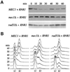
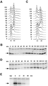
Fig. 1. Sml1 protein levels fluctuate during the cell cycle. (A) The wild-type strain (W1379-3C) was arrested in G1 by α-mating factor in YPD medium and then released into the cell cycle. Samples were collected immediately after release (time zero) and every 10 min until 130 min. Cells were fixed and DNA content was measured by FACS analysis. (B) Protein extracts were made from samples collected in (A). Sml1 protein levels were examined by a protein blot using anti-Sml1 antibody. The arrow indicates the position of Sml1. The band above Sml1 cross-reacts with anti-Sml1 serum and is used as a loading control. (C) Strain W2057-11A (GAL-SML1) was arrested in G1 by α-mating factor in YPGL medium. Sml1 was induced by the addition of 2% galactose for 30 min. Cells were next released into YPGal medium and samples were collected immediately after release (time zero) and every 10 min until 120 min. Cells were fixed and DNA content was measured by FACS analysis. Note that the 10 min delay in entry into S phase compared with (A) is due to growth conditions and is not genotype specific. (D) Sml1 levels from samples collected in (C) were examined as described in (B). (E) Strain W2057-11A (GAL-SML1) was arrested in G2/M phase by nocodazole in YPRaffinose medium (Raf). Cells were next transferred to YPGal medium containing nocodazole to induce Sml1 expression and maintain their arrest. After 45 min, GAL-SML1 expression was turned off by addition of 2% glucose. Sml1 protein levels were examined by a protein blot using anti-Sml1 antibody at the time points indicated. Zero time is immediately before the addition of 2% glucose.
None
Fig. 2. The decrease in Sml1 levels during S phase depends on MEC1 and partially on RAD53. (A) Strains U1476 (MEC1 + RNR1), U1195 (mec1Δ + RNR1) and U1198-10C (rad53Δ + RNR1) were arrested in G1 by α-mating factor in SC-LEU medium. Protein extracts were made immediately after release from G1 phase (time zero) and every 10 min for 60 min. Sml1 protein levels were examined by protein blots using anti-Sml1 antibody. The amount of Sml1 protein was quantified from the protein blot using a cross-reacting band (e.g. see Figure 1B) as a loading control. (B) DNA content was measured by FACS from samples collected in (A).
None
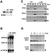
Fig. 3. Sml1 protein levels, but not its mRNA levels, diminish after treatment with DNA-damaging agents. (A and B) The wild-type strain (W1588-4C) was treated with 200 mM HU and 0.05% MMS for 80 min. Sml1 protein and SML1 RNA from these samples were examined by protein blot (A) and an RNA blot (B). In (B), actin was used as loading control and RNR2 was used as a positive control for transcriptional induction by DNA damage. (C and D) The wild-type strain (W1588-4C) was irradiated with various doses of UV light and γ-rays (C) or treated with 0.003 or 0.03% MMS (D) as indicated. Samples were harvested at different time points after treatment to examine the kinetics of Sml1 protein level changes. Samples of mock treatment (M) received no irradiation. Arrows indicate the position of the Sml1 protein.
None
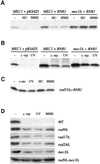
Fig. 4. The diminution of Sml1 after DNA damage depends on MEC1 and RAD53, and partially on other cell cycle checkpoint genes. Sml1 protein levels from various strains were examined after cells were treated with different types of DNA-damaging agents. The strains used are: in (A) and (B), U1475 (MEC1 + pRS425), U1476 (MEC1 + RNR1) and U1195 (mec1Δ + RNR1); in (C), U1198-10C (rad53Δ + RNR1); and in (D), W1588-4A (WT), W1518-10B (rad9Δ), W1522-11B (rad17Δ), W1519-17B (rad24Δ), W1520-10B (mec3Δ) and W2617-4A (rad9Δ mec1Δ). For HU and MMS treatments, protein extracts were made after cells were incubated with 200 mM HU or 0.05% MMS for 1 h. For UV and γ-ray treatments, protein extracts were made after cells were irradiated by UV light (120 J/m2) and γ-rays (30 krads) and grown at 30°C for 30 min.
None
Fig. 5. Wild-type and mutant Sml1 protein levels after DNA damage and in the presence of RNR1 overexpression. (A) Sml1 protein levels were examined in strains W1588-4C (SML1), W2097-63B (sml1-S87P) and W2099-63D (sml1-I76T) at various time points after incubation with HU and MMS at the concentrations indicated. Arrows show the position of wild-type or mutant Sml1 proteins. (B) Sml1 protein levels were examined in strains W1588-4C (SML1), W2097-63B (sml1-S87P) and W2099-63D (sml1-I76T) in the presence of either an empty vector pRS425 (vec.) or a 2 µm-RNR1 plasmid (RNR1). (C) W2057-11A (GAL-SML1) strains that contain either an empty vector (pRS425) or a 2 µm-RNR1 plasmid (pRS425-RNR1) were first grown in raffinose-Leu medium to early-log phase (Raf). Expression of Sml1 proteins was then induced by addition of 2% galactose for 30 min. The induction was inhibited by the addition of 2% glucose and the Sml1 proteins levels were examined at various time points. The zero time point is the time of addtion of glucose. The lower panel in (C) is an overexposure of the Sml1 portion of the protein blot from the strain containing pRS425, which illustrates the absence of Sml1 protein at later time points. The increased signal of the cross-reacting band (*) in (C) compared with (A) and (B) is due to different batches of antibody.
None
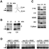
Fig. 6. Sml1 is phosphorylated in S phase and after DNA damage. (A) Wild-type strain W1588-4C was arrested in G1 phase by α-mating factor and then released into the cell cycle. Samples were taken 12 and 24 min after release. Protein extracts were made with phosphatase inhibitors included in the boiling buffer. Sml1 protein levels were examined by protein blots using anti-Sml1 antibody, and the DNA content was measured by FACS analysis. The arrow indicates the position of Sml1 protein and the star marks the position of a slower migrating Sml1 band. (B) Strain U1476 (wild-type strain containing a 2 µm-RNR1 plasmid) was irradiated by γ-rays (30 krads) or UV light (120 J/m2). Protein extracts were made from samples before (0 min) and after irradiation (times as indicated) as described in (A). Sml1 was detected by a protein blot using anti-Sml1 antibody. The arrow indicates the position of the Sml1 protein and the brackets mark the position of slower migrating Sml1 bands. (C) Cell samples from (A) and (B) or from HU- (200 mM, 1 h) and MMS- (0.05%, 1 h) treated U1476 cells were collected. Protein extracts were made using the TCA method and were incubated with calf intestinal phosphatase (CIP) at 37°C for 15 min. The phosphatase inhibitor β-glycerophosphate was added together with CIP (PPI). Mock reactions did not have either CIP or β-glycerophosphate. (D) Strains U1476 (MEC1 + RNR1), U1195 (mec1Δ + RNR1) and U1198-10C (rad53Δ + RNR1) were treated with 200 mM HU or 0.05% MMS for 1 h. Protein extracts were made using the TCA method and analyzed by protein blots using anti-Sml1 antibody. For the S phase extract (S), cells were first arrested in G1 and then released into the cell cycle. Samples were collected 25 min after release when the majority of the cells were in S phase.
None
Fig. 7. mec1 mutations cause incomplete DNA replication and exhibit synthetic lethality with rad53-1. (AC) Strains W2057-2B (mec1Δ GAL-SML1), W2057-11A (MEC1 GAL-SML1) and W2079-11B (rad53 GAL-SML1) were arrested in G1 phase by α-mating factor in YPGL medium. Sml1 was induced by addition of 2% galactose for 30 min. Cells were then released into YPGal medium and samples taken at different time points. In (A), cells were fixed in ethanol and analyzed by FACS. Note that due to the slower growth rate of the rad53 GAL-SML1 strain, longer time point intervals were used. (B) Cells were washed in YPD medium, plating units were determined by microscopic examination and appropriate dilutions were spread onto YPD plates. After incubation at 30°C for 3 days, colonies were counted and the plating efficiency was calculated using the number of colonies divided by the number of plating units. Time zero is considered as 100%. In (C), DNA plugs from 100 and 150 min were prepared from W2057-2B (mec1Δ) and W2057-11A (+) and analyzed by pulsed-field gel electrophoresis. The DNA was blotted and probed with labeled DNA from the DUN1 gene to detect chromosome IV. α-mating factor- (G1) and HU-arrested (HU-arrested) samples were used as controls for chromosome separation. (D) Six tetrads are shown for diploid strain W1986 (MATamec1-3/+ rad53-1/+ sml1Δ::HIS3/+). The genotypes of the three inviable spores are deduced from those of the sister spore clones and in each case is mec1-3 rad53-1. The arrows indicate two mec1-3 rad53-1 sml1Δ spore clones. Spore clones of the other genotypes grow equally well.
None
Fig. 8. A model for the regulation of RNR activity by the Mec1/Rad53 kinase cascade. As cells enter S phase (gray lines) or after they are challenged by DNA-damaging agents or by replication blocks (black lines), the Mec1/Rad53 kinase cascade leads to phosphorylation of the unbound form of Sml1. Note that the phosphorylation depicted here may be direct or indirect. Subsequently, phosphorylated Sml1 is targeted for protein degradation. This degradation drives the equilibrium from the inactive Sml1–RNR complex to the active form of RNR. The transcripts of the RNR genes are also induced at S phase by the Mbp1–Swi6 complex (gray lines) and by the Mec1/Rad53/Dun1 kinase cascade after DNA damage (black lines). Transcriptional up-regulation (dashed lines) in both situations probably increases the amount of the RNR enzyme.

Similar articles

Cited by

References

    1. Adams A., Gottschling,D.E., Kaiser,C.A. and Stearns,T. (1997) Methods in Yeast Genetics. Cold Spring Harbor Laboratory Press, Cold Spring Harbor, NY.
    1. Brown E.J. and Baltimore,D. (2000) ATR disruption leads to chromosomal fragmentation and early embryonic lethality. Genes Dev., 14, 397–402. - PMC - PubMed
    1. Caspari T. (2000) How to activate p53. Curr. Biol., 10, R315–R317. - PubMed
    1. Chabes A., Domkin,V. and Thelander,L. (1999) Yeast Sml1, a protein inhibitor of ribonucleotide reductase. J. Biol. Chem., 274, 36679–36683. - PubMed
    1. Christianson T.W., Sikorski,R.S., Dante,M., Shero,J.H. and Hieter,P. (1992) Multifunctional yeast high-copy-number shuttle vectors. Gene, 110, 119–122. - PubMed

Publication types

MeSH terms