. 2001 Jul 1;534(Pt 1):109-21.
doi: 10.1111/j.1469-7793.2001.00109.x.

Local response of L-type Ca(2+) current to nitric oxide in frog ventricular myocytes

Affiliations

Local response of L-type Ca(2+) current to nitric oxide in frog ventricular myocytes

M Dittrich et al. J Physiol. .

Abstract

1. The regulation of L-type Ca(2+) current (I(Ca)) by the two nitric oxide (NO) donors sodium nitroprusside (SNP, 1 microM to 1 mM) and (+/-)-S-nitroso-N-acetylpenicillamine (SNAP, 3 or 10 microM) was investigated in frog ventricular myocytes using double voltage clamp and double-barrelled microperfusion techniques. 2. SNP and SNAP depressed the isoprenaline (ISO, 10-100 nM)- or forskolin (FSK, 1 microM)-mediated stimulation of I(Ca) via cGMP activation of the cGMP-stimulated phosphodiesterase (PDE2). Complete inhibition of the ISO (100 nM) response was observed at 1 mM SNP. 3. When SNP was applied locally, i.e. to one-half of the cell, and ISO to the whole cell, the response of I(Ca) to ISO was strongly antagonized in the cell half exposed to SNP (up to 100 % inhibition at 1 mM SNP) but a relatively small depression was observed in the other half of the cell (only 20 % inhibition at 1 mM SNP). 4. The NO scavenger 2-(4-carboxyphenyl)-4,4,5,5-tetramethylimidazoline-1-oxyl 3-oxide (carboxy-PTIO, 1 mM) reversed the local effect of SNAP (3 microM) on FSK-stimulated I(Ca) when applied to the same side as the NO donor, but had no effect when applied to the other side of the cell. 5. A local application of erythro-9-(2-hydroxy-3-nonyl)adenine (EHNA, 30 microM), a selective inhibitor of PDE2, fully reversed the local effect of SNP (100 microM) or SNAP (10 microM) on I(Ca) but had no effect on the distant response. 6. When EHNA was applied on the distant side, with SNP (1 mM) and ISO (100 nM) applied locally, the distant effect of SNP was fully reversed. 7. Our results demonstrate that in frog ventricular myocytes stimulation of guanylyl cyclase by NO leads to a strong local depletion of cAMP near the L-type Ca(2+) channels due to activation of PDE2, but only to a modest reduction of cAMP in the rest of the cell. This may be explained by the existence of a tight microdomain between L-type Ca(2+) channels and PDE2.


Figures
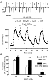
Figure 1
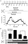
Figure 1. The NO donor SNP inhibits the ISO response via activation of PDE2
A, time course of ICa in a representative frog ventricular cell voltage clamped using standard single electrode whole-cell recording. ICa (filled circles) was recorded by applying a depolarizing pulse to 0 mV for 200 ms every 8 s from a holding potential of −80 mV. The cell was superfused with ISO (100 nm) and subsequently sodium nitroprusside (SNP) at increasing concentrations (10 μm, 100 μm and 1 mm) and EHNA (30 μm) were applied as indicated. Current traces in the upper panel were recorded at the times indicated by the corresponding letters on the main graph. The dotted line indicates the zero-current level. B, summary of seven experiments as displayed in A. The effects of SNP (open columns) and SNP+EHNA (filled columns) are expressed relative to the increase of ICa after pre-stimulation with 100 nm ISO. The columns indicate the means and the error bars the s.e.m. Significant differences determined using Student's paired t test between ISO+SNP and ISO+SNP+EHNA are indicated as: *P < 0.05; **P < 0.01, ***P < 0.001.
Figure 2
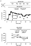
Figure 2. Half- versus whole-cell exposure to SNP after whole-cell stimulation with ISO
A frog ventricular myocyte was attached at each end to an electrode and positioned so that its approximate mid-point was gently pressed against a thin diaphragm separating channels S1 and S2 of a double-barrelled perfusion system (arrowhead, see schematic drawing). A, the myocyte was first exposed to control extracellular solutions. As shown by the horizontal bars, ISO (100 nm) was first applied to S1, and upon reaching a steady state, ISO was then applied to both sides, S1 and S2, evoking a further increase of ICa. Subsequently, SNP (100 μm) was applied to S1 and then to both sides, S1 and S2. Removal of the drugs was performed in reverse order to the sequence of application, showing half-cell application to S2 instead of S1. Current traces in the upper panel were recorded at the times indicated by the corresponding letters on the main graph. The dotted line indicates the zero-current level. B, summary of five experiments as described in A. The effects are expressed relative to the maximal ICa in the presence of ISO on both S1 and S2 (= 100 %). Because of run-down of ICa, the effects of SNP were calculated relative to an extrapolated level of the ISO response. Columns indicate the means and the error bars the s.e.m. Significant differences determined using Student's paired t test, denoted by the bracket or relative to ISO stimulation on both sides, are indicated as: *P < 0.05; **P < 0.01, ***P < 0.001.
Figure 3
Figure 3. Local and distant effects of SNP on ISO stimulation
A frog ventricular myocyte was attached at each end to an electrode and positioned so that its approximate mid-point was gently pressed against a thin diaphragm separating channels S1 and S2 of a double-barrelled perfusion system (arrowhead, see schematic drawing). A, the myocyte was first exposed to control extracellular solutions. As shown by the horizontal bars, ISO (100 nm) was first applied on S1 and then on S1 + S2. ICa shows a continuous run-down. The total current ICa at S1 +S2 (filled squares) was separated according to the contribution from both half-cells by omitting Ca2+ for S2. Thus, the remaining current is the local ICa with respect to SNP (open circles), and the difference between the two current levels is the distant ICa (see arrows). The current decreases to 55 % of the total ICa, indicating an approximately half-adjustment of the cell between S1 and S2. B, same experiment as in A, showing the effect of two increasing concentrations of SNP (1 and 100 μm) on ICa in S1 (local side, open circles) and S2 (distant side, filled circles). Note that the control current in the absence of ISO is subtracted. The dotted lines indicate spontaneous run-down.
Figure 5
Figure 5. Effect of carboxy-PTIO on the local and the distant effect of SNAP after whole-cell stimulation with FSK
A, the myocyte was first exposed to control extracellular solutions. As shown by the horizontal bars, FSK (1 μm) was applied to both cell halves, and SNAP (3 μm) was applied to S1, either in the presence or in the absence of carboxy-PTIO (cPTIO, 1 mm). Thus, S1 became the local side, and S2 became the distant side with respect to SNAP. The total current ICa in S1 +S2 (filled circles) was separated according to the contribution from both half-cells by omitting Ca2+ for S2. Thus, the remaining current is the local ICa with respect to SNP (open circles), and the difference between the two current levels is the distant ICa (see arrows). The distant effect of cPTIO was also examined by application of the drug to S2. B, summary of seven experiments performed according to the protocol described in A. As shown by the schematic representation at the bottom, FSK was used on the whole cell at 1 μm and the local (S1) and distant (S2) responses of the cell were normalized to 100 %. Subsequently, the local (open bars) and distant (filled bars) effects of FSK were measured under three different conditions: (1) when SNAP (3 μm) was applied to the local (S1) side; (2) when cPTIO (1 mm) was added to SNAP on the local (S1) side; (3) when cPTIO (1 mm) was added to the distant (S2) side. Columns indicate the mean and error bars the s.e.m. of seven experiments. Significant differences determined using Student's paired t test between the conditions tested and the effect of FSK alone are indicated as: *P < 0.05; **P < 0.01.
Figure 6
Figure 6. Estimation of the intracellular cAMP gradient upon local application of SNP
Relationship between the local and the distant effect of SNP. Percentages and error bars are taken from Fig. 4. The dotted line indicates equality of local and distant effects. The values for α given near each symbol give an estimate of the relative intracellular cAMP concentration on the distant side ([cAMP]d) compared with that on the local side ([cAMP]l): α=[cAMP]d/[cAMP]l (for further details see text).
Figure 4
Figure 4. Half-cell exposure to SNP after whole-cell stimulation with ISO: local and distant effects of SNP
Dose-response curves for the local (open circles) and distant (filled circles) effects of SNP on ISO prestimulated cells. Summary of several similar experiments as described in Fig. 3. ISO (100 nm) was applied to the whole cell, whereas SNP was applied in increasing concentrations on a half-cell. The data points show means ±s.e.m. with the number of cells indicated near each symbol. Significant differences between local and distant effects of SNP determined using Student's paired t test are indicated as: ***P < 0.001.
Figure 7
Figure 7. Effect of half-cell application of EHNA on the distant effect of SNP or SNAP after whole-cell stimulation with ISO
A, the myocyte was first exposed to control extracellular solutions. As shown by the horizontal bars, ISO (20 nm) was applied to both half-cells, and SNP (100 μm) and EHNA (30 μm) were applied to S1. Thus, S1 became the local side, and S2 became the distant side with respect to SNP. The total current ICa in S1 +S2 (filled squares) was separated according to the contribution from both half-cells by omitting Ca2+ for S2. Thus, the remaining current is the local ICa with respect to SNP (open circles), and the difference between the two current levels is the distant ICa (see arrows). B and C, summary of a number of experiments performed according to the protocol described in A with either SNP (100 μm, n = 3, B) or SNAP (10 μm, n = 3, C). ISO was applied to the whole cell at 10 or 20 nm and EHNA (30 μm) was applied on the same side as SNP (B) or SNAP (C). The currents were separated according to the local (open bars) and distant (filled bars) effects of SNP (B) or SNAP (C). Columns show means and error bars the s.e.m. relative to the response to ISO. The number of individual experiments is given above each column. The left column in each group of three shows the local (left panel) or distant (right panel) effect of SNP (B) or SNAP (C) upon ISO stimulation. The middle column in each group of three shows the effect of EHNA, which reversed the effect of SNP (B) or SNAP (C) on the local side (left panel), but had no effect on the distant side (right panel). The right column in each group of three shows the effect seen upon return to SNP (B) or SNAP (C) on the local (left panel) or distant (right panel) side immediately after removal of EHNA.
Figure 8
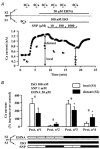
Figure 8. Effect of SNP on the distant effect of ISO
A, the myocyte was first exposed to control extracellular solutions. As shown by the horizontal bars, ISO (100 nm) was applied to S1 alone, and EHNA (30 μm) was applied to S2 for inhibition of PDE2 on that side. Thereafter, SNP was applied at three increasing concentrations (10, 100 and 1000 μm) to S1. Thus, S1 became the local side, and S2 the distant side with respect to SNP. The total current ICa in S1 +S2 (filled squares) was separated according to the contribution from both half-cells by omitting Ca2+ in S2. Thus, the remaining current is the local ICa with respect to SNP (open circles), and the difference between the two current levels is the distant ICa (see arrows). Note that, for this type of experiment, a slightly swollen cell was used in order to get a larger distant effect of ISO. B, summary of several experiments as described in A. The local (open bars) and distant (filled bars) effects of ISO (100 nm) are measured in four different protocols: ISO alone on S1 (Prot. no 1); ISO and SNP (1 mm) on S1 (Prot. no 2); ISO on S1 and EHNA (30 μm) on S2 (Prot. no 3); ISO and SNP (1 mm) on S1 and EHNA (30 μm) on S2 (Prot. no 4). Columns show means and error bars the s.e.m. of the number of experiments indicated above each pair of columns. Significant differences determined using Student's paired t test between the local and the distant effect are indicated as: *P < 0.05.

Similar articles

Cited by

References

    1. Akaike T, Yoshida M, Miyamoto Y, Sato K, Kohno M, Sasamoto K, Miyazaki K, Ueda S, Maeda H. Antagonistic action of imidazolineoxyl N-oxides against endothelium-derived relaxing factor/NO through a radical reaction. Biochemistry. 1993;32:827–832. - PubMed
    1. Beavo JA, Reifsnyder DH. Primary sequence of cyclic nucleotide phosphodiesterase isozymes and the design of selective inhibitors. Trends in Pharmacological Sciences. 1990;11:150–155. - PubMed
    1. Brodde OE, Michel MC. Adrenergic and muscarinic receptors in the human heart. Pharmacological Reviews. 1999;51:651–689. - PubMed
    1. Campbell DL, Stamler JS, Strauss HC. Redox modulation of L-type calcium channels in ferret ventricular myocytes - Dual mechanism regulation by nitric oxide and S-nitrosothiols. Journal of General Physiology. 1996;108:277–293. - PMC - PubMed
    1. Dittrich M, Georget M, Fleischmann BK, Hescheler J, Fischmeister R. Local response of cardiac L-type Ca2+ current to nitric oxide. Biophysical Journal. 1999;76:A395. - PMC - PubMed

Publication types

MeSH terms