. 2001 Jul 1;29(13):2860-74.
doi: 10.1093/nar/29.13.2860.

Amino acid-base interactions: a three-dimensional analysis of protein-DNA interactions at an atomic level

Affiliations

Amino acid-base interactions: a three-dimensional analysis of protein-DNA interactions at an atomic level

N M Luscombe et al. Nucleic Acids Res. .

Abstract

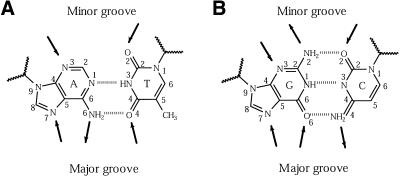
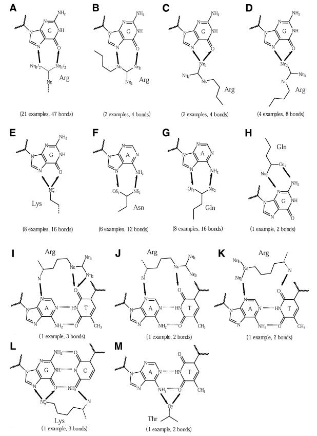
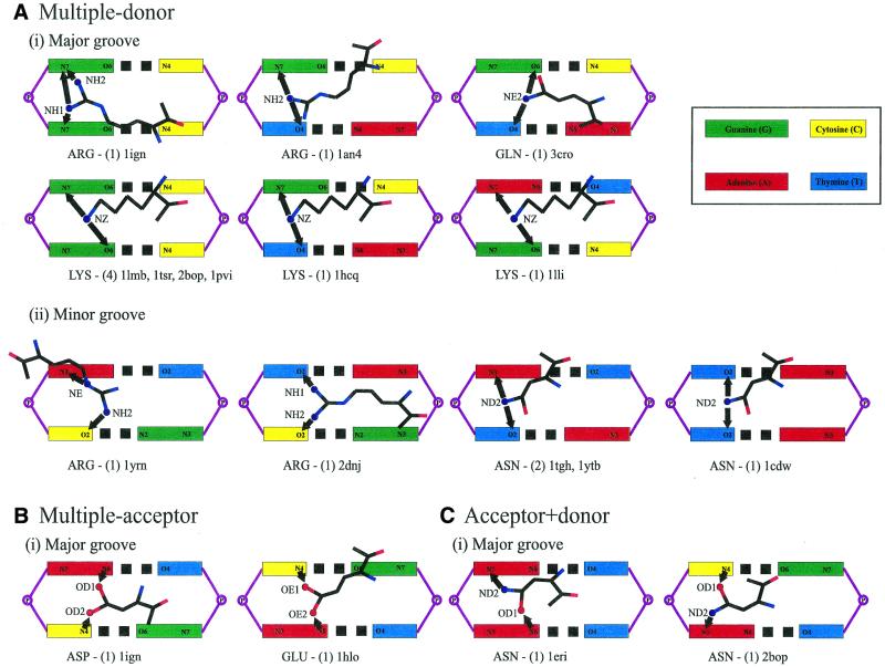
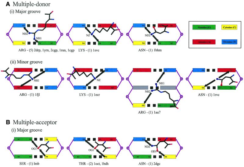
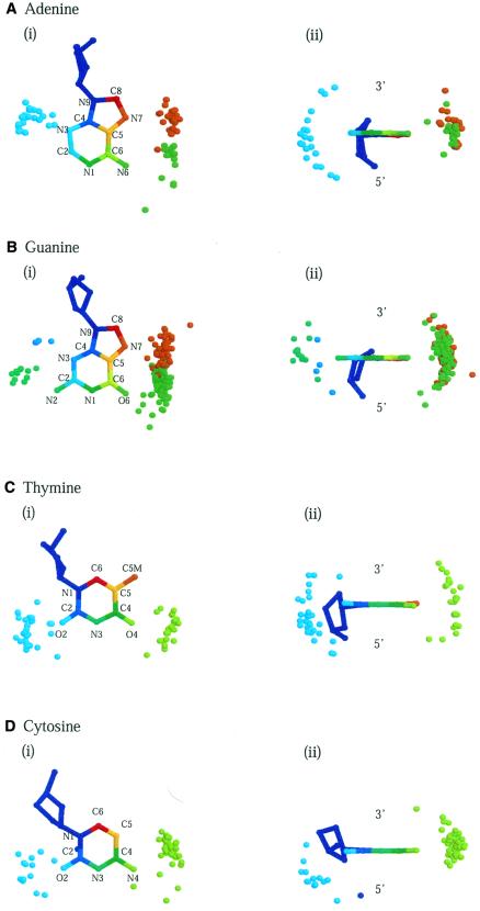
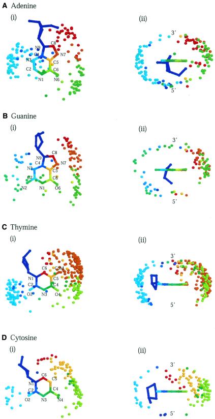
To assess whether there are universal rules that govern amino acid-base recognition, we investigate hydrogen bonds, van der Waals contacts and water-mediated bonds in 129 protein-DNA complex structures. DNA-backbone interactions are the most numerous, providing stability rather than specificity. For base interactions, there are significant base-amino acid type correlations, which can be rationalised by considering the stereochemistry of protein side chains and the base edges exposed in the DNA structure. Nearly two-thirds of the direct read-out of DNA sequences involves complex networks of hydrogen bonds, which enhance specificity. Two-thirds of all protein-DNA interactions comprise van der Waals contacts, compared to about one-sixth each of hydrogen and water-mediated bonds. This highlights the central importance of these contacts for complex formation, which have previously been relegated to a secondary role. Although common, water-mediated bonds are usually non-specific, acting as space-fillers at the protein-DNA interface. In conclusion, the majority of amino acid-base interactions observed follow general principles that apply across all protein-DNA complexes, although there are individual exceptions. Therefore, we distinguish between interactions whose specificities are 'universal' and 'context-dependent'. An interactive Web-based atlas of side chain-base contacts provides access to the collected data, including analyses and visualisation of the three-dimensional geometry of the interactions.


Figures

Figure 1
Figure 1
Schematic diagrams of bases and their pairings in a DNA molecule. Arrows mark the accessible hydrogen-bonding positions, pointing towards acceptors and away from donors. Major groove access to the bases is shown at the bottom of each diagram and minor groove access at the top. (A) Base pairing between adenine (A) and thymine (T), showing the two hydrogen bonds made between them. (B) Base pairing between guanine (G) and cytosine (C), showing the three hydrogen bonds made. The atoms are labelled according to the numbering system in PDB format. The zigzag lines represent the sugar–phosphate groups to which bases are attached.
Figure 2
Figure 2
A comparison of the number of interactions in the filtered datasets of hydrogen bonds, water-mediated bonds and van der Waals contacts (solid lines). The number of interactions in the unfiltered and ‘one representative per family’ datasets for hydrogen bonds are also shown (dotted lines). The effect of varying the resolution cut-off for the structures is depicted.
Figure 3
Figure 3
Schematic diagrams of bidentate interactions. Arrows are drawn between interacting atoms and point from the donor to the acceptor. The number of examples of each type of interaction is given in parentheses. (AD) Arg–G interactions, (E) Lys–G, (F) Asn–A, (G) Gln–A and (H) Gln–G, (IK) Arg–A:T, (L) Lys–G:C and (M) ThrA:T. The amino acid main chains are shown as dotted lines.
Figure 4
Figure 4
Schematic diagrams of complex interactions with stacked bases. The coloured boxes represent the major or minor groove base edges labelled with potential hydrogen-bonding atoms. Amino acid names are given at the bottom of each diagram, along with the number of examples and PDB structures in which they are found. Arrows are drawn between interacting atoms and point from the donor to the acceptor. Complex interactions are by (A) multiple donor amino acids in the (i) DNA major groove and (ii) minor groove, (B) by multiple acceptor and (C) acceptor + donor amino acids.
Figure 5
Figure 5
Schematic diagrams of complex interactions with diagonally-positioned bases. Complex interactions are by (A) multiple donor amino acids in the (i) major groove and (ii) minor groove, and by (B) acceptor + donor amino acids.
Figure 6
Figure 6
Three-dimensional diagrams of the spatial distribution of hydrogen-bonding atoms around bases. The interacting atoms are superposed about a central base. Protein and DNA atoms that interact with each other are identified by the same colour; those in the major groove are coloured red through green and in the minor groove green through blue. Distributions around (A) adenine, (B) guanine, (C) thymine and (D) cytosine are shown in two orientations: (i) facing onto the planes of the bases from the 3′-end and (ii) from the base-pairing edges.
Figure 7
Figure 7
Three-dimensional distributions of van der Waals-contacting atoms around bases. Diagrams are for (A) adenine, (B) guanine, (C) thymine and (D) cytosine from (i) above the planes of the bases (3′-end) and (ii) the base-pairing edges.

References

    1. Seeman N.C., Rosenberg,J.M. and Rich,A. (1976) Sequence-specific recognition of double helical nucleic acids by proteins. Proc. Natl Acad. Sci. USA, 73, 804–808. - PMC - PubMed
    1. Pabo C.O. and Sauer,R.T. (1982) Protein–DNA recognition. Annu. Rev. Biochem., 53, 293–321. - PubMed
    1. Matthews B.W. (1988) No code for recognition. Nature, 335, 294–295. - PubMed
    1. Pabo C.O. and Sauer,R.T. (1992) Transcription factors: structural families and principles of DNA recognition. Annu. Rev. Biochem., 61, 1053–1095. - PubMed
    1. Suzuki M. (1994) A framework for the DNA–protein recognition code of the probe helix in transcription factors: the chemical and stereochemical rules. Structure, 2, 317–326. - PubMed