. 2001 Jul 1;29(13):E62-2.
doi: 10.1093/nar/29.13.e62.

Exciton interaction in molecular beacons: a sensitive sensor for short range modifications of the nucleic acid structure

Affiliations

Exciton interaction in molecular beacons: a sensitive sensor for short range modifications of the nucleic acid structure

S Bernacchi et al. Nucleic Acids Res. .

Abstract

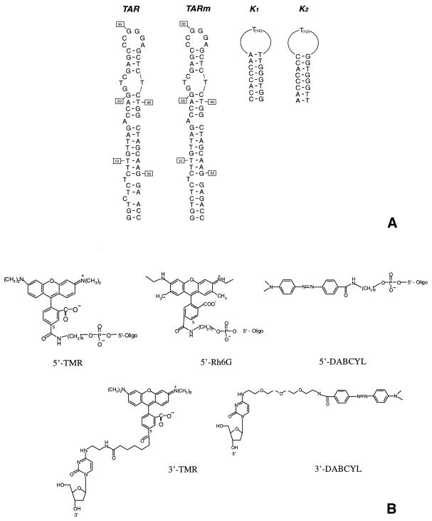
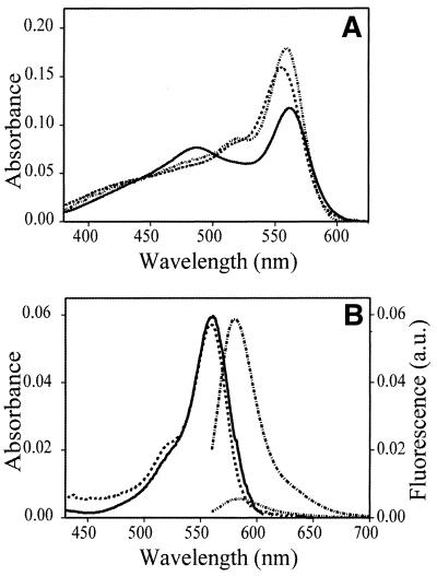
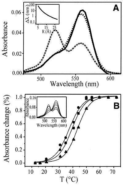
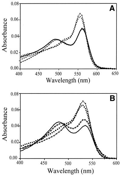
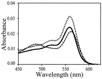
Molecular beacons are hairpin-shaped, single-stranded oligonucleotides constituting sensitive fluorescent DNA probes widely used to report the presence of specific nucleic acids. In its closed form the stem of the hairpin holds the fluorophore covalently attached to one end, close to the quencher, which is covalently attached to the other end. Here we report that in the closed form the fluorophore and the quencher form a ground state intramolecular heterodimer whose spectral properties can be described by exciton theory. Formation of the heterodimers was found to be poorly sensitive to the stem sequence, the respective positions of the dyes and the nature of the nucleic acid (DNA or RNA). The heterodimer allows strong coupling between the transition dipoles of the two chromophores, leading to dramatic changes in the absorption spectrum that are not compatible with a Förster-type fluorescence resonance energy transfer (FRET) mechanism. The excitonic heterodimer and its associated absorption spectrum are extremely sensitive to the orientation of and distance between the dyes. Accordingly, the application of molecular beacons can be extended to monitoring short range modifications of the stem structure. Moreover, the excitonic interaction was also found to operate for doubly end-labeled duplexes.


Figures

Figure 1
Figure 1
Oligonucleotide sequences (A) and chemical structures of the dyes and linkers (B) used in the present study.
Figure 2
Figure 2
Spectral properties of TMR–5′-TARm-3′–DABCYL. (A) Absorption spectra of TMR–5′-TARm-3′–DABCYL at 20 (solid line) and 65°C (dashed line). The absorption spectrum (dash-dot-dot-dashed line) of an equimolecular mixture of TMR–5′-TARm and TARm-3′–DABCYL was recorded at 20°C. (B) Excitation and emission spectrum of TMR–5′-TARm-3′–DABCYL. The excitation spectrum of TMR–5′-TARm-3′–DABCYL (dashed line) was recorded at an emission wavelength of 580 nm. The emission spectrum of TMR–5′-TARm-3′–DABCYL (dash-dot-dot-dashed line) and TMR–5′-TARm (dash-dotted line) were recorded at an excitation wavelength of 550 nm. The solid line corresponds to the absorption spectrum of TMR–5′-TARm.
Figure 3
Figure 3
Spectral properties and melting curves of TMR–5′-TARm-3′–TMR. (A) Absorption spectra of TMR–5′-TARm-3′–TMR at 20 (dash-dotted line) and 65°C (dashed line). The solid line corresponds to the absorption spectrum of an equimolecular mixture of TMR–5′-TARm and TARm-3′–TMR. The insert shows the dependence of the wavelength shift of the blue shifted peak (which results from exciton splitting) on the interchromophore distance. The orientation between the dyes is assumed to be constant. (B) Melting curve of TMR–5′-TARm-3′–TMR. The relative absorbance changes were recorded at 260 (triangle), 521 (square) and 558 nm (circle). The experimental points were fitted to equation 1. The insert shows, from top to bottom (with respect to the 558 nm peak), the absorption spectra recorded at 16, 31, 40.5, 44 and 60°C, respectively.
Figure 4
Figure 4
Dependence of exciton interaction on the oligonucleotide and xanthene dye. (A) Absorption spectra of TMR–5′-K1-3′–DABCYL at 20 (solid line) and 65°C (dashed line) and of TMR–5′-K2-3′–DABCYL at 20 (dash-dotted line) and 65°C (crosses). The melting temperature of both oligonucleotides is 54°C. (B) Absorption spectra of Rh6G–5′-TARm-3′–DABCYL at 20 (solid line) and 65°C (crosses) and of the RNA analog of Rh6G–5′-TARm-3′–DABCYL at 20 (dash-dotted line) and 65°C (dashed line).
Figure 5
Figure 5
Use of exciton interaction to show nucleic acid hybridization. Absorption spectra of an equimolecular mixture of TMR–5′-TARc and TARm-3′–DABCYL at 20°C before (dashed line) and after (dash-dotted line) hybridization. The solid line corresponds to the absorption spectrum of TMR–5′-TARm-3′–DABCYL hybridized by temperature to its complementary unlabeled sequence TARc.

References

    1. Tyagi S. and Kramer,F.R. (1996) Molecular beacons: probes that fluoresce upon hybridization. Nat. Biotechnol., 14, 303–308. - PubMed
    1. Leone G., van Schijndel,H., van Gemen,B., Kramer,F.R. and Schoen,C.D. (1998) Molecular beacon probes combined with amplification by NASBA enable homogeneous, real-time detection of RNA. Nucleic Acids Res., 26, 2150–2155. - PMC - PubMed
    1. Sokol D.L., Zhang,X., Lu,P. and Gewirtz,A.M. (1998) Real time detection of DNA.RNA hybridization in living cells. Proc. Natl Acad. Sci. USA, 95, 11538–11543. - PMC - PubMed
    1. Giesendorf B.A., Vet,J.A., Tyagi,S., Mensink,E.J., Trijbels,F.J. and Blom,H.J. (1998) Molecular beacons: a new approach for semiautomated mutation analysis. Clin. Chem., 44, 482–486. - PubMed
    1. Fang X., Liu,X., Schuster,S. and Tan,W. (1999) Designing a novel molecular beacon for surface-immobilized DNA hybridazation studies. J. Am. Chem. Soc., 121, 2921–2922.

Publication types

MeSH terms