Comparative Study
. 2001 Jul;108(1):73-81.
doi: 10.1172/JCI12466.

c-Jun N-terminal kinase is required for metalloproteinase expression and joint destruction in inflammatory arthritis

Affiliations
Comparative Study

c-Jun N-terminal kinase is required for metalloproteinase expression and joint destruction in inflammatory arthritis

Z Han et al. J Clin Invest. 2001 Jul.

Erratum in

  • J Clin Invest 2001 Dec;108(12):1883

Abstract

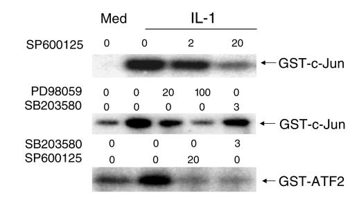
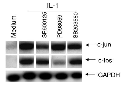
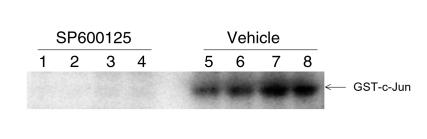
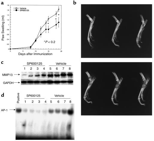
Mitogen-activated protein kinase (MAPK) cascades are involved in inflammation and tissue destruction in rheumatoid arthritis (RA). In particular, c-Jun N-terminal kinase (JNK) is highly activated in RA fibroblast-like synoviocytes and synovium. However, defining the precise function of this kinase has been difficult because a selective JNK inhibitor has not been available. We now report the use of a novel selective JNK inhibitor and JNK knockout mice to determine the function of JNK in synoviocyte biology and inflammatory arthritis. The novel JNK inhibitor SP600125 (anthra[1,9-cd]pyrazol-6(2H)-one) completely blocked IL-1--induced accumulation of phospho-Jun and induction of c-Jun transcription in synoviocytes. Furthermore, AP-1 binding and collagenase mRNA accumulation were completely suppressed by SP600125. In contrast, complete inhibition of p38 had no effect, and ERK inhibition had only a modest effect. The essential role of JNK was confirmed in cultured synoviocytes from JNK1 knockout mice and JNK2 knockout mice, each of which had a partial defect in IL-1--induced AP-1 activation and collagenase-3 expression. Administration of SP600125 modestly decreased the rat paw swelling in rat adjuvant-induced arthritis. More striking was the near-complete inhibition of radiographic damage that was associated with decreased AP-1 activity and collagenase-3 gene expression. Therefore, JNK is a critical MAPK pathway for IL-1--induced collagenase gene expression in synoviocytes and in joint arthritis, indicating that JNK is an important therapeutic target for RA.


Figures

Figure 1
Figure 1
Structure of SP600125, a selective JNK inhibitor.
Figure 2
Figure 2
Effect of SP600125 on c-Jun phosphorylating activity. Cultured FLS were stimulated with medium (Med) or 2 ng/ml of IL-1 for 15 minutes in the presence of increasing concentrations of SP600125 (0–20 μM; JNK inhibitor), PD98059 (0–100 μM; MEK/ERK inhibitor), or SB203580 (0–3 μM; p38 inhibitor). The ability of cell lysates to phosphorylate GST-c-Jun or GST-ATF2 was determined. SP600125 inhibited c-Jun and ATF2 phosphorylation in vitro.
Figure 3
Figure 3
Effect of MAP kinase inhibitors on phospho-c-Jun (P-c-Jun) levels. Cultured FLSs were stimulated with medium or 2 ng/ml of IL-1 for 15 minutes in the presence of SP600125 (20 μM), PD98059 (100 μM), or SB203580 (3 μM). Phospho-c-Jun and total c-Jun protein levels were determined by Western blot analysis. SP600125 inhibited intracellular c-Jun phosphorylation. The phospho-c-Jun/total Jun protein ratios were medium = 0.18, IL-1 = 1.00, IL-1 + SP600125 = 0.23, IL-1 + PD98059 = 0.73, IL-1 + SB203580 = 1.09.
Figure 4
Figure 4
Effect of MAP kinase inhibitors on c-jun and c-fos gene expression. Cultured FLS were stimulated with medium or 2 ng/ml of IL-1 for 15 minutes in the presence of SP600125 (20 μM), PD98059 (100 μM), or SB203580 (3 μM). The figure is representative of four FLS lines examined. The c-jun and c-fos mRNA levels were determined by Northern blot analysis. RNA loading is shown in the bottom row with GAPDH. JNK inhibition decreased c-jun expression, while ERK inhibition decreased c-fos expression.
Figure 5
Figure 5
Effect of MAP kinase inhibitors on AP-1 expression and collagenase gene. Cultured FLS were stimulated with medium or 2 ng/ml of IL-1 for 18 hours (Northern blot analysis) and 1 hour (electrophoretic mobility shift assay [EMSA]) in the presence of medium, SP600125, PD98059, or SB203580. In EMSA experiments (a), SP600125 (20 μM), PD98059 (100 μM), or SB203580 (3 μM) were tested (n = 3; two separate experiments are shown). Note that the JNK inhibitor SP600125 decreased AP-1 binding in nuclear extracts of IL-1–stimulated FLS. The ERK/MEK inhibitor had a modest effect, and the p38 inhibitor did not alter AP-1 binding. (b) Northern blot analysis in which increasing concentrations of the inhibitors were tested for the ability to inhibit collagenase gene expression: SP600125 (0–20 μM; JNK inhibitor), PD98059 (0–100 μM; MEK/ERK inhibitor), or SB203580 (0–3 μM; p38 inhibitor) (n = 3). SP600125 and, to a lesser extent, PD98059 decreased MMP1 gene expression. GAPDH shows equal loading of RNA in each lane. Here, 20 μM of SP600125 inhibited IL-1–induced MMP1 induction (GAPDH-normalized MMP1 gene expression for medium = 0.66; IL-1 = 1.70; IL-1 + SP600125 10 μM = 1.28; IL-1 + SP600125 20 μM = 0.63; IL-1 + SP600125 50 μM = 0.49). However, PD98059 did not decrease MMP1 expression to baseline (GAPDH-normalized MMP1 gene expression for medium = 0.43; IL-1 = 1.40; IL-1 + PD98059 10 μM = 1.15; IL-1 + PD 98059 100 μM = 0.86).
Figure 6
Figure 6
Metalloproteinase gene and AP-1 expression in JNK1 and JNK2 knockout (KO) FLS. Cultured FLS from JNK1 KO FLS lines, JNK2 KO FLS lines, and wild-type FLS lines were stimulated with medium or 2 ng/ml of IL-1 for 18 hours (Northern blot analysis) and 1 hour (EMSA) in the presence of medium or 20 μM SP600125. (a) MMP3 and MMP13 mRNA levels were determined by Northern blot analysis in two separate lines for each strain (line 1 and line 2). JNK1 KO and especially JNK2 KO FLS had lower MMP expression. Residual MMP expression was further suppressed by 20 μM SP600125 (see MMP3 experiment). The ethidium-stained gel shows equal loading of RNA in each lane. (b) AP-1 binding was shown for two JNK2 KO, one wild-type, and one JNK1 KO line. The far left part of the gel shows a positive control and cold competition of the positive control (cold oligo). JNK KO FLS lines have decreased basal and IL-1–stimulated AP-1 activation compared with wild-type cells.
Figure 7
Figure 7
Effect of SP600125 on JNK activity in adjuvant arthritis. Rats with adjuvant arthritis were treated with SP600125 or vehicle beginning on day 8 and sacrificed on day 14. The ability of joint lysates to phosphorylate GST-c-Jun was determined as described in Methods. Vehicle-treated rats had high levels of JNK kinase activity, while no activity was detected in the SP600125-treated rats (n = 4 each).
Figure 8
Figure 8
(a) Effect of SP600125 on adjuvant arthritis in rats. Rats were immunized with complete Freund’s adjuvant on day 0 and treated with vehicle or 30 mg/kg/d of SP600125 subcutaneously beginning on day 8. Significantly less paw swelling was observed in the treated animals. (b) Effect of SP600125 on radiographic damage in adjuvant arthritis. Representative examples of ankle radiographs demonstrate markedly less destruction in the rats treated with SP600125 (top) compared with vehicle (bottom). (c) Effect of SP600125 on synovial collagenase gene expression. Northern blot analysis was performed on joint extracts of vehicle and SP600125-treated rats. Each lane contains the extract of an ankle joint from a control or treated rat (n = 4 for each). Note the lower levels of MMP13 in the SP600125-treated animals (G3PDH-normalized MMP13 mRNA levels for SP600125 = 0.23 ± 0.086 and vehicle = 0.822 ± 0.131; P < 0.01). (d) Effect of SP600125 on synovial AP-1 activation. EMSA analysis was performed on joint extracts of vehicle and SP600125-treated rats with adjuvant arthritis. Positive control is shown on the far left lane of the gel. Note lower levels of AP-1 binding in the SP600125-treated rats (SP600125 = 2.89 ± 0.43 and vehicle = 12.6 ± 2.5, P < 0.01; data presented as arbitrary density units).

Comment in

References

    1. Robinson MJ, Cobb MH. Mitogen-activated protein kinase pathways. Curr Opin Cell Biol. 1997;9:180–185. - PubMed
    1. Seger R, Krebs EG. The MAPK signaling cascade. FASEB J. 1995;9:726–735. - PubMed
    1. Fanger GR, Gerwins P, Widmann C, Jarpe MB, Johnson GL. MEKKs, GCKs, MLKs, PAKs, TAKs, and tpls: upstream regulators of the c-Jun amino-terminal kinases? Curr Opin Genet Dev. 1997;7:67–76. - PubMed
    1. Hibi M, Lin A, Smeal T, Minden A, Karin M. Identification of an oncoprotein- and UV-responsive protein kinase that binds and potentiates the c-Jun activation domain. Genes Dev. 1993;7:2149–2160. - PubMed
    1. Firestein GS. Rheumatoid arthritis. Shaun Ruddy, editor. Scientific American Inc. New York, New York, USA. Scientific American Medicine. 1998;15:2.1–2.14.

Publication types

MeSH terms

Substances