. 2001 Jul 2;194(1):13-27.
doi: 10.1084/jem.194.1.13.

Anuria, omphalocele, and perinatal lethality in mice lacking the CD34-related protein podocalyxin

Affiliations

Anuria, omphalocele, and perinatal lethality in mice lacking the CD34-related protein podocalyxin

R Doyonnas et al. J Exp Med. .

Abstract

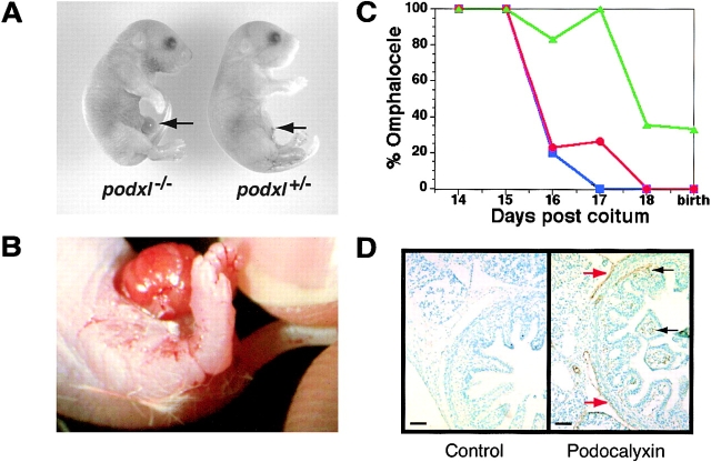
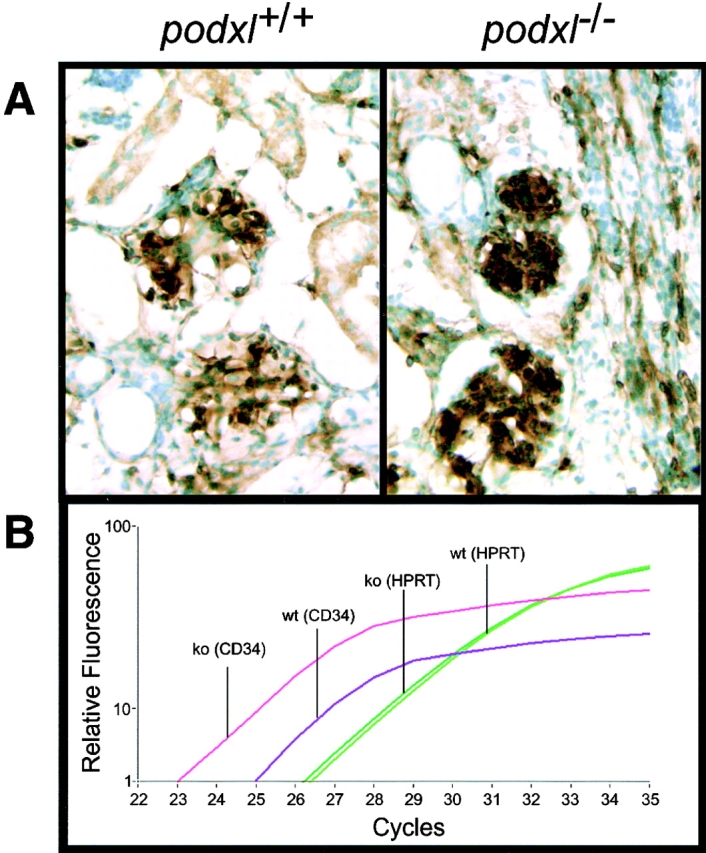
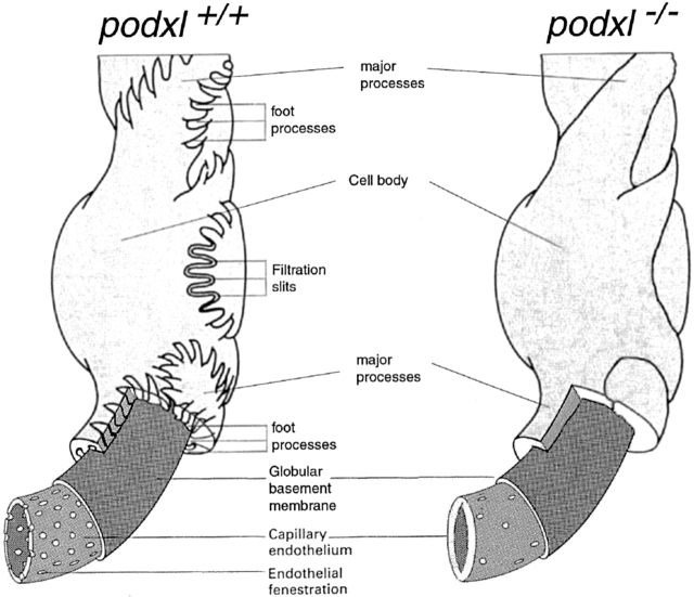
Podocalyxin is a CD34-related sialomucin that is expressed at high levels by podocytes, and also by mesothelial cells, vascular endothelia, platelets, and hematopoietic stem cells. To elucidate the function of podocalyxin, we generated podocalyxin-deficient (podxl(-/)-) mice by homologous recombination. Null mice exhibit profound defects in kidney development and die within 24 hours of birth with anuric renal failure. Although podocytes are present in the glomeruli of the podxl(-/)- mice, they fail to form foot processes and slit diaphragms and instead exhibit cell--cell junctional complexes (tight and adherens junctions). The corresponding reduction in permeable, glomerular filtration surface area presumably leads to the observed block in urine production. In addition, podxl(-/)- mice frequently display herniation of the gut (omphalocele), suggesting that podocalyxin may be required for retraction of the gut from the umbilical cord during development. Hematopoietic and vascular endothelial cells develop normally in the podocalyxin-deficient mice, possibly through functional compensation by other sialomucins (such as CD34). Our results provide the first example of an essential role for a sialomucin in development and suggest that defects in podocalyxin could play a role in podocyte dysfunction in renal failure and omphalocele in humans.


Figures

Figure 1
Figure 1
Protein structure and genomic organization of podocalyxin and the CD34 subfamily of sialomucins. (A) Schematic representation of the structure of murine CD34, podocalyxin, and endoglycan based on predicted protein sequences. White boxes, mucin domains; black boxes, cysteine-rich domains; circles, potential NH2-linked carbohydrates; horizontal bars with or without arrows, potential O-linked carbohydrates; arrows, potential sialic acid motifs on O-linked carbohydrates; protein kinase C, TK, and CK2, potential phosphorylation sites; DTHL or DTEL, potential PDZ domain docking sites. (B) Schematic showing the genomic organization of human cd34, podxl, and endgl genes based on sequence contigs identified in the human sequence database (see Materials and Methods). The location of the coding sequences for the signal peptide (S, purple), mucin domain (S-T-P, blue), cysteine-rich domain (C-C, green), transmembrane (TM, orange), and cytoplasmic tail (Cyt tail, red) are indicated. Numbers indicate exon size in base pairs. The human cd34 gene spans ∼25 kbp with a first intron of 11 kbp. The human podxl gene was found to span ∼60 kbp with a first intron of 45 kbp. The human endgl gene spans >23 kbp with a first intron of 13 kbp. The 3′ introns of all three genes are relatively small; the last four exons in each case are separated by <3 kbp.
Figure 2
Figure 2
Generation of podocalyxin-null mutation in mice. (A) Scheme showing structure of mouse podocalyxin genomic locus, targeting construct, and the predicted homologous recombination event. The targeted disruption results in the deletion of the majority of exons 5, 6, 7, and 8 encoding the 55 amino acids of the juxtamembrane (“stalk”) domain, the transmembrane region (TM), and the cytoplasmic tail (Cyt). These were replaced with the neomycin resistance cassette (NeoR) in the antisense orientation. Southern blot probe, restriction sites, and predicted sizes of targeted and wild-type alleles (HindIII digest) are indicated. (B) Southern blot and PCR analysis of podocalyxin gene in targeted ES cell clones and chimeric mice. ES lane shows a Southern blot of a mutant ES cell clone used to generate podxl −/− mice. The indicated 2.2-kbp ApaI fragment was used as probe. 4-kbp wild-type (WT) and 5-kb mutant (KO) alleles are indicated. Genomic DNA from wild-type (+/+), heterozygous (+/−), and homozygous KO mice (−/−) were analyzed by PCR using allele-specific primers (see Materials and Methods). Molecular weight markers (MW) are indicated in kbp. (C) Northern blot analysis of 16-d embryo lung RNA from wild-type (WT), podxl +/− (HT), and podxl −/− (KO) mice. 10 μg of total RNA per lane was hybridized with probes specific to the mucin domain of podocalyxin or glyceraldehyde-3-phosphate dehydrogenase as a control. (D) Analysis of podocalyxin expression by 15-d fetal liver cells. Single cell suspensions from wild-type (WT) and podxl −/− (KO) mice were stained with anti–PCLP-1 antibody followed by FITC-conjugated goat anti–rat antibodies before flow cytometry analysis. Fetal liver cells were gated on forward and side scatter to focus on the MEP21 expressing subpopulation of fetal liver.
Figure 3
Figure 3
Edema. (A) Light micrograph of day 15 podxl +/+ (+/+) embryo and one of a podxl −/− (−/−) embryo with edema. (B) Close-up color micrograph of day 15 podxl −/− embryo with edema. Arrows indicate the dorsal region of embryonic trunk where subdermal swelling occurs in −/− embryos.
Figure 5
Figure 5
Urine production and cardiac output. (A) Urine collected from the bladders of 18-d-old embryos after Caesarian section. Representative data from one of two similar experiments. White bars, wild-type embryos; black boxes, podxl +/− embryos; gray boxes, podxl −/− embryos. (B) 60-s cardiac output from 18-d-old embryos as determined by RBCs released from umbilical cord. White bars, wild-type embryos; black boxes, podxl +/− embryos; gray boxes, podxl −/−embryos.
Figure 4
Figure 4
Omphalocele. (A) Light micrograph of day 18 podxl +/+ and podxl −/− embryos. Arrow indicates umbilical cord and omphalocele. (B) Close-up color micrograph of omphalocele in podxl −/− newborn. (C) Ontogeny of omphalocele in wt, podxl +/−, and podxl −/− mice. Blue, red, and green symbols indicate wt, podxl +/−, and podxl −/− mice, respectively. Each data point represents the percentage of between 5 and 35 animals analyzed. (D) Analysis of podocalyxin expression in physiologic omphalocele of wild-type mice. Sections from 16-d embryo gut were immunostained for podocalyxin (brown stain) and counterstained with methyl green. Podocalyxin is expressed by mesothelial cells lining the outer aspect of the gut (red arrows) and by capillary ECs in the villi (black arrows).
Figure 6
Figure 6
Histological analysis of podxl −/− kidneys. (A) Immunohistological analysis of expression of the podocyte-specific tyrosine phosphatase, GLEPP1 in podxl +/+, podxl +/−, and podxl −/− kidneys. The capillary loops of the podxl +/+ and podxl +/− glomeruli are shown in black arrows. podxl −/− glomeruli had similar capillary loops but also had numerous lucent vacuoles surrounded by GLEPP1 staining (red arrows). Scale bars, 50 μm. (B) Dual-label indirect immunofluorescence of newborn mouse kidney from podxl +/+ and podxl −/− (top and bottom, respectively) with antibodies to GLEPP1 (donkey anti-chicken FITC labeled secondary antibody), and collagen α4 (type IV) (goat anti–rabbit Cy3-labeled secondary antibody). Staining for the apical membrane podocyte marker GLEPP1 and the basement membrane protein collagen α4 (type IV) is seen in mature glomeruli in podxl +/+ and podxl −/− mice. Superimposition of these images shows areas of strong overlap (orange) in the podxl +/+ mice. In the podxl −/− mice the area of overlap (orange) is diminished representing a greater distance in the localization of the apical membrane marker (GLEPP1) from the basement membrane. (C) Dual-label indirect immunofluorescence of newborn mouse kidney from podxl +/+ and podxl −/− (top and bottom, respectively) with antibodies to the basement membrane protein laminin β2 (FITC-conjugated goat anti–rat) and podocyte slit diaphragm protein nephrin (anti–rabbit Cy3-labeled secondary antibody). Strong staining is seen for both markers in podxl +/+ and podxl −/− mice. The close proximity of the slit diaphragm (nephrin staining) to the basement membrane (laminin β2 staining) can be appreciated in the superimposed images by the presence of overlapping staining (orange) in the podxl +/+ mice. In the podxl −/− glomeruli the superimposed images show diminished overlap (orange staining) of the expression of nephrin and laminin β2.
Figure 7
Figure 7
TEMs of embryonic day 18 kidneys from podxl +/+ and podxl −/− embryos. (A) podxl +/+ kidneys have normal podocytes (Pod.) with typical MPs and FPs that embrace the outer aspect of the capillary basement membrane. RBCs can be seen in the lumen of the endothelial vessels. podxl −/− show a complete loss of SDs and FPs. JCs are present between all of the podxl −/− podocytes. Podocytes in podxl −/− mice also display numerous vacuoles (Vac.) and overall the glomerular capillaries had a thicker EC layer (EC). Scale bars, 2 μm. (B) Higher magnification reveals that the SDs and FPs indicated by arrows in the podxl +/+ and podxl +/− mice are completely lost in podxl −/− glomeruli. JCs including TJs and AJs are present between podxl −/− podocytes. The endothelial fenestrae (F) within the ECs can be found in wild-type controls but the fenestrae reduced in the podxl −/− glomerular capillaries. Scale bars, 0.2 μm.
Figure 9
Figure 9
CD34 mRNA overexpression in kidney of podxl −/− 18-d embryos. (A) CD34 immunostaining of 18-d embryo kidneys. Brown stain represents CD34 antigen detected by immunoperoxidase stain; green stain represents methyl green counterstain of nuclei. (B) Relative quantification of CD34 and HPRT mRNA in 18-d embryo kidney by Real-Time reverse transcription PCR (LightCycler™; Roche). Y-axis, relative level of cDNA product as determined by SYBR Green™ fluorescence; X-axis, number of PCR cycles; Pink lines, podxl −/− mRNA with CD34 primers; purple lines, podxl +/+ mRNA with CD34 primers; light green lines, podxl −/− mRNA with HPRT primers; dark green lines, podxl +/+ mRNA with HPRT primers. One of four representative experiments with kidney mRNA from two independent mice of each genotype.
Figure 8
Figure 8
Schematic representation of the glomerular filter in wild-type and podocalyxin-deficient mice (adapted from reference 14). For ease of presentation, the mesangial cells that would normally link the capillary loops (disrupting the layer of podocytes) have been left out of the diagram. The lack of interdigitating FPs in the podxl −/− mice leads to a lack of filtration slit area. This, along with the decrease in the fenestration of the glomerular capillaries, is thought to lead to a decrease in the potential area for filtration and the anuria that occurs in the podxl −/− mice.

References

    1. Kerjaschki D., Sharkey D.J., Farquhar M.G. Identification and characterization of podocalyxin-the major sialoprotein of the renal glomerular epithelial cell. J. Cell Biol. 1984;98:1591–1596. - PMC - PubMed
    1. Dekan G., Gabel C., Farquhar M.G. Sulfate contributes to the negative charge of podocalyxin, the major sialoglycoprotein of the glomerular filtration slits. Proc. Natl. Acad. Sci. USA. 1991;88:5398–5402. - PMC - PubMed
    1. Kershaw D.B., Thomas P.E., Wharram B.L., Goyal M., Wiggins J.E., Whiteside C.I., Wiggins R.C. Molecular cloning, expression and characterization of podocalyxin-like protein 1 from rabbit as a transmembrane protein of glomerular podocytes and vascular endothelium. J. Biol. Chem. 1995;270:29439–29446. - PubMed
    1. Horvat R., Hovorka A., Dekan G., Poczewski H., Kerjaschki D. Endothelial cell membranes contain podocalyxin-the major sialoprotein of visceral glomerular epithelial cells. J. Cell Biol. 1986;102:484–491. - PMC - PubMed
    1. McNagny K.M., Pettersson I., Rossi F., Flamme I., Shevchenko A., Mann M., Graf T. Thrombomucin, a novel cell surface protein that defines thrombocytes and multipotent hematopoietic progenitors. J. Cell Biol. 1997;138:1395–1407. - PMC - PubMed

Publication types

MeSH terms

LinkOut - more resources