. 2001 Jul 2;194(1):71-8.
doi: 10.1084/jem.194.1.71.

Brain mast cells act as an immune gate to the hypothalamic-pituitary-adrenal axis in dogs

Affiliations

Brain mast cells act as an immune gate to the hypothalamic-pituitary-adrenal axis in dogs

I Matsumoto et al. J Exp Med. .

Abstract

Mast cells perform a significant role in the host defense against parasitic and some bacterial infections. Here we show that in the dog, degranulation of brain mast cells evokes hypothalamic-pituitary-adrenal responses via histamine release. A large number of mast cells were found in a circumscribed ventral region of the hypothalamus, including the pars tuberalis and median eminence. When these intracranial mast cells were passively sensitized with immunoglobulin E via either the intracerebroventricular or intravenous route, there was a marked increase in the adrenal cortisol secretion elicited by a subsequent antigenic challenge (whether this was delivered via the central or peripheral route). Comp.48/80, a mast cell secretagogue, also increased cortisol secretion when administered intracerebroventricularly. Pretreatment (intracerebroventricularly) with anti-corticotropin--releasing factor antibodies or a histamine H(1) blocker, but not an H(2) blocker, attenuated the evoked increases in cortisol. These data show that in the dog, degranulation of brain mast cells evokes hypothalamic-pituitary-adrenal responses via centrally released histamine and corticotrophin-releasing factor. On the basis of these data, we suggest that intracranial mast cells may act as an allergen sensor, and that the activated adrenocortical response may represent a life-saving host defense reaction to a type I allergy.


Figures

Figure 1
Figure 1
Mast cells in the dog brain. (A) Sagittal distribution of mast cells in the ventral hypothalamic region in serial coronal sections from 17 to 23 mm rostral to EAML (R0) in seven dogs. Inset: drawing of midline sagittal section of the brain. Shaded area was surveyed for mast cells. (B) Representative photomicrograph of a coronal section at 19.5 mm rostral to EAML in dog no. 0120. The section includes the bottom of the third ventricle (III), the PT, the ME, and the AH, and it was stained with toluidine blue. Areas indicated by rectangles are depicted at high magnification in other photomicrographs, as described below. (C) Photomicrograph of rectangular area C (see B). (D) Photomicrograph of rectangular area D (see B). (E) Photomicrograph of area E (see C). Vertical bars, 100 μm. (F) Photomicrograph of rectangular area F (see E). Arrows show numerous metachromatic red-purple granules stained with toluidine blue in each mast cell. Original magnification: ×1,000.
Figure 1
Figure 1
Mast cells in the dog brain. (A) Sagittal distribution of mast cells in the ventral hypothalamic region in serial coronal sections from 17 to 23 mm rostral to EAML (R0) in seven dogs. Inset: drawing of midline sagittal section of the brain. Shaded area was surveyed for mast cells. (B) Representative photomicrograph of a coronal section at 19.5 mm rostral to EAML in dog no. 0120. The section includes the bottom of the third ventricle (III), the PT, the ME, and the AH, and it was stained with toluidine blue. Areas indicated by rectangles are depicted at high magnification in other photomicrographs, as described below. (C) Photomicrograph of rectangular area C (see B). (D) Photomicrograph of rectangular area D (see B). (E) Photomicrograph of area E (see C). Vertical bars, 100 μm. (F) Photomicrograph of rectangular area F (see E). Arrows show numerous metachromatic red-purple granules stained with toluidine blue in each mast cell. Original magnification: ×1,000.
Figure 2
Figure 2
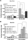
Effect of an antigenic challenge (C) delivered via the icv (A) or iv (B) route on adrenal cortisol secretion rate in dogs sensitized (S) with IgE via the icv route. (A) In the Sicv/Cicv experiment, cortisol secretion increased dramatically after a 1.6 μg/kg OVA challenge in dogs sensitized with IgE (but not in those given denatured IgE). The increase was attenuated by pretreatment with anti-CRF antiserum or an H1 blocker, but not by pretreatment with an H2 blocker. (B) In the Sicv/Civ experiment, cortisol secretion increased markedly in response to a 4.8 μg/kg OVA challenge via the iv route, but this did not occur with a 1.6 μg/kg OVA challenge via the same route. An H1 blocker significantly attenuated the increase, but anti-CRF antiserum did not. Numbers in parenthesis indicate number of animals. All data are means ± SE. **P < 0.01 versus animals sensitized with heat-inactivated IgE. P < 0.05, †† P < 0.01 versus animals without pretreatment.
Figure 4
Figure 4
Effect of Comp.48/80 on adrenal cortisol secretion rate in dogs. Comp.48/80 (Sigma-Aldrich) was given into VIII at a rate of 7.5 μg/kg/min for 5 min with or without antagonists. In response to Comp.48/80, cortisol secretion increased to four times the basal level. Pretreatment with anti-CRF antiserum or an H1 blocker significantly attenuated the increase (but an H2 blocker did not). Numbers in parenthesis indicate number of animals. All data are means ± SE. **P < 0.01 versus vehicle control. †† P < 0.01 versus animals without pretreatment.
Figure 3
Figure 3
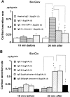
Effect of an antigen challenge (C) delivered via the icv (A) or iv (B) route on adrenal cortisol secretion rate in dogs sensitized (S) with IgE via the iv route. (A) In the Siv/Cicv experiment, a 1.6 μg/kg OVA challenge markedly increased cortisol secretion in dogs sensitized with IgE (but not in those given denatured IgE). The increase was attenuated by pretreatment with anti-CRF antiserum or an H1 blocker. (B) In the Siv/Civ experiment, an 8.0 μg/kg OVA challenge increased cortisol secretion significantly, but a 1.6 or 4.8 μg/kg OVA challenge did not. The increase after the 8.0 μg/kg OVA challenge was markedly attenuated by pretreatment with 200 μg/kg of an H1 blocker (but not by 2 μg/kg). Numbers in parenthesis indicate number of animals. All data are means ± SE. **P < 0.01 versus animals sensitized with heat-inactivated IgE. P < 0.05, †† P < 0.01 versus animals without pretreatment.

Comment in

  • The diverse roles of mast cells.
    Gurish MF, Austen KF. Gurish MF, et al. J Exp Med. 2001 Jul 2;194(1):F1-5. doi: 10.1084/jem.194.1.f1. J Exp Med. 2001. PMID: 11435478 Free PMC article. Review. No abstract available.

References

    1. Echtenacher D.N., Månnel D.N., Hültner L. Critical protective role of mast cells in a model of acute septic peritonitis. Nature. 1996;381:75–77. - PubMed
    1. Malaviya R., Ikeda T., Ross E., Abraham S.N. Mast cell modulation of neutrophil influx and bacterial clearance at sites of infection through TNF-α. Nature. 1996;381:77–80. - PubMed
    1. Chung K.F., Becher A.B., Lazarus S.C., Frick O.L., Dadel J.A., Gold W.M. Antigen-induced airway hyperresponsiveness and pulmonary inflammation in allergic dogs. J. Appl. Physiol. 1985;58:1347–1353. - PubMed
    1. Wagner E.M., Mitzner W.A., Bleecker E.R. Peripheral circulatory alterations in canine anaphylactic shock. Am. J. Physiol. 1986;251:H934–H940. - PubMed
    1. Edvinson L., Cervos-Narsson L.-I., Owman C.H., Ronnberg A.-L. Regional distribution of mast cells containing histamine, dopamine, or 5-hydroxytryptamine in the mammalian brain. Neurology. 1977;27:878–883. - PubMed

MeSH terms