. 2001 Aug;75(15):6758-68.
doi: 10.1128/JVI.75.15.6758-6768.2001.

African swine fever virus structural protein pE120R is essential for virus transport from assembly sites to plasma membrane but not for infectivity

Affiliations

African swine fever virus structural protein pE120R is essential for virus transport from assembly sites to plasma membrane but not for infectivity

G Andrés et al. J Virol. 2001 Aug.

Abstract

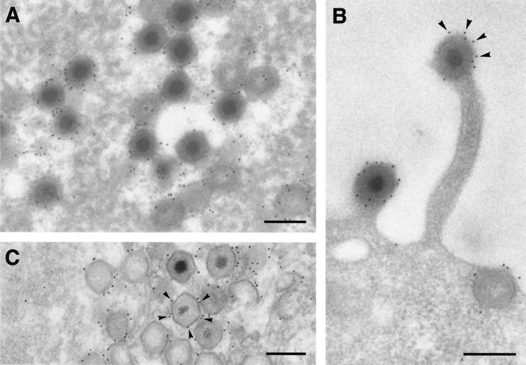
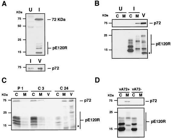
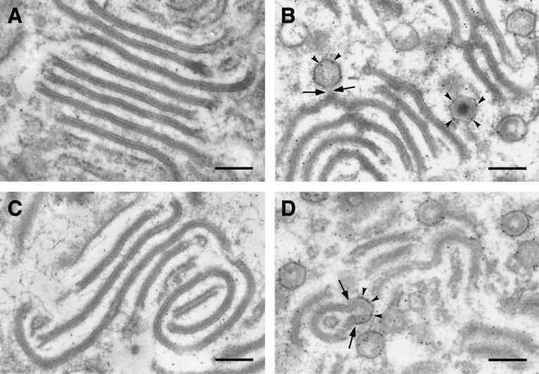
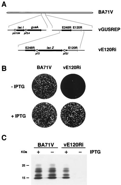
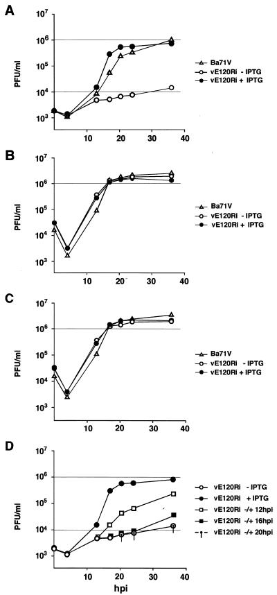
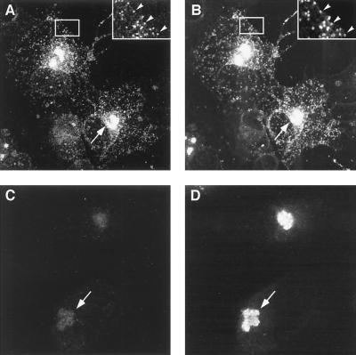
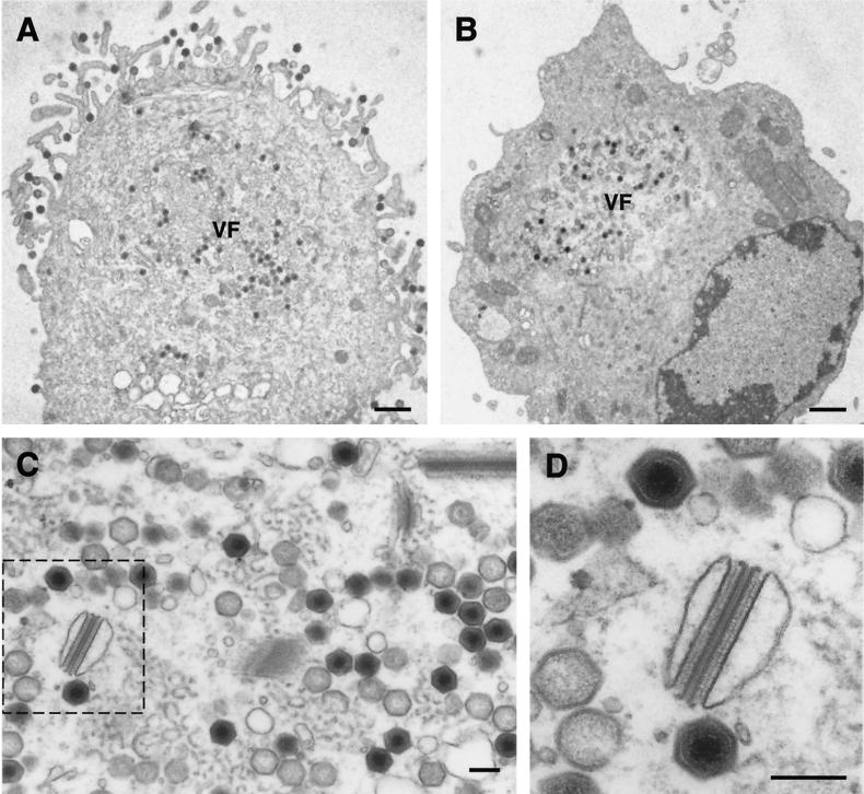
This report examines the role of African swine fever virus (ASFV) structural protein pE120R in virus replication. Immunoelectron microscopy revealed that protein pE120R localizes at the surface of the intracellular virions. Consistent with this, coimmunoprecipitation assays showed that protein pE120R binds to the major capsid protein p72. Moreover, it was found that, in cells infected with an ASFV recombinant that inducibly expresses protein p72, the incorporation of pE120R into the virus particle is dependent on p72 expression. Protein pE120R was also studied using an ASFV recombinant in which E120R gene expression is regulated by the Escherichia coli lac repressor-operator system. In the absence of inducer, pE120R expression was reduced about 100-fold compared to that obtained with the parental virus or the recombinant virus grown under permissive conditions. One-step virus growth curves showed that, under conditions that repress pE120R expression, the titer of intracellular progeny was similar to the total virus yield obtained under permissive conditions, whereas the extracellular virus yield was about 100-fold lower than in control infections. Immunofluorescence and electron microscopy demonstrated that, under restrictive conditions, intracellular mature virions are properly assembled but remain confined to the replication areas. Altogether, these results indicate that pE120R is necessary for virus dissemination but not for virus infectivity. The data also suggest that protein pE120R might be involved in the microtubule-mediated transport of ASFV particles from the viral factories to the plasma membrane.


Figures

FIG. 1
FIG. 1
Immunoelectron microscopy of protein pE120R in BA71V-infected cells. (A and B) Ultrathin Lowicryl K4M sections of infected Vero cells fixed at 24 hpi and processed by freeze substitution were incubated with anti-pE120R antibodies followed by protein A-gold (10 nm). (A) Within the virus factory, the labeling was mainly associated with mature particles and icosahedral immature virions lacking an electrondense nucleoid. In contrast, the precursor membranous structures surrounding the virus particles were weakly labeled. (B) A strong and peripheral labeling was also detected on ASFV particles budding through the plasma membrane. (C) Infected Vero cells were permeabilized with streptolysin O at 24 hpi and labeled with anti-pE120R antibody and protein A-gold (10 nm) before conventional Epon embedding. Gold particles (arrowheads) decorated the surface of ASFV particles within the virus factories. Bars, 200 nm.
FIG. 2
FIG. 2
Protein pE120R interacts with the major capsid protein p72. (A) Uninfected (U) Vero cells or cells infected with BA71V virus (I) were pulse-labeled with [35S]methionine-[35S]cysteine from 16 to 18 hpi. The cell extracts were immunoprecipitated with anti-pE120R serum and analyzed by SDS-polyacrylamide gel electrophoresis and autoradiography (upper panel). The immunoprecipitated material from infected cell extracts was further analyzed, together with highly purified extracellular ASFV (V), by Western immunoblotting with anti-p72 MAb 17L.D3 (lower panel). (B) Western immunoblotting with MAb anti-p72 (upper panel) and anti-pE120R (lower panel) antibodies of cytosolic (C) and membrane-particulate (M) fractions from uninfected cells (U) or cells infected with parental BA71V (I). Analysis of highly purified extracellular virions (V) is also shown. The asterisk indicates the position of the major structural pE120R form of 12 kDa. (C) Infected Vero cells were pulse-labeled with [35S]methionine-[35S]cysteine from 11 to 12 hpi (P1), chased for 3 h (C3) and 24 h (C24), and then fractionated into soluble cytosolic (C), membrane-particulate (M), and extracellular virus (V) fractions. Equivalent amounts of the fractions were immunoprecipitated with anti-pE120R antibodies and analyzed by SDS-polyacrylamide gel electrophoresis. The asterisk indicates the position of the major structural pE120R form of 12 kDa. (D) Immunoblotting with anti-p72 (upper panel) and anti-pE120R (lower panel) antibodies of cytosolic (C) and membrane-particulate (M) fractions from extracts of vA72-infected cells grown in the presence (vA72+) or in the absence (vA72−) of IPTG. The migration position of molecular mass markers is indicated on the left. The bands corresponding to protein p72 and the different forms of protein pE120R are indicated on the right.
FIG. 3
FIG. 3
Immunoelectron microscopy of protein pE120R on vA72-infected cells. Lowicryl sections of vA72-infected cells maintained 16 h in the absence of IPTG (A and C) or treated with the inducer at 16 hpi during an 8-h period (B and D) were incubated with anti-pE120R antibody (A and B) or anti-p72 antibody (C and D), followed by protein A-gold (10 nm). In the absence of IPTG the aberrant zipper-like structures were poorly labeled by both sera while, in the presence of the inducer, anti-pE120R and anti-p72 antibodies strongly labeled (arrowheads) icosahedral particles as well as polyhedral forms derived from previously assembled zipper-like structures. The arrows indicate icosahedral forms emerging from zipper-like structures. Bars, 200 nm.
FIG. 4
FIG. 4
(A) Genomic structure of the recombinant ASFV virus vE120Ri. Recombinant virus vE120Ri was obtained from recombinant vGUSREP, which contains the lacI gene encoding the lac repressor inserted into the nonessential tk locus. In vE120Ri virus, the gene E120R is under the transcriptional control of an inducible promoter p72.1, which is composed by the strong late promoter p72.4 and the lac operator sequence (●). The reporter genes lacZ and gusA, used for selection and purification of the recombinants, are also represented. (B) Plaque phenotype of vE120Ri. Monolayers of Vero cells were infected in the absence or presence of 1 mM IPTG with parental BA71V or recombinant vE120Ri virus. Plaques were visualized with 1% crystal violet 5 days after infection. (C) Inducible expression of protein pE120R. Vero cells were infected with BA71V or recombinant vE120Ri in the presence (+) or absence (−) of 1 mM IPTG. At 24 hpi, samples were electrophoresed and analyzed by Western immunoblotting with a serum anti-pE120R. The electrophoretic mobility of molecular weight markers is indicated on the left.
FIG. 5
FIG. 5
One-step growth curves of vE120Ri. Vero cells were infected with 10 PFU of BA71V or vE120Ri virus per cell in the presence or absence of 1 mM IPTG. Virus from the culture supernatants (A) and the infected cells (B) were collected at the indicated times of infection and titrated separately by plaque assay on fresh Vero cells in the presence of the inducer. (C) Curves of total virus yield were deduced from the intracellular and extracellular virus yields shown in panels A and B. (D) In the same experiment, recombinant virus vE120Ri was grown under nonpermissive conditions for the indicated times and then induced with IPTG. At different times postinfection, extracellular virus from the culture supernatant was titrated as described above in the presence of inducer. As a control, the extracellular virus yields of recombinant vE120Ri grown in the absence or presence of IPTG throughout the infection are shown.
FIG. 6
FIG. 6
Immunofluorescence microscopy of vE120Ri-infected cells. Vero cells infected with recombinant vE120Ri virus in the presence (A and B) or absence (C and D) of IPTG were fixed at 18 hpi and double-labeled with rabbit serum anti-pE120R (A and C) and mouse MAb 17L.D3 anti-p72 (B and D). Labeling was revealed with Alexa 488 goat anti-rabbit rabbit IgG and with Alexa 594 goat anti-mouse IgG. Insets in panels A and B show enlarged images of the delimited cytoplasmic areas. Viral factories and virions spread throughout the cytoplasm are indicated by arrows and arrowheads, respectively.
FIG. 7
FIG. 7
Electron microscopy of vE120Ri-infected cells. Ultrathin Epon sections of vE120Ri-infected Vero cells incubated for 18 h in the presence (A) or in the absence (B, C, and D) of IPTG. While in the presence of inducer (A) ASFV particles move from the virus factories (VF) to the plasma membrane to be released by budding, in the absence of IPTG (B) virus particles are assembled within the virus factories but neither transport nor budding occur. Note also that, under restrictive conditions, the factories (panel C) contain all normal virus assembly stages, including large amounts of intracellular mature particles. Additionally, some aberrant virus structures can be detected. Panel D shows a higher magnification of the region delimited by the dashed line in panel C, which contains an aberrant structure and two apparently normal intracellular mature particles. Bars: A and B, 1 μm; C and D, 200 nm.

References

    1. Alcamí A, Angulo A, López-Otín C, Muñoz M, Freije J M P, Carrascosa A L, Viñuela E. Amino acid sequence and structural properties of protein p12, an African swine fever virus attachment protein. J Virol. 1992;66:3860–3868. - PMC - PubMed
    1. Almazán F, Rodríguez J M, Andrés G, Pérez R, Viñuela E, Rodríguez J F. Transcriptional analysis of multigene family 110 of African swine fever virus. J Virol. 1992;66:6649–6654. - PMC - PubMed
    1. Alves de Matos A P, Carvalho Z G. African swine fever virus interaction with microtubules. Biol Cell. 1993;78:229–234. - PubMed
    1. Andrés G, Simón-Mateo C, Viñuela E. Assembly of African swine fever virus: role of polyprotein pp220. J Virol. 1997;71:2331–2341. - PMC - PubMed
    1. Andrés G, García-Escudero R, Simón-Mateo C, Viñuela E. African swine fever virus is enveloped by a two-membraned collapsed cisterna derived from the endoplasmic reticulum. J Virol. 1998;72:8988–9001. - PMC - PubMed

Publication types

Substances