. 2001 Aug;75(15):6850-6.
doi: 10.1128/JVI.75.15.6850-6856.2001.

Second-site suppressors of Rous sarcoma virus Ca mutations: evidence for interdomain interactions

Affiliations

Second-site suppressors of Rous sarcoma virus Ca mutations: evidence for interdomain interactions

J B Bowzard et al. J Virol. 2001 Aug.

Abstract

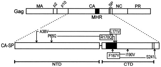
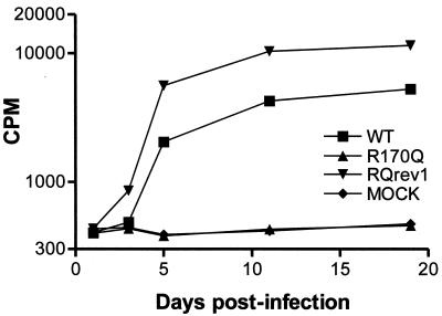
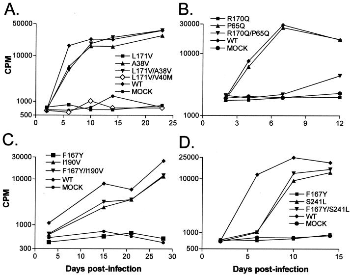
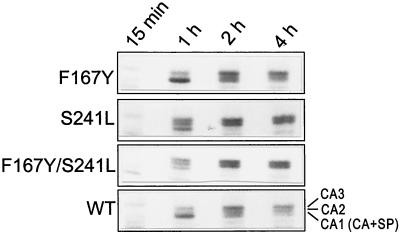
The capsid (CA) protein, the major structural component of retroviruses, forms a shell that encases the ribonucleoprotein complex in the virion core. The most conserved region of CA, approximately 20 amino acids of the major homology region (MHR), lies within the carboxy-terminal domain of the protein. Structural and sequence similarities among CA proteins of retroviruses and the CA-like proteins of hepatitis B virus and various retrotransposons suggest that the MHR is involved in an aspect of replication common to these reverse-transcribing elements. Conservative substitutions in this region of the Rous sarcoma virus protein were lethal due to a severe deficiency in reverse transcription, in spite of the presence of an intact genome and active reverse transcriptase in the particles. This finding suggests that the mutations interfered with normal interactions among these constituents. A total of four genetic suppressors of three lethal MHR mutations have now been identified. All four map to the sequence encoding the CA-spacer peptide (SP) region of Gag. The F167Y mutation in the MHR was fully suppressed by a single amino acid change in the alpha helix immediately downstream of the MHR, a region that forms the major dimer interface in human immunodeficiency virus CA. This finding suggests that the F167Y mutation indirectly interfered with dimerization. The F167Y defect could also be repaired by a second, independent suppressor in the C-terminal SP that was removed from CA during maturation. This single residue change, which increased the rate of SP cleavage, apparently corrected the F167Y defect by modifying the maturation pathway. More surprising was the isolation of suppressors of the R170Q and L171V MHR mutations, which mapped to the N-terminal domain of the CA protein. This finding suggests that the two domains, which in the monomeric protein are separated by a flexible linker, must communicate with each other at some unidentified point in the viral replication cycle.


Figures

FIG. 1
FIG. 1
Positions of MHR and suppressor mutations in Gag. The wild-type RSV Gag protein is illustrated with the locations of the major cleavage products indicated along the top. Expanded below Gag is the CA-SP protein with the two domains of CA (NTD and CTD) separated by the flexible linker and the MHR (black box). The three MHR alleles are indicated by boxes and are connected by arrows to their respective suppressors.
FIG. 2
FIG. 2
Growth kinetics of infectious R170Qrev1 (RQrev1) virus. A cell line releasing RQrev1 virus was created by passage of RT-positive culture medium from a pJD.R170Q-transfected TEF culture on fresh TEFs and selection of individual transformed cell clones by focus formation (see Materials and Methods). To confirm the infectivity of the RQrev1 virus released by the cells, culture medium was collected and used to infect fresh TEFs. Virus particles bearing the original R170Q mutation but which had not had the opportunity to undergo reversion were included for comparison. These, as well as the wild-type (WT) control particles, were produced by transfection of QT6 cells and normalized for RT activity against the RQrev1 virus preparation prior to initiation of infection in TEFs.
FIG. 3
FIG. 3
Assay of potential MHR suppressors. Virus particles bearing the indicated single or double mutations were produced in QT6 cells by transfection with the corresponding proviral plasmids. At 48 h posttransfection, medium was collected from the plates, normalized for RT activity, and used to initiate infections of TEFs (see Materials and Methods). Potential suppressors tested were A38V and V40M (A), P65Q (B), I190V (C), and S241L (D). WT, wild type.
FIG. 4
FIG. 4
Kinetics of maturation of S241L CA protein. QT6 cells transfected with RCAN plasmids bearing each of the indicated mutations or pair of mutations were labeled as described in Materials and Methods. At 15 min, most of the CA protein remained in the Gag precursor (data not shown). CA-SP, the precursor to the CA2 and CA3 proteins, ran more rapidly on the gel than predicted from its molecular mass (28). Unusually rapid processing in the S241L and F167Y/S241L mutants, compared to that in the wild type (WT), is evident from the appearance of abundant CA2 and CA3 proteins by 1 h.
FIG. 5
FIG. 5
Positions of relevant residues on the RSV CA protein structure. Models of the CA NTD (top) and the CA CTD (below) are combined in this diagram using PDB files 1EM9 and 1EOQ for the NTD and the CTD, respectively. The flexible interdomain linker is represented by the broken line. MHR residues F167, R170, and I171 in the CTD are marked with boxes and are connected by arrows to their respective suppressors. The S241L mutation, which suppresses the F167 MHR substitution, is not shown since it occurs in the SP and is not represented in the CA model.

References

    1. Accola M A, Strack B, Gottlinger H G. Efficient particle production by minimal Gag constructs which retain the carboxy-terminal domain of human immunodeficiency virus type 1 capsid-p2 and a late assembly domain. J Virol. 2000;74:5395–5402. - PMC - PubMed
    1. Berthet-Colominas C, Monaco S, Novelli A, Sibai G, Mallet F, Cusack S. Head-to-tail dimers and interdomain flexibility revealed by the crystal structure of HIV-1 capsid protein (p24) complexed with a monoclonal antibody Fab. EMBO J. 1999;18:1124–1136. - PMC - PubMed
    1. Borsetti A, Ohagen A, Gottlinger H G. The C-terminal half of the human immunodeficiency virus type 1 Gag precursor is sufficient for efficient particle assembly. J Virol. 1998;72:9313–9317. - PMC - PubMed
    1. Cairns T M, Craven R C. Viral DNA synthesis defects in assembly-competent Rous sarcoma virus CA mutants. J Virol. 2001;75:242–250. - PMC - PubMed
    1. Campos-Olivas R, Newman J L, Summers M F. Solution structure and dynamics of the Rous sarcoma virus capsid protein and comparison with capsid proteins of other retroviruses. J Mol Biol. 2000;296:633–649. - PubMed

Publication types

LinkOut - more resources