. 2001 Aug;75(15):6969-76.
doi: 10.1128/JVI.75.15.6969-6976.2001.

Extrachromosomal recombinant adeno-associated virus vector genomes are primarily responsible for stable liver transduction in vivo

Affiliations

Extrachromosomal recombinant adeno-associated virus vector genomes are primarily responsible for stable liver transduction in vivo

H Nakai et al. J Virol. 2001 Aug.

Abstract

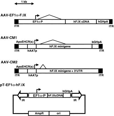
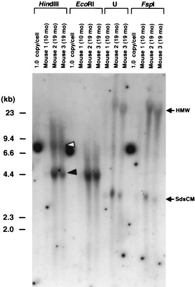
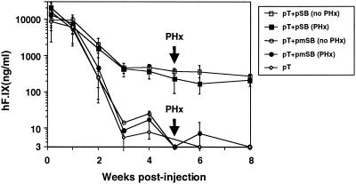
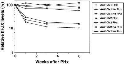
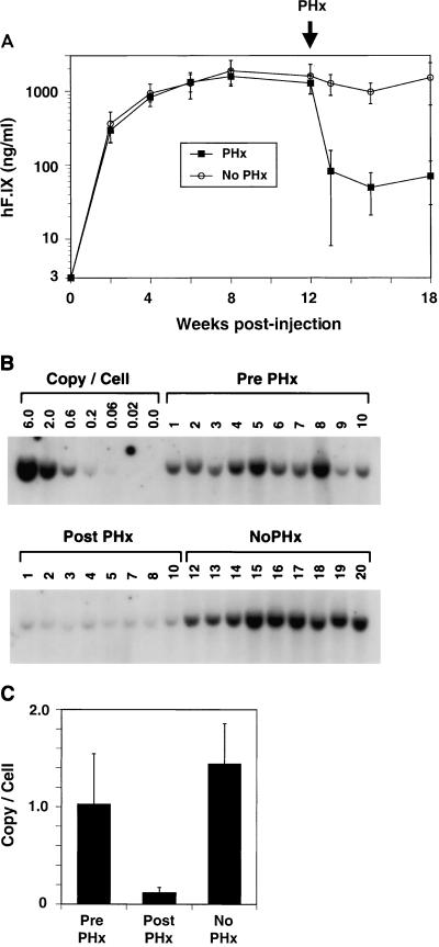
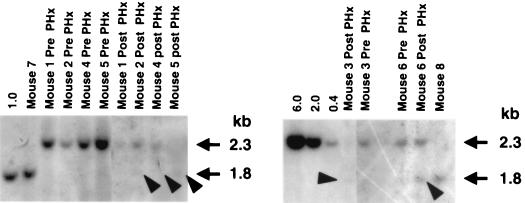
Recombinant adeno-associated virus (rAAV) vectors stably transduce hepatocytes in experimental animals. Although the vector genomes are found both as extrachromosomes and as chromosomally integrated forms in hepatocytes, the relative proportion of each has not yet been clearly established. Using an in vivo assay based on the induction of hepatocellular regeneration via a surgical two-thirds partial hepatectomy, we have determined the proportion of integrated and extrachromosomal rAAV genomes in mouse livers and their relative contribution to stable gene expression in vivo. Plasma human coagulation factor IX (hF.IX) levels in mice originating from a chromosomally integrated hF.IX-expressing transposon vector remained unchanged with hepatectomy. This was in sharp contrast to what was observed when a surgical partial hepatectomy was performed in mice 6 weeks to 12 months after portal vein injection of a series of hF.IX-expressing rAAV vectors. At doses of 2.4 x 10(11) to 3.0 x 10(11) vector genomes per mouse (n = 12), hF.IX levels and the average number of stably transduced vector genomes per cell decreased by 92 and 86%, respectively, after hepatectomy. In a separate study, one of three mice injected with a higher dose of rAAV had a higher proportion (67%) of integrated genomes, the significance of which is not known. Nevertheless, in general, these results indicate that, in most cases, no more than approximately 10% of stably transduced genomes integrated into host chromosomes in vivo. Additionally, the results demonstrate that extrachromosomal, not integrated, genomes are the major form of rAAV in the liver and are the primary source of rAAV-mediated gene expression. This small fraction of integrated genomes greatly decreases the potential risk of vector-related insertional mutagenesis associated with all integrating vectors but also raises uncertainties as to whether rAAV-mediated hepatic gene expression can persist lifelong after a single vector administration.


Figures

FIG. 1
FIG. 1
Map of rAAV vectors and plasmid: AAV-EF1α-F.IX, AAV-CM1, AAV-CM2, and pT-EF1α-hF.IX. The 1-kb scale is valid for the three vectors but not for the plasmid. EF1α-P, human elongation factor 1α gene enhancer-promoter; hGHpA, the human growth hormone gene polyadenylation signal; ApoEHCR(s), a shorter fragment of the HCR from the apolipoprotein E gene (ApoE) (25); hAATp, the human α1-antitrypsin gene promoter; hF.IX minigene, hF.IXcDNA containing 1.4-kb truncated intron A from the hF.IX gene; hF.IX minigene + 3′UTR, hF.IX cDNA containing 0.3-kb truncated intron A and 1.7-kb 3′ untranslated region (UTR) including the polyadenylation signal; IR, SB transposon inverted repeat; ori, plasmid origin of replication from pUC; Ampr, ampicillin resistance gene.
FIG. 2
FIG. 2
Southern blot analysis of liver DNA from three mice injected via the portal vein with AAV-EF1α-F.IX at a dose of 2.7 × 10¹¹ vg per animal. Twenty micrograms of total mouse liver DNA was digested with the enzymes indicated above the lanes or undigested (U), separated on an 0.8% agarose gel, blotted on a nylon membrane, and hybridized with a vector-specific PpuMI EF1α-hF.IX probe (the same as probe C in reference 27). The time of sacrifice is shown above each lane. The 1.0-copy/cell standard is 20 μg of naive mouse liver DNA mixed with the appropriate number of plasmid pV4.1e-hF.IX molecules. This control plasmid is linearized when cut with HindIII, EcoRI, or FspI, generating a 7.6-kb band. HindIII and EcoRI cut the vector genome once at the 3′ side and the center of the vector genome, respectively, while FspI does not cut within the vector genome. Head-to-tail and head-to-head molecules are denoted by closed and open arrowheads, respectively. The positions of high-molecular-weight (HMW) species and supercoiled ds circular monomers (SdsCM) of vector genomes are shown. Ethidium bromide staining of the gel showed no lane-to-lane variations of the amount of loaded DNA among digestions. Note that extrachromosomal forms of the vector genomes (SdsCM) are readily detectable. The Southern blot results for mice sacrificed 3 months postinjection were previously published (27).
FIG. 3
FIG. 3
Effects of partial hepatectomy on integrated plasmid vector genomes in liver. The figure shows plasma hF.IX levels of mice injected with 25 μg of an SB-based transposon plasmid (pT-EF1α-hF.IX) via the tail vein together with 1 μg of a helper plasmid encoding active SB transposase (pCMV-SB) (group 2, n = 10) or inactive mutated SB (pCMV-mSB) (group 3, n = 10) or without any helper plasmid (group 4, n = 5). A two-thirds partial hepatectomy (PHx) was performed 5 weeks postinjection. pT, pT-EF1α-hF.IX; pSB, pCMV-SB; pmSB, pCMV-mSB. Vertical bars indicate standard deviations.
FIG. 4
FIG. 4
rAAV-mediated gene expression in partially hepatectomized mice. The figure shows serum hF.IX levels after partial hepatectomy (PHx) performed a year after portal vein injection of 3.0 × 10¹¹ vg of AAV-CM1 or AAV-CM2. Results for control mice that were injected at the same time but did not receive partial hepatectomy are also shown. Each line represents an individual mouse. hF.IX levels are shown as percentages relative to their levels at the time of hepatectomy.
FIG. 5
FIG. 5
rAAV-mediated gene expression and quantification of rAAV genomes in partially hepatectomized mice. (A) Plasma hF.IX levels of 20 mice injected with AAV-EF1α-F.IX at a dose of 2.4 × 10¹¹ vg per mouse. Half of the mice underwent partial hepatectomy (PHx) 12 weeks postinjection. Vertical bars indicate standard deviations. (B) Southern blot analysis of liver DNAs from the mice in panel A. All animals were sacrificed 18 weeks postinjection (6 weeks after partial hepatectomy for the partial hepatectomy group), and 20 μg of total genomic liver DNA was subjected to Southern blot analysis with BglII digestion and hybridized to a BglII F.IX probe (28). Copy number standards are indicated as 0.0 to 6.0 copies/cell. The numbers above each lane indicate individual mouse numbers. Ethidium bromide staining of the gel showed that all the lanes had the same amount of digested DNA (data not shown). Both blots were from the same membrane. (C) Comparison of rAAV vector genome copy numbers per cell before and after partial hepatectomy. The intensity of each band in panel B was determined by densitometry. A Student t test revealed a statistical difference between “Pre PHx” and “Post PHx” values (P < 0.0002). Vertical bars indicate standard deviations.
FIG. 6
FIG. 6
The XhoI resistance assay of vector genomes in liver from mice injected with AAV-EF1α-F.IX.PMT vector at doses of 2.4 × 10¹¹ or 7.2 × 10¹¹ vg, before and after partial hepatectomy. Twenty micrograms of total liver DNA was digested with BglII and XhoI (except for the 0.4, 2.0, and 6.0 copy number standards, which were digested with only BglII) and subjected to Southern blot analysis with an XhoI F.IX probe (28). Copy number standards are 20 μg of naive mouse liver DNA mixed with the appropriate number of plasmid pV4.1e-hF.IX molecules followed by restriction enzyme digestion and shown above each lane as 1.0, 6.0, 2.0, and 0.4 copies/cell. The vector doses and the times of the analyses for mice 1 to 6 are summarized in Table 2. Mice 7 and 8 were injected with a nonmethylated AAV-EF1α-F.IX vector at a dose of 2.4 × 10¹¹ vg, and liver DNA was harvested 18 weeks postinjection (mouse 7) and at the end of this study (mouse 8). XhoI-resistant genomes migrate at the 2.3-kb position, while XhoI-digestible genomes migrate at the 1.8-kb position. Closed arrowheads indicate XhoI-digestible genomes. The vector copy numbers were determined by another Southern blotting with BglII digestion and a BglII F.IX probe (data not shown).

References

    1. Baiker A, Maercker C, Piechaczek C, Schmidt S B, Bode J, Benham C, Lipps H J. Mitotic stability of an episomal vector containing a human scaffold/matrix-attached region is provided by association with nuclear matrix. Nat Cell Biol. 2000;2:182–184. - PubMed
    1. Budker V, Zhang G, Knechtle S, Wolff J A. Naked DNA delivered intraportally expresses efficiently in hepatocytes. Gene Ther. 1996;3:593–598. - PubMed
    1. Burton M, Nakai H, Colosi P, Cunningham J, Mitchell R, Couto L. Coexpression of factor VIII heavy and light chain adeno-associated viral vectors produces biologically active protein. Proc Natl Acad Sci USA. 1999;96:12725–12730. - PMC - PubMed
    1. Chao H, Samulski R, Bellinger D, Monahan P, Nichols T, Walsh C. Persistent expression of canine factor IX in hemophilia B canines. Gene Ther. 1999;6:1695–1704. - PubMed
    1. Chen S J, Tazelaar J, Moscioni A D, Wilson J M. In vivo selection of hepatocytes transduced with adeno-associated viral vectors. Mol Ther. 2000;1:414–422. - PubMed

Publication types