. 2001 Aug;75(15):7009-17.
doi: 10.1128/JVI.75.15.7009-7017.2001.

Minute virus of mice initiator protein NS1 and a host KDWK family transcription factor must form a precise ternary complex with origin DNA for nicking to occur

Affiliations

Minute virus of mice initiator protein NS1 and a host KDWK family transcription factor must form a precise ternary complex with origin DNA for nicking to occur

J Christensen et al. J Virol. 2001 Aug.

Abstract

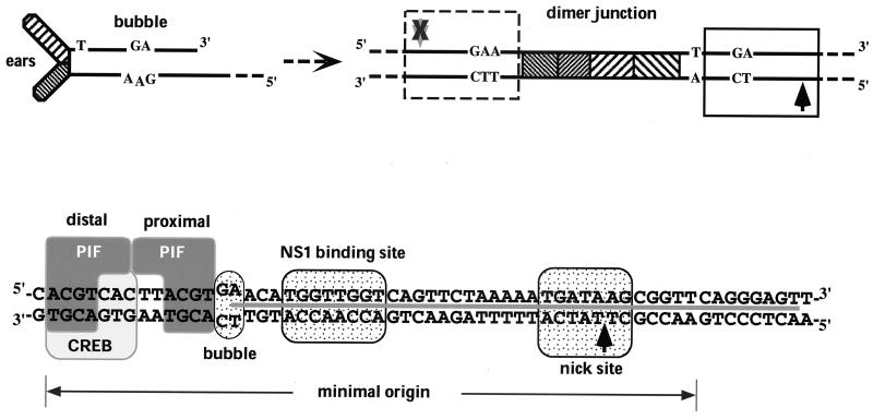
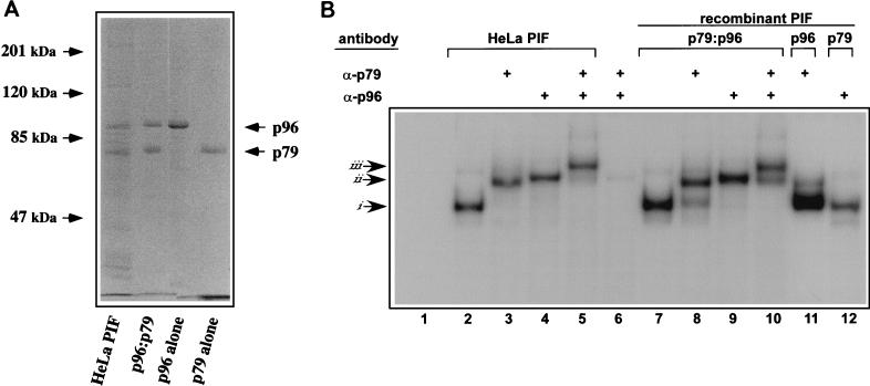
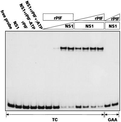
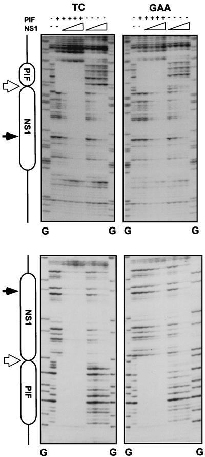
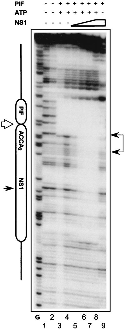
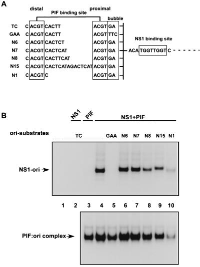
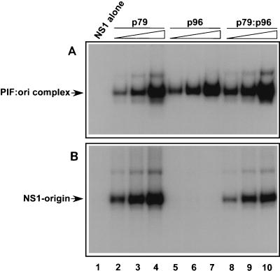
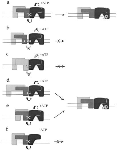
Parvoviral rolling hairpin replication generates palindromic genomic concatemers whose junctions are resolved to give unit-length genomes by a process involving DNA replication initiated at origins derived from each viral telomere. The left-end origin of minute virus of mice (MVM), oriL, contains binding sites for the viral initiator nickase, NS1, and parvovirus initiation factor (PIF), a member of the emerging KDWK family of transcription factors. oriL is generated as an active form, oriL(TC), and as an inactive form, oriL(GAA), which contains a single additional nucleotide inserted between the NS1 and PIF sites. Here we examined the interactions on oriL(TC) which lead to activation of NS1 by PIF. The two subunits of PIF, p79 and p96, cooperatively bind two ACGT half-sites, which can be flexibly spaced. When coexpressed from recombinant baculoviruses, the PIF subunits preferentially form heterodimers which, in the presence of ATP, show cooperative binding with NS1 on oriL, but this interaction is preferentially enhanced on oriL(TC) compared to oriL(GAA). Without ATP, NS1 is unable to bind stably to its cognate site, but PIF facilitates this interaction, rendering the NS1 binding site, but not the nick site, resistant to DNase I. Varying the spacing of the PIF half-sites shows that the distance between the NS1 binding site and the NS1-proximal half-site is critical for nickase activation, whereas the position of the distal half-site is unimportant. When expressed separately, both PIF subunits form homodimers that bind site specifically to oriL, but only complexes containing p79 activate the NS1 nickase function.


Figures

FIG. 1
FIG. 1
Formation and organization of MVM oriL. (Top left) Structure of the left-end hairpin showing the 3′ OH used for priming replication and the mismatched bubble sequence as present in the parental single-stranded viral genome. (Top right) Organization of the left-end hairpin sequences within the duplex dimer junction generated by replication through the hairpin. Hatched boxes represent the palindromic sequences which were originally folded to give the ears of the hairpin form. The boxed sequence (expanded below) represents the minimum active replication origin on the outboard arm, oriLTC, while the sequence in the dashed box represents the corresponding origin on the inboard arm, oriLGAA, which is inactive. The potential nick sites on each side of the junction are indicated by arrows (black arrow, active site; grey arrow with X, inactive site). (Bottom panel) Sequence of oriLTC, showing the different elements involved in replication. The PIF binding site (see the text) overlaps a consensus binding site for the CREB/ATF family of host transcription factors. The two tetranucleotide motifs bound by PIF are designated “distal” and “proximal” to indicate their positions relative to the NS1 binding site. Other boxes indicate sequences involved in the bubble dinucleotide (or trinucleotide) spacer element described in the text, the (ACCA)2 NS1 binding site, and the nick site, a specific sequence required for nicking and covalent attachment of NS1. The heavy line between the DNA strands indicates sequences protected by NS1 from DNase I digestion.
FIG. 2
FIG. 2
Characterization of rPIF subunits expressed from separate baculovirus vectors. (A) SDS-polyacrylamide gel electrophoresis analysis of purified rPIF. DNA-affinity purified PIF from HeLa S3 cells was compared to purified rPIF subunits, either coexpressed (p96-p79) or expressed alone in insect cells using baculovirus vectors. The gel was stained with Coomassie brilliant blue. (B) Autoradiograph of an EMSA gel of HeLa cell derived- and rPIF complexes showing complexes generated when 32P-labeled duplex oligonucleotides covering the PIF binding region in the MVM 3′ origin (5′-T C A T C A C G T C A C T T A C G T G A A-3′) were incubated with PIF purified from HeLa S3 cells by DNA-affinity chromatography (lanes 2 to 5) or with human PIF complexes purified from High Five insect cells infected with recombinant baculoviruses (lanes 7 to 12). PIF p79-p96 heterocomplexes purified from coinfected cells are shown in lanes 7 to 10, while lanes 11 and 12 contain p79 and p96 complexes purified from cells singly infected with the appropriate baculovirus. Preformed complexes were incubated with rabbit antisera specific for one of the PIF subunits, as indicated at the top of each lane. Lanes 6, 11, and 12 are negative controls (lane 6 contains both antibodies and the probe, but no PIF). Arrows at the side mark the positions of shifted (i), supershifted (ii), and super-duper-shifted (iii) PIF-oriL probe complexes.
FIG. 3
FIG. 3
rPIF activates NS1 to nick the viral origin. A 32P-labeled DNA fragment containing the oriLTC sequence (TC) was incubated in the absence or presence of NS1 (100 ng), rPIF (25 ng), and ATP (3 mM) or γ-S-ATP (3 mM). In the titrations, a constant amount of rPIF (25 ng) or NS1 (100 ng) was incubated with increasing amounts of NS1 (10, 25, 50, and 100 ng) or rPIF (0.5 2.5, 12.5, and 50 ng). Only covalent NS1-DNA complexes are retarded in the SDS-polyacrylamide gel system used for this assay (see Materials and Methods). GAA, reactions performed with oriLGAA with NS1 and rPIF at inputs of 100 ng and 2.5 to 50 ng.
FIG. 4
FIG. 4
PIF enhances NS1 binding to oriLTC but not to oriLGAA. Diagrams of the major elements in oriL are shown on the left. Sequences protected from DNase I digestion by PIF and NS1 are indicated by boxes. Positions of the nick site and bubble sequence are indicated by filled and open arrows, respectively. TC and GAA denote which form of oriL was used as the substrate. PIF (top), 50 ng of purified rPIF p79-p96 complex; NS1 (top), increasing amounts of purified His-tagged NS1 (25, 50, 100, and 225 ng). All assays were performed in the presence of 2 mM γ-S-ATP. Upper and lower pairs of autoradiographs show the protection patterns obtained when the DNA strands were 32P-, 3′-end-labeled on the upper and lower strands, respectively, of the oriL diagrammed in Fig. 1. Lanes G, products of a G-specific chemical sequencing reaction of each labeled substrate (25), used for aligning each autoradiograph with its relevant protection diagram.
FIG. 5
FIG. 5
PIF stabilizes NS1 binding the to oriLTC in the absence of ATP. An autoradiograph of DNase I protection analysis of rPIF and NS1 binding to an oriLTC probe in the presence or absence of γ-S-ATP is shown. The major elements of oriL are indicated on the left, as described in the legend to Fig. 4. PIF (top), 50 ng of purified rPIF p79-p96 complex; NS1 (top), increasing amounts of purified His-tagged NS1 (25, 50, 100, and 225 ng). Samples were digested with 0.8 (lanes 2 and 4 through 9) or 0.4 (lanes 1 and 3) U of DNase I. Lane G, G-specific digest of the probe, as described in the legend to Fig. 4.
FIG. 6
FIG. 6
Only the position of the NS1 proximal PIF half-site is important for oriL activation. (A) Sequences of the PIF site in the top strand of 32P-labeled, double-stranded, minimal origin probes used as substrates in nicking and gel mobility shift assays. The proximal and distal PIF half-sites are boxed, and the nucleotides between the half-sites are indicated. (B, top) Autoradiograph of nicking assays performed in the presence of purified GST-tagged NS1 (100 ng) and purified recombinant p79-p96 complex (10 ng). 32P-labeled double-stranded DNA substrates used in each lane are indicated, and the nicking assays were analyzed as described in the legend to Fig. 3. The reaction products (NS1-ori) are covalent NS1-DNA complexes. Lanes 1 and 2, samples in which the probe was incubated alone or in the presence of only NS1, respectively. (B, bottom) Autoradiograph of an EMSA gel demonstrating the binding of purified recombinant p79-p96 complex (2 ng) to the 32P-labeled mutant origin substrates. Under these assay conditions, the shifted reaction products (PIF:ori complex) are not covalently modified.
FIG. 7
FIG. 7
The PIF p79 subunit activates the NS1 nickase. (A) Autoradiograph of EMSA gels demonstrating the binding of purified GST-tagged NS1 (100 ng) alone and of increasing amounts of purified recombinant p79 (0.9, 2.7, and 8.1 ng [lanes 2 to 4, respectively]), p96 (0.9, 2.7, and 8.1 ng [lanes 5 to 7, respectively]), or p79-p96 heterocomplexes (0.9, 2.7, and 8.1 ng [lanes 8 to 10, respectively]) to a 32P-labeled duplex oligonucleotide probe containing the PIF recognition site in oriL. Shifted reaction products (PIF:ori complex) are not covalently modified. (B) Autoradiograph of gels from nicking assays performed in the presence of purified GST-tagged NS1 (100 ng) alone and in the presence of increasing amounts of purified recombinant homodimers and heterodimers, as in the top panel. The nicking assay was performed as described in the legend to Fig. 3, using a 32P-labeled DNA fragment containing oriLTC. Reaction products (NS1-origin) represent covalent NS1-DNA complexes.
FIG. 8
FIG. 8
Proposed interactions of PIF and NS1 at different forms of oriL. PIF subunits p79 and p96 are shown as dark and light gray boxes, respectively, while the subunits of the putative NS1 dimer are depicted in black. Functional intersubunit interactions are depicted as curved arrows (black arrows, interactions which occur; grey arrows with X, interactions which fail to occur). Potential substrate complexes are shown on the left, and the nicked strand with its covalent NS1-oriL product is shown on the right.

References

    1. Astell C R, Chow M B, Ward D C. Sequence analysis of the termini of virion and replicative forms of minute virus of mice DNA suggests a modified rolling hairpin model for autonomous parvovirus DNA replication. J Virol. 1985;54:171–177. - PMC - PubMed
    1. Baldauf A Q, Willwand K, Mumtsidu E, Nüesch J P, Rommelaere J. Specific initiation of replication at the right-end telomere of the closed species of minute virus of mice replicative-form DNA. J Virol. 1997;71:971–980. - PMC - PubMed
    1. Christensen J, Cotmore S F, Tattersall P. Minute virus of mice transcriptional activator protein NS1 binds directly to the transactivation region of the viral P38 promoter in a strictly ATP-dependent manner. J Virol. 1995;69:5422–5430. - PMC - PubMed
    1. Christensen J, Cotmore S F, Tattersall P. A novel cellular site-specific DNA-binding protein cooperates with the viral NS1 polypeptide to initiate parvovirus DNA replication. J Virol. 1997;71:1405–1416. - PMC - PubMed
    1. Christensen J, Cotmore S F, Tattersall P. Parvovirus initiation factor PIF: a novel human DNA-binding factor which coordinately recognizes two ACGT motifs. J Virol. 1997;71:5733–5741. - PMC - PubMed

Publication types

MeSH terms

Substances

LinkOut - more resources