. 2001 Aug;75(15):7018-29.
doi: 10.1128/JVI.75.15.7018-7029.2001.

The vaccinia virus superoxide dismutase-like protein (A45R) is a virion component that is nonessential for virus replication

Affiliations

The vaccinia virus superoxide dismutase-like protein (A45R) is a virion component that is nonessential for virus replication

F Almazán et al. J Virol. 2001 Aug.

Abstract

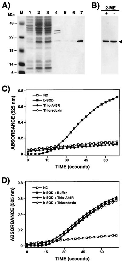
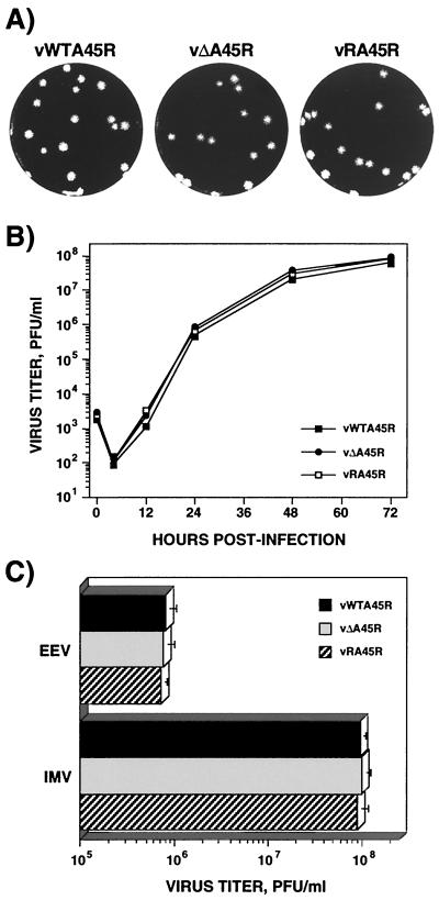
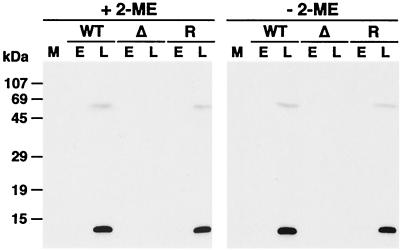
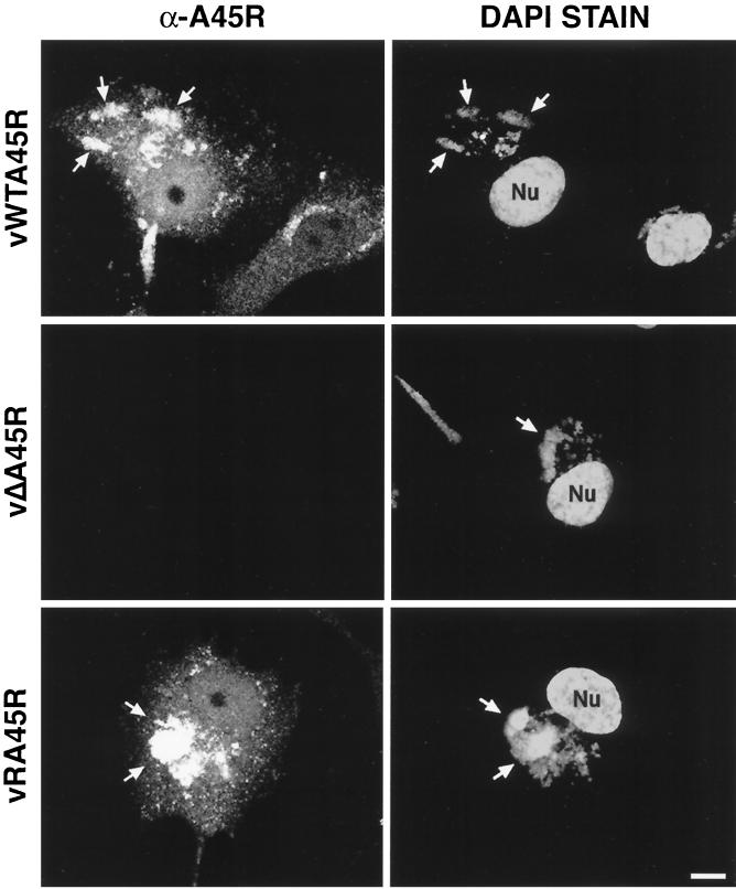
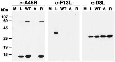
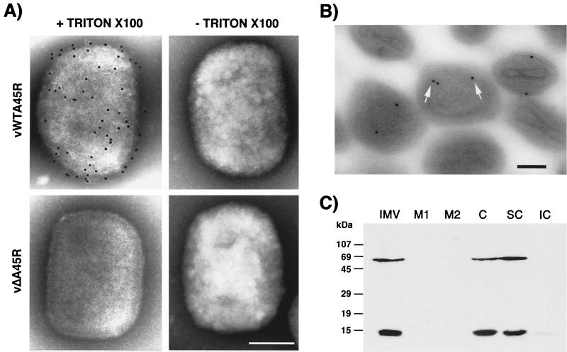
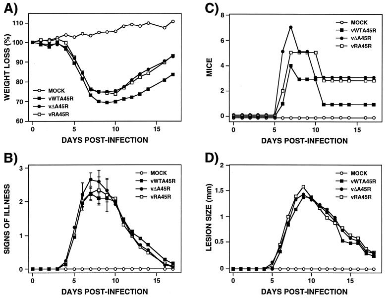
A characterization of the A45R gene from vaccinia virus (VV) strain Western Reserve is presented. The open reading frame is predicted to encode a 125-amino-acid protein (M(r), of 13,600) with 39% amino acid identity to copper-zinc superoxide dismutase (Cu-Zn SOD). Sequencing of the A45R gene from other orthopoxviruses, here and by others, showed that the protein is highly conserved in all viruses sequenced, including 16 strains of VV, 2 strains of cowpox virus, camelpox virus, and 4 strains of variola virus. In all cases the protein lacks key residues involved in metal ion binding that are important for the catalytic activity. The A45R protein was expressed in Escherichia coli, purified, and tested for SOD activity, but neither enzymatic nor inhibitory SOD activity was detected. Additionally, no virus-encoded SOD activity was detected in infected cells or purified virions. A monoclonal antibody raised against the A45R protein expressed in E. coli identified the A45R gene product as a 13.5-kDa protein that is expressed late during VV infection. Confocal microscopy of VV-infected cells indicated that the A45R protein accumulated predominantly in cytoplasmic viral factories. Electron microscopy and biochemical analyses showed that the A45R protein is incorporated into the virion core. A deletion mutant lacking the majority of the A45R gene and a revertant virus in which the deleted gene was restored were constructed and characterized. The growth properties of the deletion mutant virus were indistinguishable from those of wild-type and revertant viruses in all cell lines tested, including macrophages. Additionally, the virulence and pathogenicity of the three viruses were also comparable in murine and rabbit models of infection. A45R is unusual in being the first VV core protein described that affects neither virus replication nor virulence.


Figures

FIG. 1
FIG. 1
Expression, purification, and biochemical characterization of A45R protein. (A) Expression of A45R in E. coli. The A45R protein was expressed in E. coli as a thioredoxin fusion protein with the His-Patch ThioFusion Expression System (Invitrogen, BV) and purified by affinity chromatography on a nickel column as described in Materials and Methods. Samples were analyzed by SDS-PAGE (12% gel), and proteins were visualized by staining with Coomassie brilliant blue. Lanes: 1, uninduced whole-cell extract; 2, induced whole-cell extract; 3, induced soluble lysate; 4, induced insoluble lysate; 5, 6, and 7, proteins eluted from the Ni2+ column with 350 or 500 mM imidazole or 50 mM EDTA, respectively. Molecular size markers (M) are shown in kilodaltons. (B) Analysis of the purified fusion protein under reducing and nonreducing conditions. Before electrophoresis on a 12% polyacrylamide gel, samples were boiled for 3 min in Laemmli buffer with (+) or without (−) 2% 2-ME. Proteins were detected by staining with Coomassie brilliant blue. The arrowhead indicates the position of the fusion protein. (C and D) Investigation of possible enzymatic and inhibitory activity of the A45R protein expressed in E. coli. The SOD activity assay (C) was carried out with 5 μg of the purified fusion protein (Thio-A45R) as described in Materials and Methods. The same assay was done without protein (NC) or with 5 μg of thioredoxin as a negative control and with 5 μg of bovine Cu-Zn SOD (b-SOD) as a positive control. The inhibitory effect of the purified protein on the enzymatic activity of the bovine Cu-Zn SOD was also analyzed (D). In this case, before the SOD assay, 5 μg of bovine Cu-Zn SOD was incubated for 30 min at 37°C with either buffer, 5 μg of the purified fusion protein, or 5 μg of thioredoxin. Mean values from three experiments are shown.
FIG. 2
FIG. 2
Growth properties of vΔA45R in cell culture. (A) Plaque phenotype. vWTA45R, vΔA45R, and vRA45R were plated onto monolayers of BSC-1 cells and were overlaid with MEM supplemented with 2.5% FBS and 1.5% carboxymethylcellulose for 2 days before being stained with 0.1% crystal violet in 15% ethanol. (B) Growth curves. BSC-1 cells were infected at 0.01 PFU/cell with vWTA45R, vΔA45R, or vRA45R. At different times p.i. cells were scraped into the growth medium, and the production of infectious virus was determined by plaque assay on BSC-1 cells. Data shown are mean values from triplicate samples. The standard deviation was lower than 20% in all cases (not shown). (C) IMV and EEV production. Monolayers of BSC-1 cells were infected with 10 PFU of either vWTA45R, vΔA45R, or vRA45R/cell. After 24 h, the yields of IMV and EEV were determined by plaque assay on BSC-1 cells. Data are means from three experiments. Error bars, standard deviations.
FIG. 3
FIG. 3
Identification of the A45R protein in VV-infected cells. BSC-1 cells were mock infected (M) or infected with vWTA45R (WT), vΔA45R (Δ), or vRA45R (R) at 10 PFU/cell in the presence (E) or absence (L) of 40 μg of araC/ml. At 18 h p.i. cell lysates were resolved by SDS-PAGE (15% gel) under reducing (+ 2-ME) or nonreducing (− 2-ME) conditions, transferred to nitrocellulose, and incubated with MAb 2.B.11 (diluted 1/10,000). Bound antibodies were detected by using horseradish peroxidase-conjugated goat α-mouse Ig (Sigma) and ECL reagents as described in Materials and Methods. Positions of molecular size markers are shown.
FIG. 4
FIG. 4
Localization of VV A45R protein in infected cells by immunofluorescence. Subconfluent BSC-1 cells were infected with the indicated viruses at 1 PFU/cell. At 14 h p.i., cells were fixed, permeabilized, and incubated with MAb 2.B.11. Bound antibody was detected with FITC-conjugated secondary antibody as described in Materials and Methods. Representative cells are shown. Viral and cellular DNA were stained with DAPI. Arrows indicate viral factories. Nu, nucleus. Bar, 10 μm.
FIG. 5
FIG. 5
The A45R protein is incorporated into virions. IMVs from vWTA45R (WT), vΔA45R (Δ), or vRA45R (R) were analyzed by SDS-PAGE together with extracts from BSC-1 cells mock infected (M) or infected with vWTA45R at 10 PFU/cell for 18 h (L). After transfer to nitrocellulose filters, proteins were detected by immunoblotting with MAb 2.B.11. As controls, the blot was reprobed with a rabbit α-F13L antibody or with the mouse α-D8L MAb AB1.1. Positions of molecular size markers are shown.
FIG. 6
FIG. 6
The A45R protein is located in the viral core. (A) Immunogold labeling and negative staining electron microscopy of purified IMV particles. IMV purified by sucrose gradient centrifugation from RK13 cells infected with either vWTA45R or vΔA45R was adsorbed to carbon-coated nickel 400-mesh grids, permeabilized (+ Triton X-100) or unpermeabilized (− Triton X-100), and incubated with MAb 2.B.11. Bound antibody was detected with 5-nm gold particle-conjugated goat α-mouse Ig. Representative viral particles are shown. Bar, 100 nm. (B) Immunogold labeling of thin sections of purified IMV particles. Purified vWTA45R IMV particles were pelleted by high-speed centrifugation, fixed with 8% PFA, and embedded in LR White (London Resin Company). Ultrathin sections were immunolabeled with MAb 2.B.11, followed by 10-nm gold particle-conjugated goat α-mouse Ig, and were examined by electron microscopy. Some gold particles are indicated by arrows. Bar, 100 nm. (C) Fractionation of purified IMV by detergent treatment. Purified vWTA45R IMV particles (IMV) were incubated in lysis buffer containing 1% NP-40 in the absence or presence of 50 mM DTT, and soluble envelopes (M1 and M2, respectively) were removed from viral cores (C) by centrifugation. Cores were partitioned into soluble (SC) and insoluble (IC) fractions by treatment with 0.5% sodium deoxycholate and 0.1% SDS. Fractions were then subjected to SDS-PAGE (15% gel), transferred to nitrocellulose, blotted with MAb 2.B.11, and developed with ECL reagents. Molecular size markers are shown.
FIG. 7
FIG. 7
Virulence of vΔA45R in mice. (A, B, and C) Murine intranasal model. Groups of 10 female BALB/c mice, 5 to 6 weeks old, were mock infected or infected intranasally with 104 PFU of the indicated virus per animal. Percent weight changes (A), signs of illness (B), and the number of animals with illness scores of ≥2.5 (C) were analyzed daily. Animals that had lost more than 30% of their body weight were sacrificed. The mean weight of each group of mice was expressed as a percentage of the mean weight of that group of animals immediately prior to infection. Signs of illness were scored from zero to four, and the mean value for each group is shown. The number of animals that presented strong signs of illness (scores of ≥2.5) is represented at different days p.i. Standard deviations in panel B are given as error bars for days 6 to 9. (D) Murine intradermal model. Groups of six 8 to 9-week-old female BALB/c mice were mock infected or infected intradermally in the left ear pinnae with 104 PFU of the indicated virus. The diameters of lesions produced were determined daily using a micrometer. Each data point represents the mean for a group.

References

    1. Aguado B, Selmes I P, Smith G L. Nucleotide sequence of 21.8 kbp of variola major virus strain Harvey and comparison with vaccinia virus. J Gen Virol. 1992;73:2887–2902. - PubMed
    1. Alcamí A, Smith G L. Vaccinia, cowpox, and camelpox viruses encode soluble gamma interferon receptors with novel broad species specificity. J Virol. 1995;69:4633–4639. - PMC - PubMed
    1. Alcamí A, Symons J A, Collins P D, Williams T J, Smith G L. Blockade of chemokine activity by a soluble chemokine binding protein from vaccinia virus. J Immunol. 1998;160:624–633. - PubMed
    1. Alcamí A, Khanna A, Paul N L, Smith G L. Vaccinia virus strains Lister, USSR and Evans express soluble and cell-surface tumour necrosis factor receptors. J Gen Virol. 1999;80:949–959. - PubMed
    1. Antoine G, Scheiflinger F, Dorner F, Falkner F G. The complete genomic sequence of the modified vaccinia Ankara strain: comparison with other orthopoxviruses. Virology. 1998;244:365–396. - PubMed

Publication types

MeSH terms

LinkOut - more resources