. 2001 Jul;159(1):321-7.
doi: 10.1016/S0002-9440(10)61698-7.

Matrix metalloproteinase-2 is associated with tenascin-C in calcific aortic stenosis

Affiliations

Matrix metalloproteinase-2 is associated with tenascin-C in calcific aortic stenosis

B Jian et al. Am J Pathol. 2001 Jul.

Abstract

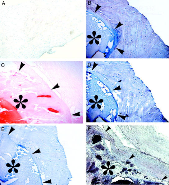
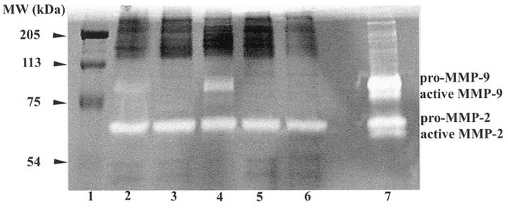
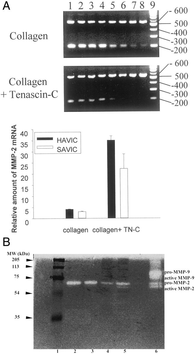
We previously showed that the expression of tenascin (TN-C), an extracellular matrix glycoprotein found in developing bone and atherosclerotic plaque, and matrix metalloproteinase-2 (MMP-2) are coordinated and interdependent in cultured vascular smooth muscle cells. In this study, we hypothesized that TN-C and MMP-2 are mechanistically involved in the pathobiology of calcific aortic stenosis. Human calcific aortic stenosis cusps demonstrated immunohistochemically prominent deposition of TN-C, MMP-2, and alkaline phosphatase activity, as well as MMP-2 gelatinolytic activity. Although far lesser amounts of TN-C were noted in several of the grossly non-calcified valve cusps, MMP-2 and AP were never detected. Further, when aortic valve interstitial cells (both sheep and human) were cultivated on collagen supplemented with TN-C, both MMP-2 mRNA expression and MMP-2 gelatinolytic activity (both pro and active forms), were up-regulated compared to control. These observations support the view that accumulation of first TN-C and then MMP-2 are associated with progression of calcification. The residual presence of these proteins in severe calcifications is indicative of their involvement in the pathogenesis.


Figures

Figure 1.
Figure 1.
Representative photomicrographs of normal, noncalcific aortic valve cusps with faint, widespread TN-C by immunohistochemistry (brown staining) and hematoxylin counterstain (case 4 in Table 1 ▶ ) (A); Calcific aortic stenosis (* denotes calcified region, with arrows denoting the margin of the calcification) with intense TN-C immunopositive brown staining (case 10 in Table 1 ▶ ) (B); calcific aortic stenosis with Alizarin Red S (calcifications staining red, case 10 in Table 1 ▶ ) (C); calcific aortic stenosis with intense MMP-2 immunopositive staining (case 10 in Table 1 ▶ ) (D); IgG negative control for b and d shows a absence of peroxidase reactivity (case 10 in Table 1 ▶ ) (E); calcific aortic stenosis with intense alkaline phosphatase activity (purple stained region) localized within the calcific deposits (F), but not in the non-calcified extracellular matrix (case 7 in Table 1 ▶ ). Original magnification, ×200
Figure 2.
Figure 2.
MMP-2 activity was present in individual extracts of five explanted aortic valve cusps obtained at surgery for calcific aortic stenosis. Gel zymography shows MMP-2 (pro form only) in all cases (lanes 2–6), and qualitatively lesser amounts MMP-9 in cases 1, 2, and 3 (lanes 2-4) as compared to MMP standards (lane 7). Lane 1 contains molecular weight standards.
Figure 3.
Figure 3.
The results of aortic valve interstitial cell culture studies in which expression of matrix metalloproteinase-2 (MMP-2) is up-regulated by tenascin-C (TN-C). A: relative amounts of mRNA in sheep (SAVIC) and human aortic valve interstitial cells (HAVIC), evaluated by competitive RT-PCR. Cells were grown on a substrate of either bovine type I collagen or collagen with TN-C (15 μg/ml). A representative agarose gel (see Materials and Methods) is shown for SAVIC demonstrating greater amounts of MMP-2 RNA in the TN-C cultures. The top bands are derived from MMP-2 reverse transcriptase (RT) products, while the bottom bands are due to the MMP-2M13 competitors. The lanes from left to right represent PCRs with a series of dilutions of the MMP-2M13 competitor. The reduction in MMP-2M13 band intensity (bottom bands) occurs in conjunction with an increase in the upper bands (MMP-2 RT). The last lane is a 100 bp DNA ladder. Bars represent the mean ± SE of 3 independent measurements. TN-C significantly increased MMP-2 expression (P < 0.05). B: gel zymograms, demonstrating the results of SAVIC cultures, with greater MMP-2 activity, both pro-, and active-form, in the TN-C cell culture lysates (lane 5) compared to control (lane 4). MMP-2 levels in the conditioned media (detected only as the pro form) were comparable in the TN-C group (lane 3) versus control group (lane 2). No MMP-9 activity was detected in these cell culture studies. Lanes 1 and 6 are molecular marker and MMP gelatinase zymography standards, respectively.

References

    1. Kim KM: Calcification of matrix vesicles in human aortic valve and aortic media. Fed Proc 1976, 35:156-162 - PubMed
    1. Kim KM: Apoptosis and calcification. Scanning Microsc 1995, 9:1137-1178 - PubMed
    1. Srivatsa SS, Harrity PJ, Maercklein PB, Kleppe L, Veinot J, Edwards WD, Johnson CM, Fitzpatrick LA: Increased cellular expression of matrix proteins that regulate mineralization is associated with calcification of native human and porcine xenograft bioprosthetic heart valves. J Clin Invest 1997, 99:996-1009 - PMC - PubMed
    1. Bini A, Mann KG, Kudryk BJ, Schoen FJ: Noncollagenous bone matrix proteins, calcification, and thrombosis in carotid artery atherosclerosis. Arterioscler Thromb Vasc Biol 1999, 19:1852-1861 - PubMed
    1. Mori K, Shioi A, Jono S, Nishizawa Y, Morii H: Expression of matrix Gla protein (MGP) in an in vitro model of vascular calcification. FEBS Lett 1998, 433:19-22 - PubMed

Publication types

LinkOut - more resources