. 2001 Jul;159(1):329-38.
doi: 10.1016/S0002-9440(10)61699-9.

Ethanol-induced apoptosis in mouse liver: Fas- and cytochrome c-mediated caspase-3 activation pathway

Affiliations

Ethanol-induced apoptosis in mouse liver: Fas- and cytochrome c-mediated caspase-3 activation pathway

Z Zhou et al. Am J Pathol. 2001 Jul.

Abstract

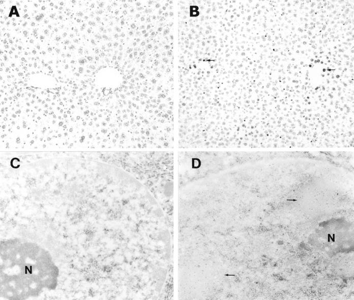
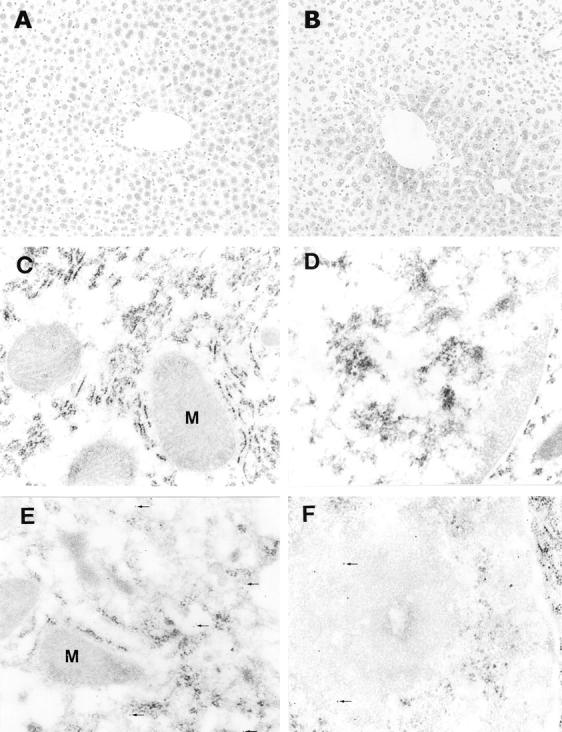
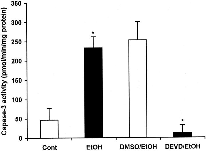
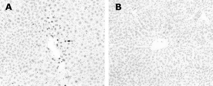
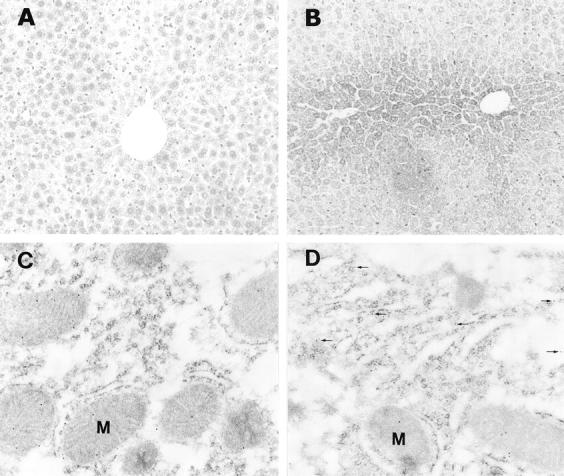
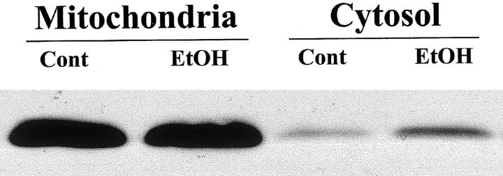
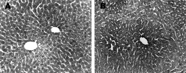
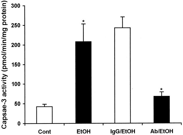
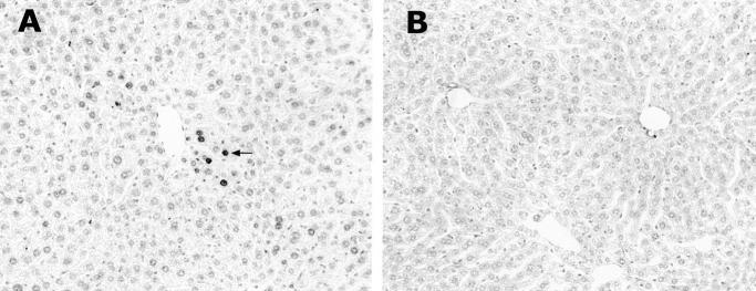
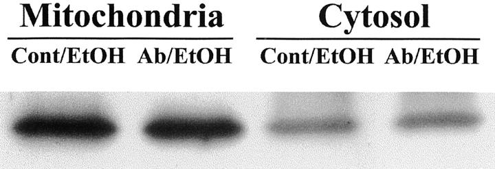
Hepatic apoptosis has been shown to occur in both experimental and clinical alcoholic liver disease, but the signaling pathway remains unknown. This study was undertaken to examine specifically the involvement of the upstream signals, Fas and cytochrome c, in alcohol-induced caspase-3 activation and apoptosis in the liver. Male FVB mice were administrated intragastrically a single dose of alcohol at 6 g/kg, which has been shown to represent binge drinking in humans. Hepatic apoptosis was detected by a terminal deoxynucleotidyl transferase dUTP nick-end labeling assay. Active form of caspase-3 was identified by immunoperoxidase staining and confirmed by immunogold labeling and was found to be in the cytosol and nucleus. Enzymic assay further confirmed caspase-3 activation and nucleus localization. Systemic administration of caspase-3 inhibitor, Ac-DEVD-FMK, inhibited caspase-3 activity and abrogated apoptosis. Elevation of cytosolic cytochrome c was found by immunoperoxidase staining, immunogold labeling, and Western blot. Increased Fas ligand expression was detected by immunoperoxidase staining. Intravenous administration of a neutralizing Fas ligand monoclonal antibody resulted in suppression of caspase-3 activation and attenuation of apoptosis, but did not inhibit mitochondrial cytochrome c release. The results thus demonstrate that Fas/Fas ligand system-mediated caspase-3 activation plays a central role in the ethanol-induced hepatic apoptosis.


Figures

Figure 1.
Figure 1.
Acute ethanol administration of an accumulative dose of 6 g/kg body weight induced apoptosis in the mouse liver as detected by TUNEL assay. Under light microscope, the control liver showed negative staining (A), and the ethanol-treated liver showed positive cells (arrows) around the central vein (B). Using immunogold TUNEL under electron microscope, only a few gold particles were seen in the nucleus of hepatocytes in control liver (C), whereas numerous gold particles (arrows) in the nucleus of hepatocytes in ethanol-treated liver were found (D), indicating severe DNA fragmentation. N, nucleolus. Original magnifications: ×130 (A and B); ×24,000 (C and D).
Figure 2.
Figure 2.
Detection of active form of caspase-3 in the liver of ethanol-treated mice. Using light microscopic immunoperoxidase staining, no active caspase-3 was detectable in the control liver (A), and moderate staining was found in the liver of ethanol-treated mice (B). Under electron microscope, immunogold labeling of active caspase-3 showed negative results in both cytoplasm (C) and nucleus (D) of hepatocytes in the control liver. However, active caspase-3 was observed not only in the cytosol (arrows, E) but also in the nucleus (arrows, F) of hepatocytes in the ethanol-treated liver. M, mitochondria. Original magnifications: ×130 (A and B); ×43,000 (C–F).
Figure 3.
Figure 3.
Colorimetric assay of caspase-3 activity in the liver of mice treated with ethanol with or without systemic administration of caspase-3 inhibitor. Before administration of ethanol, a caspase-3 inhibitor, Z-DEVD-FMK (dissolved in 1% DMSO) was intravenously injected at a dosage of 20 μg/g body weight, and the same volume of 1% DMSO was injected as control. Ethanol induced a fivefold increase in caspase-3 activity in comparison to the control. Intravenous injection of Z-DEVD-FMK abrogated the ethanol-induced caspase-3 activation. EtOH, ethanol; DMSO, 1% DMSO; DEVD, Z-DEVD-FMK/1% DMSO. *, Significant difference between treatments and controls at P < 0.05 by Student’s t-test (n = 5).
Figure 4.
Figure 4.
Attenuation of hepatic apoptosis by systemic administration of caspase-3 inhibitor. Light microscopic TUNEL demonstrated apoptotic cells (arrow) in the liver of control mice treated with 1% DMSO and ethanol (A), but not in the liver treated with intravenous injection of caspase-3 inhibitor and ethanol (B). Original magnifications, ×130.
Figure 5.
Figure 5.
Detection of cytosolic cytochrome c elevation in the liver of ethanol-treated mice. Light microscopic immunoperoxidase showed moderate and uniform staining in the liver of control mice (A), but strong staining around central veins in the ethanol-treated liver (B). Using electron microscopic immunogold labeling, cytochrome c was found to be localized mainly in mitochondria and little in the cytosol of hepatocytes in the control liver (C), however, it was obviously apparent in the cytosol (arrows, D) in the ethanol-treated liver. M, mitochondria. Original magnifications: ×130 (A and B); ×43,000 (C and D).
Figure 6.
Figure 6.
Western blot analysis of cytochrome c elevation in the liver of ethanol-treated mice. Densitometric analysis indicated that a threefold elevation of the cytosolic cytochrome c was induced by ethanol. However, there was no detectable decrease in the mitochondrial cytochrome c content in ethanol-treated liver.
Figure 7.
Figure 7.
Detection of Fas ligand expression in the liver of ethanol-treated mouse. Light microscopic immunoperoxidase staining demonstrated a weak reactivity in the normal liver (A). Ethanol treatment markedly increased the Fas ligand reactivity in the liver (B). Original magnifications, ×130.
Figure 8.
Figure 8.
Suppression of caspase-3 activity by systemic administration of a neutralizing Fas ligand antibody. Before administration of ethanol, the neutralizing Fas ligand monoclonal antibody was intravenously injected at a dosage of 5 mg/kg body weight, and normal hamster IgG was injected as control. High levels of caspase-3 activity in the ethanol-treated mouse liver with or without hamster IgG was observed. Intravenous injection of Fas ligand antibody significantly inhibited the caspase-3 activity. *, Significant difference between the treatments and controls at P < 0.05 by Student’s t-test (n = 5).
Figure 9.
Figure 9.
Attenuation of hepatic apoptosis by systemic administration of a neutralizing Fas ligand antibody. Light microscopic TUNEL demonstrated apoptotic cells (arrow) in the liver of control mouse treated with normal hamster IgG and ethanol (A), but not in the mice treated with Fas ligand antibody and ethanol (B). Original magnifications, ×130.
Figure 10.
Figure 10.
Western blot analysis of cytochrome c elevation after systemic administration of a neutralizing Fas ligand antibody. Ethanol induced cytosolic elevation of cytochrome c in the livers of mice pretreated with both normal hamster IgG and Fas ligand antibody. Densitometric analysis showed no detectable differences in either mitochondrial or cytosolic cytochrome c contents between the two groups. Cont, normal hamster IgG; EtOH, ethanol; Ab, Fas ligand antibody.

References

    1. Nanji AA: Apoptosis and alcoholic liver disease. Semin Liver Dis 1998, 18:187-190 - PubMed
    1. Slomiany BL, Piotrowski J, Piotrowski E, Slomiany A: Induction of buccal mucosal apoptosis with chronic alcohol ingestion. Biochem Mol Biol Int 1998, 44:381-389 - PubMed
    1. Slomiany BL, Piotrowski J, Slomiany A: Chronic alcohol ingestion enhances tumor necrosis factor-alpha expression and salivary gland apoptosis. Alcohol Clin Exp Res 1997, 21:1530-1533 - PubMed
    1. Piotrowski J, Piotrowski E, Slomiany A, Slomiany BL: Gastric mucosal apoptosis induced by ethanol: effect of antiulcer agents. Biochem Mol Biol Int 1997, 42:247-254 - PubMed
    1. Zhang FX, Rubin R, Rooney TA: Ethanol induces apoptosis in cerebellar granule neurons by inhibiting insulin-like growth factor 1 signaling. J Neurochem 1998, 71:196-204 - PubMed

Publication types

MeSH terms