. 2001 Jul 15;21(14):5169-81.
doi: 10.1523/JNEUROSCI.21-14-05169.2001.

Stages of synapse development defined by dependence on F-actin

Affiliations

Stages of synapse development defined by dependence on F-actin

W Zhang et al. J Neurosci. .

Erratum in

  • J Neurosci 2001 Sep 1;21(17):6991

Abstract

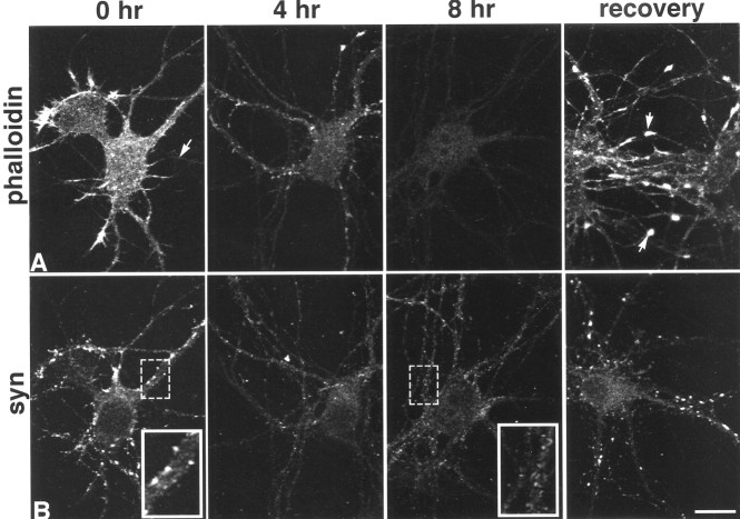
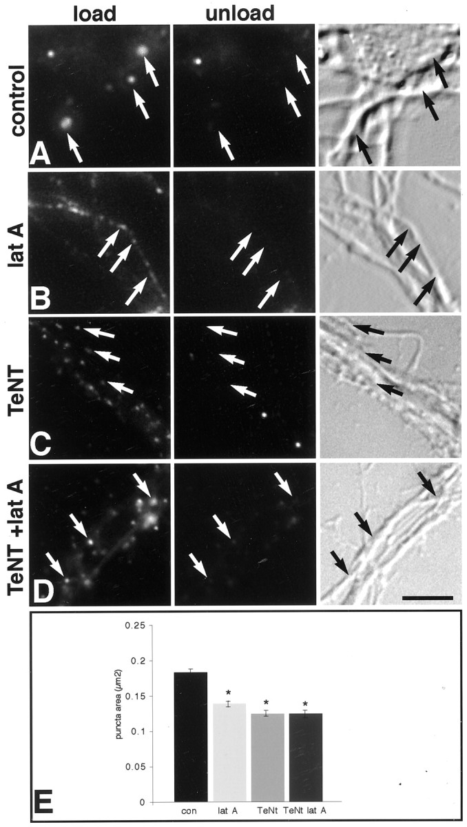
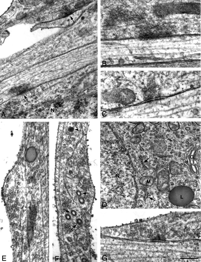
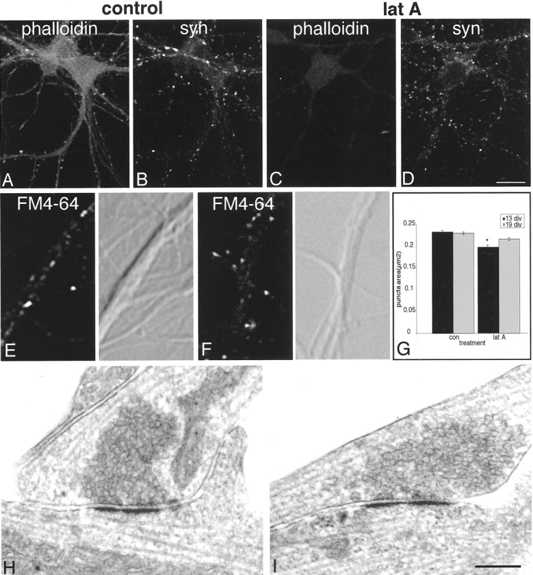
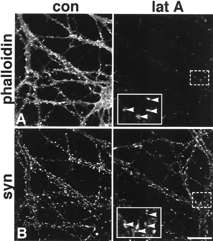
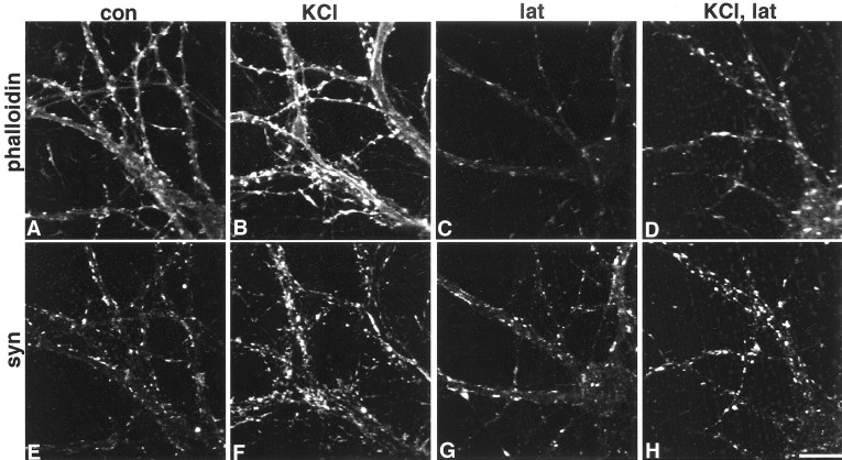
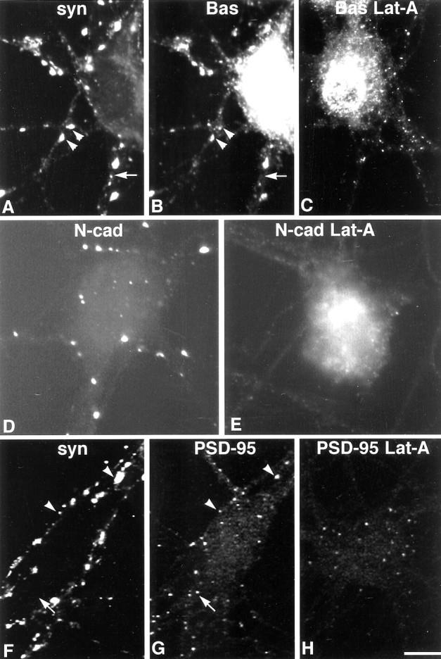
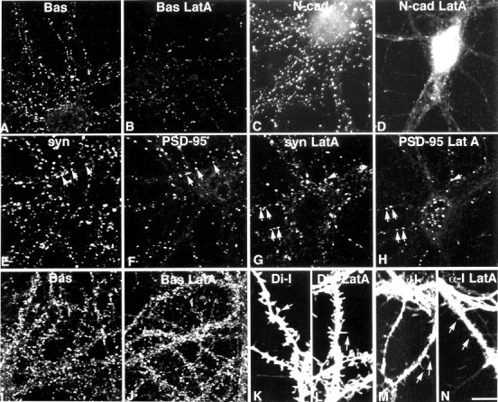
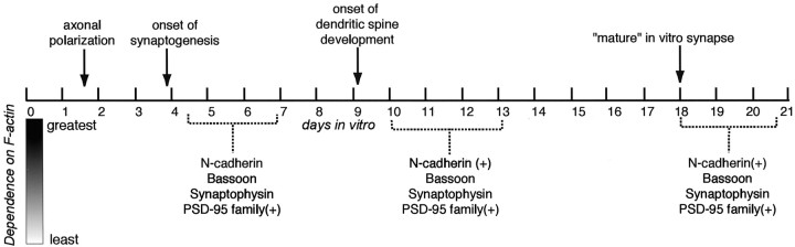
It has been widely speculated that actin plays a central role in CNS synapse assembly, but such a requirement for actin filaments (F-actin) has not yet been demonstrated experimentally. We used hippocampal neurons grown in culture and the actin depolymerizing agent, latrunculin A, to examine directly the relationship between F-actin and synapse formation and maturation. During the first week in culture, actin depolymerization results in a near complete loss of synapses defined by synaptophysin-labeled vesicle clusters, synaptic vesicle recycling, and ultrastructure. Over the second week in culture, F-actin becomes increasingly stable, but actin depolymerization no longer disrupts basic synaptic structure. There is, however, a reduction in the number and size of synaptophysin-labeled clusters and in the size of vesicle clusters undergoing FM4-64 recycling, suggesting that synaptic vesicle anchoring remains partially dependent on F-actin. By 18 d in culture, synaptophysin clusters and synaptic vesicle recycling are largely resistant to F-actin depolymerization. The decrease in synapse dependence on F-actin correlates well with the acquisition and retention of presynaptic scaffolding proteins such as Bassoon and postsynaptic scaffolding proteins such as those of the postsynaptic density-95 family. Increased activity stabilizes F-actin and its associated proteins at synaptic sites, suggesting a correlation between active synapses, actin stability, and synapse stability. Our findings demonstrate that F-actin is essential for the development and maintenance of young synapses. Because F-actin is also highly regulatable, we propose that F-actin may be a principal target for stabilizing or destabilizing signals that ultimately result in synapse maintenance or elimination.


Figures

Fig. 1.
Fig. 1.
Time course of F-actin depolymerization and loss of synaptophysin (syn) clusters in 5-d-old cultured hippocampal neurons. Phalloidin labeling shows that latrunculin A depolymerizes most F-actin by 4 hr and essentially all of it by 8 hr (A, arrow at 0 hr indicates axon). After incubation in fresh medium overnight, F-actin levels are similar to those seen at 0 hr, but growth cones become particularly enriched (arrows in A, recovery). Synaptophysin labeling (B) over the same time course reveals a progressive loss of synaptic clusters along dendrites that is replaced by lightly labeled, small granules that extend through most processes (compare insets). After recovery, synaptophysin clusters are indistinguishable from those in control cultures. Scale bar, 20.2 μm; insets, 10.1 μm.
Fig. 2.
Fig. 2.
Actin depolymerization disrupts synaptic vesicle recycling in presynaptic boutons, whereas recycling in axons is unperturbed. Fluorescent (left and middle panels) and bright-field images (right panels) show activity-dependent recycling indicated by activity-dependent FM4–64 dye uptake in left panels (load), and subsequent activity-induced destaining in middle panels (unload). Arrows indicate recycling sites. Most recycling sites in control neurons are larger and associated with somata and dendrites (A), whereas in latrunculin A (latA)-treated neurons they are smaller and mostly associated with axons (B). After treatment with TeNT alone (C) or TeNT and latrunculin A (D, TeNT +lat A), all of the cycling boutons are small and mostly associated with axons that often fasciculate together (C, D).E, Quantitative analysis of the area of recycling puncta shows control puncta to be significantly larger than any of the treatment groups, which are not significantly different from one another. An asterisk indicates a significant difference from control (ANOVA, p < 0.0001; Scheffe'spost hoc analysis). Scale bar, 7.9 μm.
Fig. 3.
Fig. 3.
Actin depolymerization results in loss of synaptic ultrastructure. Electron photomicrographs of 6-d-old neurons from control (AC) and latrunculin A-treated (DG) cultures. In untreated 6-d-old cultures, synapses are relatively common (A,arrows) and range from mature morphology having a large number of vesicles and a well formed postsynaptic density (B) to those having much smaller numbers of vesicles and only slight postsynaptic densities (C). After latrunculin A treatment, neurons look surprisingly normal as demonstrated by the clearly defined nuclear membrane, endoplasmic reticulum (arrows), and Golgi apparatus in a cell soma (D). Processes have increased numbers of small vesicles (E), clathrin-coated vesicles (arrows), tubulovesicular bodies, and large dense-core vesicles (F, arrowhead). Rare structures can be seen resembling synapses (G), but in the example shown the presynaptic density is unusually broad, cleft material is sparse, and synaptic vesicles are not apposed to the active zone. Scale bar: A, D, 500 nm;B, C, EG, 250 nm. N, Nucleus; G, Golgi;M, mitochondria; L, lysosome.
Fig. 4.
Fig. 4.
In 12-d-old neurons, the effects of actin depolymerization are modest. Phalloidin-labeled F-actin (A) requires 16–20 hr of incubation in latrunculin A (lat A) to depolymerize (C). Synaptophysin (syn) labeling remains largely intact (B vs D), but individual puncta are reduced in size and number (Table 1). In both control (E) and latrunculin A-treated (F) 12-d-old neurons, FM4–64 uptake (left panels) is largely restricted to clusters on somata and dendrites (compare with differential interference contrast images in right panels). Cluster area is slightly but significantly reduced in size (G, theasterisk indicates a significant difference from control, p < 0.0001). Electron microscopy of 12-d-old neurons reveals that synapses in control cultures cover a broad range of maturity (H) and are not distinguishably different in latrunculin A-treated cultures (I). Scale bar:AD, 26.9 μm;EH, 250 nm.
Fig. 5.
Fig. 5.
In 20-d-old neurons, some synaptic F-actin is exceedingly stable. Most F-actin is depolymerized in 24 hr (A). However, even at 24 hr, some F-actin clusters remain (A). Synaptophysin (syn) labeling (B) of the same neurons shown in A is unaltered by the treatment.Boxed regions are shown at higher magnification ininsets and illustrate that many of the most stable F-actin puncta are codistributed with synaptophysin puncta (arrowheads). con, Control; lat A, latrunculin A. Scale bar, 26.9 μm; insets, 10.8 μm.
Fig. 6.
Fig. 6.
Activity can reorganize and stabilize F-actin. In 17-d-old neurons, 5 min exposure to 75 mm KCl results in an increase in phalloidin-labeled F-actin in the cortex and dendritic protrusions [control (A, con) vs stimulated (B)], and intensity of synaptophysin (syn)-labeled puncta appears to increase and spread slightly in the same neurons [control (E) vs stimulated (F)]. When neurons incubated in latrunculin A (lat) for 24 hr (C,G) are compared with those that were first stimulated for 5 min with 75 mm KCl (D,H), there are more phalloidin-labeled F-actin puncta (D vs C) that codistribute with synaptophysin-labeled puncta (G,H) (compare C and Gwith D and H). Scale bar, 20.2 μm.
Fig. 7.
Fig. 7.
In 6-d-old neurons, the distribution of synaptic proteins is disrupted by latrunculin A. Bassoon (Bas) labeling (B) mostly codistributes with synaptophysin (A, syn) in control cultures (A, B,arrowheads), although small synaptophysin puncta sometimes lack detectable Bassoon labeling (A,B, arrow). Like synaptophysin, clusters of Bassoon labeling are greatly diminished after actin depolymerization (C). Very small Bassoon-labeled puncta remain, perhaps denoting sites of synapse loss or the vesicles seen in processes after depolymerization. Clusters of synaptic N-cadherin (N-cad) labeling (D) are lost after exposure to latrunculin A (E,Lat-A). Synaptophysin (F) and PSD-95 (G) are partially codistributed at this age (arrowheads), but a number of PSD-95 puncta are synaptophysin negative (F, G,arrow). After latrunculin A exposure, most PSD-95-labeled puncta are found within cell somata (H). Scale bar, 11.3 μm.
Fig. 8.
Fig. 8.
In 12-d-old neurons (AH), Bassoon-labeled clusters (A, Bas) are diminished in size and number after latrunculin A treatment (B, Bas LatA), but are still readily detected. N-cadherin-labeled clusters (C, N-cad) are lost after latrunculin A treatment (D, N-cad LatA), but a few tiny puncta remain (D). In control neurons, PSD-95 labeling (F) codistributes with a subpopulation of synaptophysin boutons (E,syn, arrows). After latrunculin A treatment (G, H), many synaptic PSD-95 puncta are retained (G, H,arrows), but many PSD-95 puncta are nonsynaptic, which is particularly evident in the cell soma (H). In 20-d-old neurons, latrunculin A treatment does not affect the distribution of Bassoon (J) compared with untreated control neurons (I). Di-I labeling reveals both spine-like and filopodia-like protrusions in control neurons (K). After latrunculin A treatment, nearly all protrusions appear filopodia-like (L). α-internexin (α-I) labeling is concentrated in axons, but also appears in dendrites where it can be seen to invade protrusions (M). After latrunculin A treatment, α-internexin labeling remains at the base of dendritic protrusions (N). Scale bar:AJ, 20.2 μm;KN, 11.3 μm.
Fig. 9.
Fig. 9.
Juxtaposed against a time line summarizing major developmental events in cultured hippocampal neurons are synaptic proteins that are written with black, medium gray, or light gray to depict their dependence on F-actin for their synaptic localization (black = most dependent). Molecules restricted to excitatory synapses are indicated by (+).

References

    1. Adams CL, Nelson WJ, Smith SJ. Quantitative analysis of cadherin-catenin-actin reorganization during development of cell-cell adhesion. J Cell Biol. 1996;135:1899–1911. - PMC - PubMed
    1. Adam G, Matus A. Role of actin in the organization of brain postsynaptic densities. Brain Res Mol Brain Res. 1996;43:246–250. - PubMed
    1. Ahmari SE, Buchanan J, Smith SJ. Assembly of presynaptic active zones from cytoplasmic transport packets. Nat Neurosci. 2000;3:445–451. - PubMed
    1. Allison DW, Gelfand VI, Spector I, Craig AM. Role of actin in anchoring postsynaptic receptors in cultured hippocampal neurons: differential attachment of NMDA versus AMPA receptors. J Neurosci. 1998;18:2423–2436. - PMC - PubMed
    1. Allison DW, Chervin AS, Gelfand VI, Craig AM. Postsynaptic scaffolds of excitatory and inhibitory synapses in hippocampal neurons: maintenance of core components independent of actin filaments and microtubules. J Neurosci. 2000;20:4545–4554. - PMC - PubMed

Publication types

MeSH terms

LinkOut - more resources