. 2001 Jul 15;21(14):5358-66.
doi: 10.1523/JNEUROSCI.21-14-05358.2001.

Local injection of endothelin-1 produces pain-like behavior and excitation of nociceptors in rats

Affiliations

Local injection of endothelin-1 produces pain-like behavior and excitation of nociceptors in rats

A P Gokin et al. J Neurosci. .

Abstract

Neurobehavioral and neurophysiological actions of the peptide endothelin-1 (ET-1) were investigated after subcutaneous plantar hindpaw injections in adult male Sprague Dawley rats. Hindpaw flinching developed within minutes after ET-1 (8-16 nmol) injection, peaked at 30 min, lasted for 60 min, and was strongly inhibited by the endothelin-A (ET(A)) receptor antagonist, BQ-123 (3.2 m). In separate experiments, impulse activity of single, physiologically characterized sensory C-, Adelta-, and Abeta-fibers was recorded from the sciatic nerve in anesthetized rats after subcutaneous injections of endothelin-1 (1-20 nmol), alone or together with BQ-123 (3.2 m), into the plantar hindpaw receptive fields of these units. All nociceptive C-fibers (31 of 33 C-fibers studied) were excited by ET-1 (1-20 nmol) in a dose-dependent manner. For doses of 16-20 nmol, the mean latency for afferent activation after injection of ET-1 was 3.16 +/- 0.31 min, and the mean and maximum response frequency were 2.02 +/- 0.48 impulses (imp)/sec and 14.0 +/- 3.2 imp/sec, respectively. All 10 nociceptive Adelta-fibers (of 12 Adelta-fibers studied) also responded to 1-20 nmol of ET-1 in a dose-dependent manner with a mean latency of 3.5 +/- 0.12 min and mean response frequency of 3.3 +/- 2.3 imp/sec. In contrast, most Abeta-fibers (9 of 12) did not respond to ET-1. BQ-123, when coinjected with ET-1, blocked ET-1-induced activation in all C- and Adelta-fibers tested. These data demonstrate that subcutaneous administration of ET-1 to the rat plantar hindpaw produces pain-like behavior and selective excitation of nociceptive fibers through activation of ET(A) receptors.


Figures

Fig. 1.
Fig. 1.
Hindpaw flinching induced by the injection of 8, 12, and 16 nmol (n = 10 animals for each dose) ET-1 (equivalent to 200, 300, and 400 μm, respectively) subcutaneously into the rat plantar hindpaw. Termination of ET-1 administration was at t = −5 min; animals were allowed 5 min to recover before assessments began. The frequency of hindpaw flinching occurring every 5 min, measured fromt = 0, over a period of 60 min was determined. Differences between 400 μm ET-1 and PBS were present from 10 to 40 min after observations began (*p < 0.05; f-ANOVA, StatView).
Fig. 2.
Fig. 2.
Blockade of ET-1-induced hindpaw flinching by subcutaneous BQ-123 (3.2 mm) applied together with ET-1 (400 μm) over a 9–10 min period. Termination of ET-1 + BQ-123 administration was at t = −5 min; animals were allowed 5 min to recover before assessments began. Differences between ET-1/BQ-123-treated and ET-1-treated animals are significant at all time points (*p < 0.05; f-ANOVA, StatView).
Fig. 3.
Fig. 3.
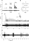
Excitation of an HTMr C-fiber after subcutaneous injection of ET-1 into the rat plantar hindpaw. A, Physiological characterization of an HTMr C-fiber (CV 0.72 m/sec) showing the fiber response to graded mechanical stimulation (below), presented also as a bin histogram (above). B, Procedure for confirming single unit activity. Top and bottom traces, Spikes are evoked by transcutaneous electrical nerve stimulation (ES) that has a consistent latency of 63 msec; middle trace, the electrically evoked spike fails to appear, presumably because of the refractory period generated by the spike evoked by mechanical stimulation of the receptive field.C, Representative C-fiber response (CV 1.8 m/sec) to the subcutaneous injection of 2 nmol (10 μl of 200 μm) of ET-1 (below), presented also as a bin histogram (above). Note the short latency to onset of spiking, and the “bursting” pattern of this spike activity occurring within the first 10 min of the response. Inset shows the location of the receptive field of this unit. D, Expanded time scale of the early response to ET-1 (shown in C) illustrating the approach to latency measurement. Thearrow points to the beginning of injection, whereas thetwin arrows and intervening line denote the end of injection (needle withdrawal) and latency (LAT) to the first measured spike response.
Fig. 4.
Fig. 4.
Dose dependence of excitation of C-fibers induced by subcutaneous ET-1 2 nmol (n = 10), 8 nmol (n = 5), and 16–20 nmol (n = 17). Shown are the averaged MRF (A), mean duration of responses (B), and mean latency of responses (C). *p < 0.05 when compared with 16–20 nmol and p< 0.05 when compared with 2 nmol.
Fig. 5.
Fig. 5.
Blockade of HTMr C-fiber (CV 1.18 m/sec) activation by coinjection of BQ 123 (3.2 mm) together with 8 nmol of ET-1. The physiological responses of this unit to graded mechanical stimulation (von Frey hairs) before (A) and after (B) ET-1/BQ-123 are shown. C, Four successive injections of ET-1/BQ 123 (10 μl each) failed to induce spike responses for a total period of 60 min), although the unit maintained its responsiveness to graded mechanical stimulation (Stim RF).D, Spike activity appeared at ∼50 min after the coinjection of ET-1/BQ-123 and was augmented by successive injections of ET-1 (2 nmol) alone.
Fig. 6.
Fig. 6.
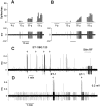
Spike responses in two different HTMr Aδ-fibers after subcutaneous injection of 16 nmol of ET-1. A, In the first unit (CV 3.08 m/sec), a bin histogram illustrates the increasing frequency of spiking within the first 5 min after completion of injections, followed by a gradual decline in spike frequency. Noxious mechanical stimulation performed 9 min after ET-1 injection demonstrates continued responsiveness of the RF of this unit.Inset shows the location of the receptive field of this unit. B, In the second unit (CV 5.7 m/sec), the characteristic bursting pattern of the spike response is shown in a single episode extracted from the full response after injection with 16 nmol of ET-1.
Fig. 7.
Fig. 7.
Lack of response of an Aβ-fiber (hair follicle afferent, CV 36.4 m/sec) to subcutaneous injection of 16 nmol of ET-1.A, Record of the response of this fiber to hair bending, brush, and von Frey hair (3.5 gm) stimulation of its RF located at the border between hairy and glabrous plantar skin (seeinset). B, Identification of the unit with the collision method (see Fig. 3B).C, Failure of this same unit to respond to 16 nmol of ET-1 (total dose) into its RF.

References

    1. Carducci MA, Bowling MK, Rogers T, Leahy TL, Janus TJ, Padley RJ, Nelson JB (1998) Endothelin receptor antagonist, ABT-627, for prostate cancer: initial trial results. Presented at American Association for Cancer Research Meeting, Indian Wells, CA, December. Abstr. C-24.
    1. Crossman DC, Brain SD, Fuller RW. Potent vasoactive properties of endothelin 1 in human skin. J Appl Physiol. 1991;70:260–266. - PubMed
    1. Dahlof B, Gustafsson D, Hedner T, Jern S, Hannsson L. Regional hemodynamic effects of endothelin-1 in rat and man: unexpected adverse effects. J Hypertens. 1990;8:811–818. - PubMed
    1. Davar G, Hans G, Fareed MU, Sinnott C, Strichartz G. Behavioral signs of acute pain produced by application of endothelin-1 to rat sciatic nerve. NeuroReport. 1998;9:2279–2283. - PubMed
    1. De-Melo JD, Tonussi CR, D'Orleans-Juste P, Rae GA. Articular nociception induced by endothelin-1, carrageenan and LPS in naive and previously inflamed knee-joints in the rat: inhibition by endothelin receptor antagonists. Pain. 1998;77:261–269. - PubMed

Publication types

MeSH terms