. 2001 Aug;21(15):4929-37.
doi: 10.1128/MCB.21.15.4929-4937.2001.

c-Myc is necessary for DNA damage-induced apoptosis in the G(2) phase of the cell cycle

Affiliations

c-Myc is necessary for DNA damage-induced apoptosis in the G(2) phase of the cell cycle

S Adachi et al. Mol Cell Biol. 2001 Aug.

Abstract

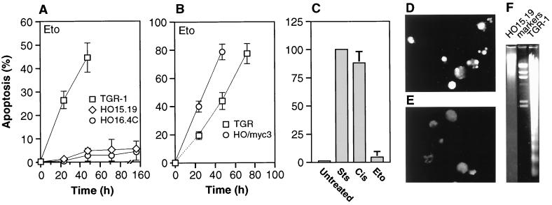
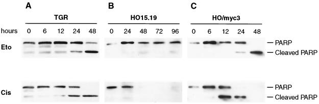
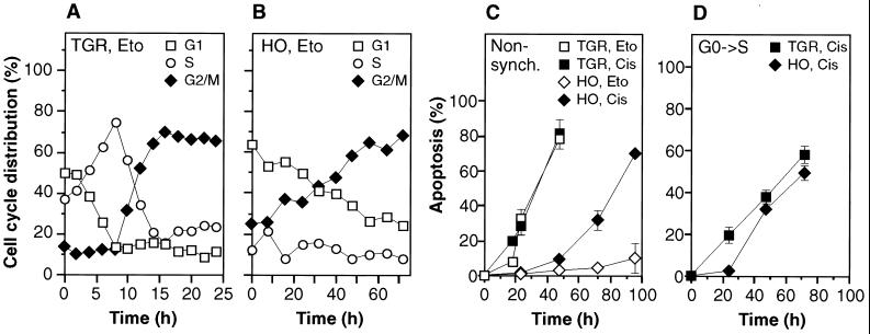
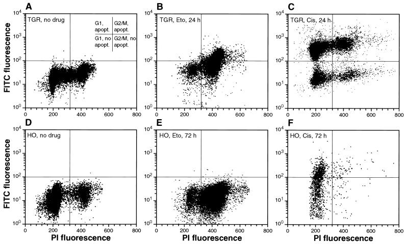
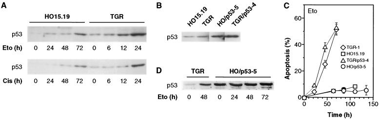
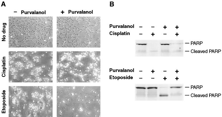
The c-myc proto-oncogene encodes a transcription factor that participates in the regulation of cellular proliferation, differentiation, and apoptosis. Ectopic overexpression of c-Myc has been shown to sensitize cells to apoptosis. We report here that cells lacking c-Myc activity due to disruption of the c-myc gene by targeted homologous recombination are defective in DNA damage-initiated apoptosis in the G(2) phase of the cell cycle. The downstream effector of c-Myc is cyclin A, whose ectopic expression in c-myc(-/-) cells rescues the apoptosis defect. The kinetics of the G(2) response indicate that the induction of cyclin A and the concomitant activation of Cdk2 represent an early step during commitment to apoptosis. In contrast, expression of cyclins E and D1 does not rescue the apoptosis defect, and apoptotic processes in G(1) phase are not affected in c-myc(-/-) cells. These observations link DNA damage-induced apoptosis with cell cycle progression and implicate c-Myc in the functioning of a subset of these pathways.


Figures

FIG. 1
FIG. 1
(A) Apoptosis elicited in c-myc+/+ and c-myc−/− cells by treatment with etoposide (Eto). (B) Rescue of apoptosis by reconstitution of c-Myc activity. (C) Apoptosis elicited in c-myc−/− cells by a variety of agents. Sts, staurosporine; Cis, cisplatin; Eto, etoposide. Cells were treated for 48 h. (D and E) TUNEL assay of c-myc+/+ (D) and c-myc−/− (E) cells. TGR-1 and HO15.19 cells were harvested after 24 and 96 h of treatment with etoposide, respectively. (F) Nucleosomal laddering assay of c-myc+/+ and c-myc−/− cells. Cells were treated as in panels D and E, and 3 μg of DNA was loaded into each lane. Cell lines: TGR-1, c-myc+/+; HO15.19, HO16.4, c-myc−/−; HO/myc3, c-myc−/− expressing ectopic c-myc. HO/myc3 cells express wild-type murine c-Myc at a level two- to threefold above that found in TGR-1 cells.
FIG. 2
FIG. 2
PARP cleavage elicited by drug treatment of c-myc+/+ and c-myc−/− cells. Exponentially growing cultures of the indicated cell lines were treated with drugs at the zero time point, collected at the indicated times, and analyzed by immunoblotting. Eto, etoposide treatment; Cis, cisplatin treatment. See Fig. 1 for a description of the cell lines.
FIG. 3
FIG. 3
(A and B) Cell cycle progression of c-myc+/+ (A) and c-myc−/− (B) cells following treatment with etoposide (Eto). Samples were collected concurrently with the experiment shown in Fig. 1A and analyzed by flow cytometry. The G2/M plateau remained constant until the termination of the experiments (48 h for TGR-1 cells, 96 h for HO15.19 cells). (C) Apoptosis elicited by cisplatin (Cis). (D) Apoptosis elicited in synchronized cells by cisplatin. Quiescent cultures were stimulated with serum at the zero time point and treated with cisplatin 2 h later.
FIG. 4
FIG. 4
Concurrent analysis of apoptosis and cell cycle progression. Exponentially growing cultures of c-myc+/+ (A, B, and C) and c-myc−/− (D, E, and F) cells were treated with etoposide (Eto) or cisplatin (Cis) as indicated, harvested at the indicated time points, processed for TUNEL using the Apo-Direct kit (Pharmingen), counterstained with propidium iodide, and analyzed by two-parameter flow cytometry.
FIG. 5
FIG. 5
Relationship of p53 expression and apoptosis. (A) Expression of p53 after DNA damage. Exponentially growing cells were treated with drugs at the zero time point, collected at the indicated times, and analyzed by immunoblotting. (B) Expression of p53 in cells ectopically expressing a temperature-sensitive mutant of p53. Subconfluent cells at 37°C in the absence of drugs were analyzed by immunoblotting. (C) Influence of p53 overexpression on apoptosis elicited by etoposide. All cells were grown at 37°C prior to the experiment. Exponentially growing subconfluent cells were treated with etoposide at the zero time point and shifted to 32.5°C. Cell cycle profiles, monitored by flow cytometry, were similar to those shown in Fig. 2A and B. (D) p53 expression in the experiment shown in panel C. Samples were collected at the indicated times and analyzed by immunoblotting.
FIG. 6
FIG. 6
Effect of the Cdk inhibitor aminopurvalanol on apoptosis induced by etoposide and cisplatin. Exponentially growing c-myc+/+ cells were treated with drugs as indicated and incubated for 48 h. At the end of this incubation the cultures were photographed (A), and the cells were recovered and processed for immunoblotting (B) with anti-PARP antibody.
FIG. 7
FIG. 7
Relationship of cyclin A expression and apoptosis. (A) Expression of cyclin A after etoposide treatment. Exponentially growing cells were treated with drugs at the zero time point, collected at the indicated times, and analyzed by immunoblotting. (B) Expression of cyclin A in cells ectopically expressing cyclin A. Subconfluent cells in the absence of drugs were analyzed by immunoblotting. (C) Cyclin A expression in the experiment shown in panel E analyzed by immunoblotting. (D) Cyclin A-associated Cdk activity after etoposide treatment. Exponentially growing cells were treated with drugs at the zero time point, collected at the indicated times, immunoprecipitated with anti-cyclin A antibody, and assayed for histone H1 kinase activity. (E) Influence of cyclin A overexpression on apoptosis elicited by etoposide. Exponentially growing subconfluent cells were treated with etoposide at the zero time point. (F) Influence of cyclin D and cyclin E overexpression on apoptosis elicited by etoposide. The experiment was performed as indicated in panel E. (G) Cell cycle progression profiles in the experiment shown in panel E.

References

    1. Adachi S, Ito H, Tamamori-Adachi M, Ono Y, Nozato T, Abe S, Ikeda M, Marumo F, Hiroe M. Cyclin A/cdk2 activation is involved in hypoxia-induced apoptosis in cardiomyocytes. Circ Res. 2001;88:408–414. - PubMed
    1. Anderson J A, Lewellyn A L, Maller J L. Ionizing radiation induces apoptosis and elevates cyclin A1-Cdk2 activity before but not after the midblastula transition in Xenopus. Mol Biol Cell. 1997;8:1195–1206. - PMC - PubMed
    1. Askew D S, Ashmun R A, Simmons B C, Cleveland J L. Constitutive c-myc expression in an IL-3-dependent myeloid cell line suppresses cell cycle arrest and accelerates apoptosis. Oncogene. 1991;6:1915–1922. - PubMed
    1. Canelles M, Delgado M D, Hyland K M, Lerga A, Richard C, Dang C V, Leon J. Max and inhibitory c-Myc mutants induce erythroid differentiation and resistance to apoptosis in human myeloid leukemia calls. Oncogene. 1997;14:1315–1327. - PubMed
    1. Chang Y T, Gray N S, Rosania G R, Sutherlin D P, Kwon S, Norman T C, Sarohia R, Leost M, Meijer L, Schultz P G. Synthesis and application of functionally diverse 2,6,9-trisubstituted purine libraries as CDK inhibitors. Chem Biol. 1999;6:361–375. - PubMed

Publication types

MeSH terms

LinkOut - more resources