. 2001 Jul 17;98(15):8525-30.
doi: 10.1073/pnas.131213698. Epub 2001 Jul 3.

BeF(3)(-) acts as a phosphate analog in proteins phosphorylated on aspartate: structure of a BeF(3)(-) complex with phosphoserine phosphatase

Affiliations

BeF(3)(-) acts as a phosphate analog in proteins phosphorylated on aspartate: structure of a BeF(3)(-) complex with phosphoserine phosphatase

H Cho et al. Proc Natl Acad Sci U S A. .

Abstract

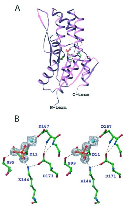
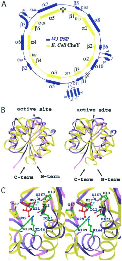
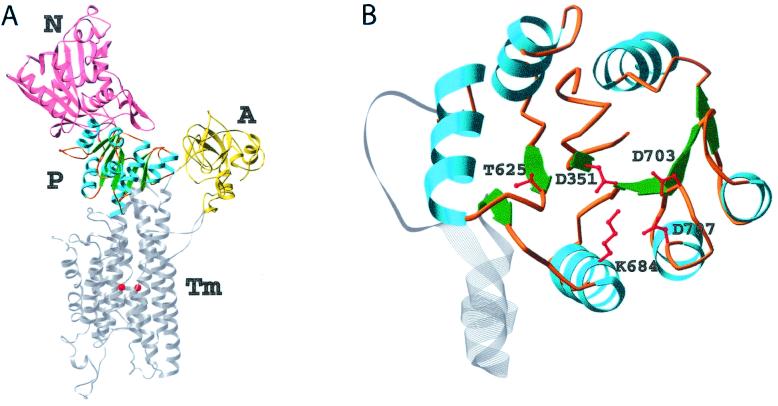
Protein phosphoaspartate bonds play a variety of roles. In response regulator proteins of two-component signal transduction systems, phosphorylation of an aspartate residue is coupled to a change from an inactive to an active conformation. In phosphatases and mutases of the haloacid dehalogenase (HAD) superfamily, phosphoaspartate serves as an intermediate in phosphotransfer reactions, and in P-type ATPases, also members of the HAD family, it serves in the conversion of chemical energy to ion gradients. In each case, lability of the phosphoaspartate linkage has hampered a detailed study of the phosphorylated form. For response regulators, this difficulty was recently overcome with a phosphate analog, BeF(3)(-), which yields persistent complexes with the active site aspartate of their receiver domains. We now extend the application of this analog to a HAD superfamily member by solving at 1.5-A resolution the x-ray crystal structure of the complex of BeF(3)(-) with phosphoserine phosphatase (PSP) from Methanococcus jannaschii. The structure is comparable to that of a phosphoenzyme intermediate: BeF(3)(-) is bound to Asp-11 with the tetrahedral geometry of a phosphoryl group, is coordinated to Mg(2+), and is bound to residues surrounding the active site that are conserved in the HAD superfamily. Comparison of the active sites of BeF(3)(-) x PSP and BeF(3)(-) x CeY, a receiver domain/response regulator, reveals striking similarities that provide insights into the function not only of PSP but also of P-type ATPases. Our results indicate that use of BeF(3)(-) for structural studies of proteins that form phosphoaspartate linkages will extend well beyond response regulators.


Figures

Figure 1
Figure 1
1H-15N FHSQC spectrum of BeFformula image⋅PSP. PSP (0.5 mM) was incubated with 5 mM MgCl2, 3 mM NaF, and 0.6 mM BeCl2. Red crosses represent the crosspeak positions in the absence of NaF and BeCl2. Spectra were recorded at 308 K and pH 6.5.
Figure 2
Figure 2
Ribbon diagram of BeFformula image⋅PSP (A) and stereoview of the active site (B). Ball-and-stick representations of the sidechain carbon, nitrogen, and oxygen atoms are colored green, blue, and red, respectively. In A, the sidechains of highly conserved active site residues, Asp-11, Ser-99, Lys-144, Asp-167, and Asp-171, are shown in ball-and-stick form. BeFformula image (red balls and orange bonds) is bonded to Asp-11 Oδ in the center of the active site (33). In B, the 2FoFc electron density map covering the magnesium (cyan) and fluorine (red) atoms was calculated in the absence of these atoms and is shown contoured at 1σ. The dashed lines represent hydrogen bonds, salt bridges, and metal–ligand interactions. Three of the Mg2+ ligands (two water molecules and Asp-13-C = O) are not shown.
Figure 3
Figure 3
Alignment and stereoviews showing the superposition of BeFformula image⋅PSP and BeFformula image⋅CheY. Structurally homologous β-strand residues (PSP: 7–11, 94–98, 116–120, 162–167, 180–182, and CheY: 53–57, 82–86, 104–108, 8–13, 34–36) were used for the superposition. PSP and CheY are colored purple and yellow, respectively. (A) Diagram of the circular permutation required to align the conserved active site residues of PSP and CheY (29). (B) Ribbon structures of PSP and CheY. The molecules are oriented with the H4-β5-H5 face of CheY directed toward the reader. The N and C termini of CheY are denoted. Some PSP residues that have no structural counterparts in CheY are not shown. (C) A top-down view of the superimposed active sites. This orientation was obtained by applying a −90° rotation around the x-axis starting from the orientation in B. The active site residues of PSP and CheY are labeled in purple and black, respectively. The sidechains are shown in ball-and-stick form with carbon, nitrogen, and oxygen atoms colored green, blue, and red, respectively. BeFformula image is represented by red balls with orange bonds. The cyan- and blue-green-colored balls, lower and upper, respectively, adjacent to BeFformula image represent the Mg2+ ions in the PSP and CheY structures, respectively. The purple arrow points to D167 of PSP.
Figure 4
Figure 4
Ribbon diagrams of the Ca2+-ATPase from rabbit sarcoplasmic reticulum (A) and its catalytic P domain (B) [drawn from coordinates of Toyoshima et al. (32)]. (A) The N (nucleotide-binding), A (actuator), and M (Ca2+-binding) domains are shown in pink, yellow, and gray, respectively. The latter traverses the membrane of the sarcoplasmic reticulum. The two Ca2+ ions are shown in red. The catalytic P domain is shown in blue (helices), green (β-strands), and orange (loops). (B) This top-down view was obtained by applying a −90° rotation around the x-axis starting from the orientation in A. The quintet of conserved residues in the P domain is shown in ball-and-stick form in red. Mg2+ was apparently not present in this domain.

Comment in

References

    1. Post R L, Kume S. J Biol Chem. 1973;248:6993–7000. - PubMed
    1. Weiss V, Magasanik B. Proc Natl Acad Sci USA. 1988;85:8919–8923. - PMC - PubMed
    1. Hoch J A, Silhavy T J. Two-Component Signal Transduction. Washington, DC: Am. Soc. Microbiol.; 1995.
    1. Djordjevic S, Stock A M. J Struct Biol. 1998;124:189–200. - PubMed
    1. Falke J J, Bass R B, Butler S L, Chervitz S A, Danielson M A. Annu Rev Cell Dev Biol. 1997;13:457–512. - PMC - PubMed

Publication types

MeSH terms

Associated data