. 2001 Jul 17;98(15):8650-5.
doi: 10.1073/pnas.151236498. Epub 2001 Jul 3.

Subnuclear targeting of Runx/Cbfa/AML factors is essential for tissue-specific differentiation during embryonic development

Affiliations

Subnuclear targeting of Runx/Cbfa/AML factors is essential for tissue-specific differentiation during embryonic development

J Y Choi et al. Proc Natl Acad Sci U S A. .

Abstract

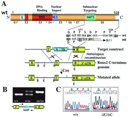
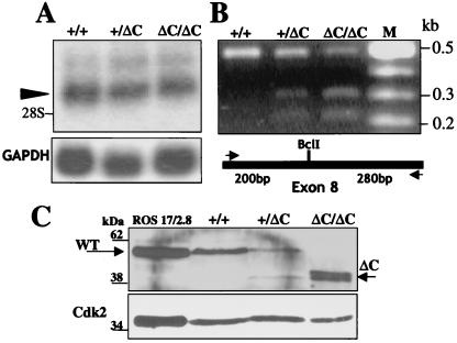
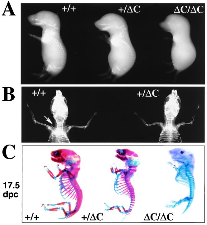
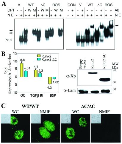
Runx (Cbfa/AML) transcription factors are critical for tissue-specific gene expression. A unique targeting signal in the C terminus directs Runx factors to discrete foci within the nucleus. Using Runx2/CBFA1/AML3 and its essential role in osteogenesis as a model, we investigated the fundamental importance of fidelity of subnuclear localization for tissue differentiating activity by deleting the intranuclear targeting signal via homologous recombination. Mice homozygous for the deletion (Runx2 Delta C) do not form bone due to maturational arrest of osteoblasts. Heterozygotes do not develop clavicles, but are otherwise normal. These phenotypes are indistinguishable from those of the homozygous and heterozygous null mutants, indicating that the intranuclear targeting signal is a critical determinant for function. The expressed truncated Runx2 Delta C protein enters the nucleus and retains normal DNA binding activity, but shows complete loss of intranuclear targeting. These results demonstrate that the multifunctional N-terminal region of the Runx2 protein is not sufficient for biological activity. We conclude that subnuclear localization of Runx factors in specific foci together with associated regulatory functions is essential for control of Runx-dependent genes involved in tissue differentiation during embryonic development.


Figures

Figure 1
Figure 1
Gene replacement for Runx2ΔC. (A) Generation of the mutant locus. The schematic at the top shows the functional domains of Runx proteins and Runx2 exon organization. Domains in the N terminus (N) conserved among Runx transcription factors are indicated: the runt homology/DNA binding domain (RHD) and the nuclear localization signal (NLS). The letters Q and A designate homopolymeric stretches of glutamine and alanine residues unique to Runx2. The C terminus (C) defined by exon 8 includes the nuclear matrix targeting signal (NMTS). Below this schematic is indicated the sequence of the WT (wt) and mutated (mt) allele, and ** denotes the premature stop codon. The bottom portion shows a diagram of target constructs used for introduction of the mutation at the start of exon 8. The C-terminal genomic organization of the WT and mutated alleles are also illustrated. The restriction sites (B, BamHI; E, EcoRI) in the Runx2 genomic locus and the regions of homologous recombination are indicated. Removal of the neo-thymidine kinase cassette by Cre recombinase results in the mutant allele. The loxP sites are shown by filled triangles. The Runx2 gene encodes two major isoforms with two distinct N termini (p56/Type I and p57/Type II) that are generated from two different promoters. Our strategy produces a C-terminal deletion in both isoforms. UTR, untranslated region. (B) Genotyping by PCR analysis of mouse tail genomic DNA from WT (+/+), heterozygous (+/ΔC), and homozygous mutant (ΔC/ΔC) mice. Locations of the forward and reverse primers (which span the lox P site) used as probes for genotyping are indicated by arrows. The presence of the lox P site generates a slower migrating PCR band that represents the Runx2ΔC mutant allele carrying the premature stop codon. (C) Sequence analysis of the PCR products amplified from genomic DNA of WT (+/+) and homozygous mutant (ΔC/ΔC) mice confirms in vivo incorporation of the premature stop codon (denoted by *).
Figure 2
Figure 2
Runx2ΔC mRNA expression and protein synthesis in vivo. (A) Northern blot analysis of total cellular RNA (10 μg/lane) prepared from the heads of WT (+/+), heterozygous (+/ΔC), and homozygous (ΔC/ΔC) embryos at 17.5 dpc. The Runx2 major transcript (arrowhead) migrates above the 28S ribosomal RNA. (Lower) Glyceraldehyde-3-phosphate dehydrogenase (GAPDH) mRNA as a control for RNA loading. (B) Detection of WT and Runx2ΔC mRNAs in +/+, +/ΔC, and ΔC/ΔC mouse embryos by RT-PCR and BclI restriction enzyme digestion. Lane M represents size markers. Schematic shows the locations of the primers (arrows) used for PCR amplification of the WT and mutant mRNAs. The BclI site present in the mutant generates the 0.2- and 0.3-kb digestion products. (C) Western blot analysis of nuclear lysates from cultured calvarial cells isolated from +/+, +/ΔC, and ΔC/ΔC Runx2 mice at 17.5 dpc and cultured for 2 wk. Lysates from ROS 17/2.8 osteoblast-like cells serve as a positive control for full-length Runx2. Standard immunoblotting was performed with the monoclonal Runx2 antibody. The expected molecular masses for Runx2 WT and ΔC proteins are indicated by arrows below the 62-kDa and above the 38-kDa markers, respectively. Relative marker positions are indicated.
Figure 3
Figure 3
The Runx2ΔC knock-in results in embryonic lethality in homozygotes and absence of clavicles in heterozygotes. (A) Whole animal contact radiographs of 17.5 dpc mice (Faxitron X-Ray, Wheeling, IL). At this age no skeletal elements are detected by soft x-rays in the ΔC/ΔC compared with +/+ and +/ΔC mice. (B) Radiography of 6-wk mice to visualize phenotype of the heterozygote. Shown is a dorsal view of a +/+ mouse with arrow pointing to the clavicle that is missing in the +/ΔC mouse. (C) Staining of skeletons from (17.5 dpc and 18.5 dpc) embryos with Alizarin red to detect bone and Alcian blue to detect cartilage by standard procedures (60).
Figure 4
Figure 4
Absence of bone tissues in the ΔC/ΔC knock-in mutant mouse. Histologic appearance of limbs from WT and Runx2ΔC homozygous mutant embryos (ΔC/ΔC) at 17.5 dpc. Tibial bone paraffin sections stained with Safranin O-fast green (ad) from WT (a and c) and homozygous ΔC/ΔC (b and d) mice at ×4 (a and b) and ×10 (c and d) magnifications to show growth plate zones. Cartilage (red) and bone, marrow, blood vessels (blue) are distinguished. Brackets in a and b show regions at higher magnification in c and d. Undecalcified tibial bone sections with hematoxylin and eosin/von Kossa silver staining (to detect mineral) for WT (e) and ΔC/ΔC (f) at ×10 magnification. (g) Alkaline phosphatase and (h) tartrate-resistant acid phosphatase histochemistry of the ΔC/ΔC bone (×20). (i) The central portion of the limb from ΔC/ΔC bone and indicates accumulation of blood vessels (hematoxylin and eosin stain) (×40 magnification). Arrow indicates one osteoclast (Ocl). V designates blood vessels. P shows periochondrium.
Figure 5
Figure 5
Functional properties of the Runx2ΔC protein. (A) Runx2ΔC protein retains DNA binding activity. (Left) HeLa cell nuclear extracts (NE) were prepared 24 h after transfection with empty vector (V), WT, or Runx2ΔC mutant protein expression plasmids. Nuclear extracts from Ros 17/2.8 cells that endogenously express Runx2 provided a positive control. An oligonucleotide containing the Runx consensus sequence was used as labeled probe in all lanes. Competitor lanes (CPT) contained 100-fold excess of either WT (W) or mutant (M) Runx binding site oligonucleotides compared with control without specific competitor (−) are indicated. The arrows indicate the specific protein-DNA complexes for full-length (solid arrow) and truncated (open arrow) Runx2; NS represent a nonspecific band. (Right) Gel mobility-immunoshift analysis with (+) or without (−) the addition of 1 μl polyclonal Runx2 antibody (Ab) to detect WT and Runx2ΔC (ΔC) mutant proteins transiently expressed in HeLa cells. CON designates two control lanes for probe alone and probe plus antibody. V represents HeLa cell nuclear extracts transfected with the empty vector. The arrow indicates the supershifted complex. (B) Functional activity of the WT and ΔC Runx proteins on Runx-dependent promoters. (Left) HeLa cells were cotransfected with −1,097/+23 rat osteocalcin (OC), −1.0-kb transforming growth factor β R1, or −620/+25 bone sialoprotein (BSP) promoter-chloramphenicol acetyltransferase constructs together with either X-press-tagged WT Runx2 or Runx2ΔC expression plasmids (–42). Promoter activities were determined 24 h posttransfection. Studies were repeated twice with n = 6 replicates with two different DNA preparations. Data were normalized to luciferase values. (Right) Western blot analysis of lysates from transfected cells from the same experiment. The X-press-tagged WT and mutant Runx proteins were detected by using α-X-press antibody from Invitrogen. Lamin B shows equal protein loading. (C) In vivo targeting of Runx2ΔC to subnuclear sites is compromised. Cells were obtained from calvarial tissue of WT and homozygous ΔC mutant mice at 17.5 dpc cultured for 2 days on glass coverslips and prepared for in situ immunohistochemistry of WT and Runx2ΔC proteins. WT cells (×100) show punctate staining of Runx2 in whole cells (WC), which is retained in the nuclear scaffold/NMIF preparations. Homozygous (ΔC/ΔC) cells (×100) also show distinct punctate foci in whole cells, but complete absence of Runx2ΔC in the NMIF preparations. The antibody control (no primary antibody) shows background fluorescence for comparison (Fig. 7, which is published as supplemental material for this data). Phase microscopy (Insets) shows cellular and nuclear morphology in WC and NMIF. Nuclei were stained with 4′,6-diamidino-2-phenylindole (0.5 μg/ml) to verify removal of DNA during the NMIF extraction procedure (see Fig. 7 for this data).

References

    1. Blencowe B J, Nickerson J A, Issner R, Penman S, Sharp P A. J Cell Biol. 1994;127:593–607. - PMC - PubMed
    1. van Steensel B, Jenster G, Damm K, Brinkmann A O, van Driel R. J Cell Biochem. 1995;57:465–478. - PubMed
    1. Penman S. Proc Natl Acad Sci USA. 1995;92:5251–5257. - PMC - PubMed
    1. Lamond A I, Earnshaw W C. Science. 1998;280:547–553. - PubMed
    1. Tang Y, Getzenberg R H, Vietmeier B N, Stallcup M R, Eggert M, Renkawitz R, DeFranco D B. Mol Endocrinol. 1998;12:1420–1431. - PubMed

Publication types

Substances

LinkOut - more resources