. 2001 Jul 3;98(14):7742-7.
doi: 10.1073/pnas.141126698.

Mechanistic coupling of protease signaling and initiation of coagulation by tissue factor

Affiliations

Mechanistic coupling of protease signaling and initiation of coagulation by tissue factor

M Riewald et al. Proc Natl Acad Sci U S A. .

Abstract

The crucial role of cell signaling in hemostasis is clearly established by the action of the downstream coagulation protease thrombin that cleaves platelet-expressed G-protein-coupled protease activated receptors (PARs). Certain PARs are cleaved by the upstream coagulation proteases factor Xa (Xa) and the tissue factor (TF)--factor VIIa (VIIa) complex, but these enzymes are required at high nonphysiological concentrations and show limited recognition specificity for the scissile bond of target PARs. However, defining a physiological mechanism of PAR activation by upstream proteases is highly relevant because of the potent anti-inflammatory in vivo effects of inhibitors of the TF initiation complex. Activation of substrate factor X (X) by the TF--VIIa complex is here shown to produce enhanced cell signaling in comparison to the TF--VIIa complex alone, free Xa, or Xa that is generated in situ by the intrinsic activation complex. Macromolecular assembly of X into a ternary complex of TF--VIIa--X is required for proteolytic conversion to Xa, and product Xa remains transiently associated in a TF--VIIa--Xa complex. By trapping this complex with a unique inhibitor that preserves Xa activity, we directly show that Xa in this ternary complex efficiently activates PAR-1 and -2. These experiments support the concept that proinflammatory upstream coagulation protease signaling is mechanistically coupled and thus an integrated part of the TF--VIIa-initiated coagulation pathway, rather than a late event during excessive activation of coagulation and systemic generation of proteolytic activity.


Figures

Figure 1
Figure 1
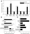
Protease signaling-mediated MAPK phosphorylation in TF-transfected CHO cells. (A–C) Confluent CHO/TF- (solid bars) or PAR-2-transfected CHO/TF/PAR2 (open bars) cells were serum starved and stimulated for 10 min with the indicated concentrations of agonists. Where indicated, specific inhibitors NAP5 (N5) or NAPc2 (C2) and active site-mutated VIIa (iVIIa) were added 2 min before agonist stimulation. MAPK phosphorylation was quantified by Western blotting, and fold induction over unstimulated control was calculated on the basis of densitometric scans. Means and standard deviations are shown (A, n = 4; B and C, n = 3).
Figure 2
Figure 2
(A) Dose response of Xa-induced MAPK phosphorylation. Serum-starved CHO/TF cells (solid symbols) or CHO/TF/PAR2 cells (open symbols) were stimulated (10 min) with the indicated concentrations of Xa alone (squares) or Xa in the presence of iVIIa (10 nM) and NAPc2 (100 nM) (circles). (B) Effect of inhibitors of ternary TF–VIIa–Xa complex formation on iVIIa/Xa/NAPc2-induced signaling in CHO/TF/PAR2 cells. Serum-starved cells were preincubated (10 min) with no antibody (control) or monoclonal anti-VIIa 12C7 (50 μg/ml), anti-VIIa 12D10-Fab (25 μg/ml), anti-TF 5G9 (50 μg/ml), or anti-TF 6B4 (25 μg/ml)/9C3 (25 μg/ml). Cells were stimulated (10 min) with iVIIa/Xa/NAPc2 (10/50/100 nM) (solid bars) or 10 nM thrombin (open bars). Induction of MAPK phosphorylation is shown in A and B as fold induction over control (mean ± SD, n = 3).
Figure 3
Figure 3
PAR-2-dependent induction of EGR-1 promoter activity. (A) Serum-starved PAR-1-deficient fibroblasts, transfected with human PAR-2 and TF, as well as an EGR-1 promoter luciferase reporter construct, were incubated for 5 h in the presence of agonists. PAR-2-activating peptide SLIGRL (100 μM) induced luciferase activity 3.9 ± 0.6-fold (mean ± SD, n = 3) over control. Gene induction by the indicated agonist and in the presence of inhibitors NAP5 (N5) or NAPc2 (C2) is shown as percent of the SLIGRL response (mean ± SD, n = 3). (B) Dose response of Xa in transfected fibroblasts. Fold induction of luciferase activity is shown for cells stimulated (5 h) with the indicated concentrations of Xa alone (open bars), Xa in the presence of 10 nM iVIIa (gray bars), or Xa with 10 nM iVIIa/50 nM NAPc2 (solid bars) (mean ± SD, n = 3). (C) Time requirement for protease signaling input in transfected cells stimulated with 10 nM VII (●), 10 nM VII/100 nM X (□), 10 nM VII/100 nM X in the presence of 1 μM NAP5 (○), or 10 nM iVIIa/2 nM Xa/5 nM NAPc2 (■). Proteases were inhibited at the indicated times with 1 μM 5L15 (added to all reactions with VII) and 1 μM NAP5 (added to all reactions with X/Xa), followed by determination of luciferase activity after 5 h (mean ± SD, n = 3). Fold induction by SLIGRL was 3.1 ± 0.6 in these experiments. (D) Dose response of EGR-1 promoter induction by in situ-generated Xa versus exogenously added Xa. On cells incubated with 10 nM VII and 100 nM X, the amount of Xa generated in situ was manipulated by blocking VIIa with 1 μM 5L15 after 0, 2, 5, 10, 20, or 30 min. Xa concentrations were determined by amidolytic assay for all reactions at 30 min, followed by termination of signaling input with 1 μM NAP5 at 30 min. The plots show gene induction in dependence of the concentration of exogenously added Xa (■) or in situ-generated Xa (●). A typical experiment of three is shown. Control experiments with Xa generated by the intrinsic activation complex did not show enhanced signaling. Xa (12 ± 1 nM) generated by 2 nM VIIIa–IXa in 20 min produced a 1.3 ± 0.2-fold induction of luciferase activity, as compared with a 1.6 ± 0.7-fold induction by 12 ± 2 nM free Xa (n = 3).
Figure 4
Figure 4
Upstream coagulation protease signaling in endothelial cells. (A) MAPK phosphorylation in endothelial cells on ternary TF–VIIa–Xa complex signaling. HUV-EC-Cs were serum starved for 5 h in the absence (open bars) or presence (solid bars) of 5 nM TNFα followed by 10-min stimulation with the indicated agonists. Fold induction of phosphorylated MAPK is shown (mean ± SD, n = 3). (B) Role of PAR-1 in TF–VIIa–Xa complex signaling in TNFα-stimulated endothelial cells. MAPK phosphorylation on thrombin, SLIGRL, or TF–iVIIa–Xa–NAPc2 activation for 10 min is shown in the absence of inhibitors (control; black bars), the presence of 1 μM NAP5 (open bars), or with 10 μg/ml of ATAP2 and 25 μg/ml of WEDE15 (anti-PAR-1; gray bars). (C) Gene induction by the coagulation initiation complex in endothelial cells. HUV-EC-C cells were serum starved for 12 h in the presence of 5 nM TNFα and stimulated for 60 min with the indicated agonists. Induction of connective tissue growth factor mRNA expression by Northern blotting was normalized to the housekeeping gene GAPDH (7).
Figure 5
Figure 5
Model for mechanistic coupling of PAR signaling to the initiation of coagulation by TF.

References

    1. Coughlin S R. Nature (London) 2000;407:258–264. - PubMed
    1. Camerer E, Rottingen J A, Iversen J G, Prydz H. J Biol Chem. 1996;271:29034–29042. - PubMed
    1. Poulsen L K, Jacobsen N, Sorensen B B, Bergenhem N C, Kelly J D, Foster D C, Thastrup O, Ezban M, Petersen L C. J Biol Chem. 1998;273:6228–6232. - PubMed
    1. Camerer E, Rottingen J A, Gjernes E, Larsen K, Skartlien A H, Iversen J G, Prydz H. J Biol Chem. 1999;274:32225–32233. - PubMed
    1. Pendurthi U R, Allen K E, Ezban M, Rao L V. J Biol Chem. 2000;275:14632–14641. - PubMed

Publication types

LinkOut - more resources