. 2001 Jul 3;98(14):7910-5.
doi: 10.1073/pnas.141223398.

A mouse model for adenovirus gene delivery

Affiliations

A mouse model for adenovirus gene delivery

T Tallone et al. Proc Natl Acad Sci U S A. .

Abstract

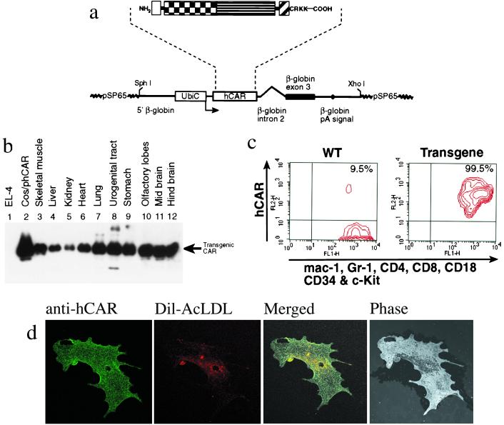
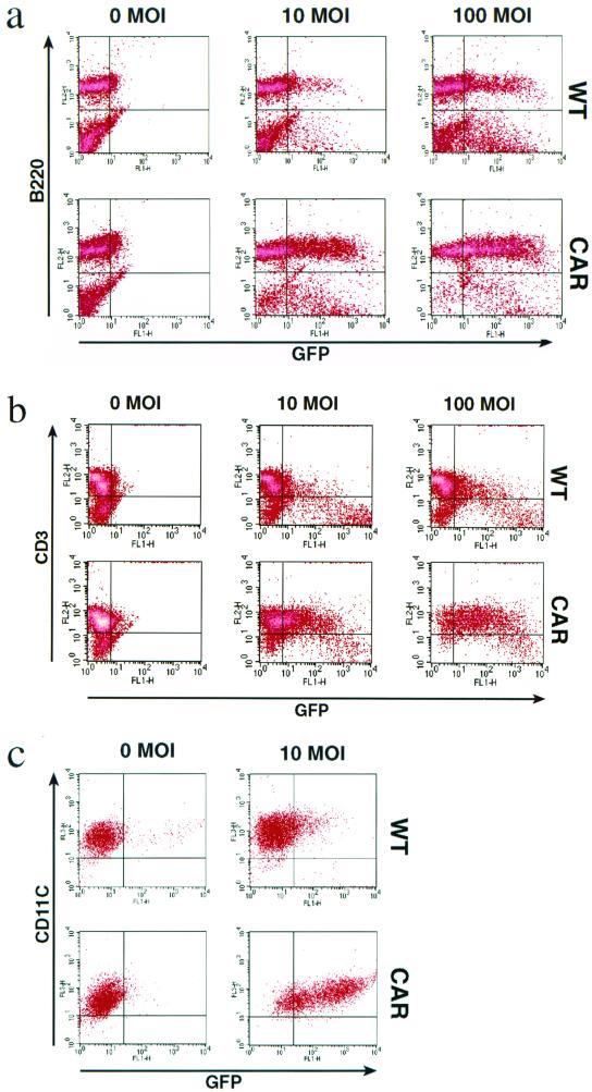
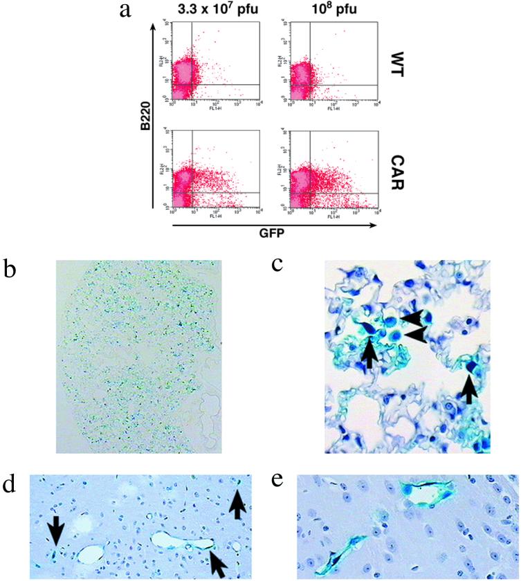
The cellular attachment receptor for adenovirus (Ad), Coxsackie adenovirus receptor (CAR), required for delivery of Ad into primary cells, is not present on all cell types, thus restricting Ad-gene delivery systems. To circumvent this constrain, a transgenic mouse has been generated that expresses a truncated human CAR in all tissues analyzed. These mice allowed efficient in vitro infections at low multiplicities into lymphoid, myeloid, and endothelial cells. Furthermore, in vivo administration of Ad-vectors results in infection of macrophages, lymphocytes, and endothelial cells. In addition, tail vein injection resulted in targeting of virus into previously inaccessible areas, such as the lung and the capillaries of the brain. The CAR transgenic mice will be useful for rapid functional genomic analysis in vivo, for testing the efficacy of gene therapy procedures or as a source of easily transducible cells.


Figures

Figure 1
Figure 1
Structure of the transgene construct and hCAR protein expression pattern in different organs. (a) Outlined is the truncated hCAR (SP, signal peptide; IG1 and IG2, Ig-like domain 1 and 2, respectively; TM, transmembrane region). Note that the cytoplasmic domain has only the first four amino acids (CRKK) C-terminal to TM. Below is a schematic map of the pβUbiC-hCAR(1–262) plasmid. The human ubiquitin-C promoter and the truncated hCAR are shown as open boxes. The black box and the thick line denote the rabbit β-globin sequences, as indicated. The polyadenylation signal is indicated by a black dot. (b) The expression patterns of the hCAR transgene in different tissues was analyzed by Western blot using a mouse monoclonal antibody (RmcB) specific for the transgenic hCAR. An arrow indicates the signal corresponding to the transgenic hCAR. Lane 1, negative control of the CAR-deficient EL-4 mouse thymoma cell line. Lane 2, positive control of COS cells transfected with the hCAR expression plasmid pβUbiC-hCAR(1–262). (c) Detection of hCAR expression in transgenic bone marrow cells by flow cytometry. Bone marrow (BM) cells were depleted of the B lymphocytes by B220 immunomagnetic beads (Miltenyli Biotech, Germany). The mouse monoclonal anti-hCAR antibody RmcB and a PE-conjugated secondary rabbit anti-mouse immunoglobulin antibody were used to detect the transgene. The cells were also stained with FITC-labeled antibodies for the markers indicated at the bottom of the diagram. These markers stain all hematopoietic lineages in BM. The hCAR+ population (9.5%) detected with wild-type BM cells are B lymphocyte contaminants that escaped the immunomagnetic depletion step. (d) Indirect immunostaining of transgenic aorta endothelial cells. (Left) hCAR staining. (Left Center) Specific staining of aorta endothelial cells after receptor mediated uptake of fluorochrome-labeled acetylated low-density lipoprotein (DIl-Ac-LDL). (Right Center) Merged picture (hCAR and DIl-Ac-LDL staining). The control staining, with only FITC-conjugated anti-mouse Ig (Amersham Pharmacia) secondary antibody, was negative, as were the stainings with wild-type endothelial cells (data not shown). (Right) Same field, phase-contrast.
Figure 2
Figure 2
The expression of the transgenic hCAR in B lymphocytes, T lymphocytes, and dendritic cells confers enhanced susceptibility to Ad transduction. (a) PMA-stimulated splenocytes were infected with the indicated MOI (0, 10, or 100) of AdGFP. After 48 h, the live cells were analyzed by flow cytometry for the expression of GFP and for the presence of the B lymphocyte marker B220. (Upper) wild type (WT). (Lower) hCAR transgene (CAR). Percentage of GFP-positive cells: WT; 10 MOI, 20%; 100 MOI, 38%. CAR; 10 MOI, 55%; 100 MOI, 75%. (b) IL-2-stimulated splenic T cells were infected with the indicated MOI (0, 10, or 100) of AdGFP. The live cells were analyzed by flow cytometry for the expression of GFP and for the presence of the T lymphocyte marker CD3. Percentage of GFP positive cells: WT; 10 MOI, 24%; 100 MOI, 28%. CAR; 10 MOI, 52%; 100 MOI, 85%. (c) Mature in vitro generated dendritic cells were infected with the indicated MOI (0 or10) of AdGFP. After 48 h, the live cells were analyzed by flow cytometry for the expression of GFP and for the presence of the CD11c as a marker for mature DCs. Percentage of GFP-positive cells: WT; 10 MOI, 16%. CAR; 10 MOI, 81%.
Figure 3
Figure 3
In vivo administration of Ad vectors. (a) Infection of B1a lymphocytes after injection of AdGFP into the peritoneal cavity. The mice were injected with the indicated amount of AdGFP. Nonadherent cells harvested from the injected mice were analyzed by flow cytometry for the expression of GFP and for the presence of the B lymphocyte marker B220. Percentage of GFP positive: 7% and 15% (Left); 5% and 28% (Right). (b–e) Histological sections of lung (b and c) and brain (d and e) after tail-vein injection of AdLacZ in the transgenic hCAR mice. (b) An overview of the distribution of β-galactosidase-expressing cells in the lung is shown. Positive, infected cells can be seen throughout the section, indicating widespread Ad uptake. (c) Higher-resolution image demonstrating cell types infected. Arrows indicate endothelial cells of septal capillaries and arrowheads indicate alveolar macrophages. (d) The blue staining for β-galactosidase expression in the endothelium of many of the cerebral capillaries (arrows) is shown. (e) Magnification of two capillaries in the plexus choroideus showing a β-galactosidase-positive signal lining the inside of the vessels. (Section b is unstained, sections c to e are counterstained with hematoxylin and eosin.)

References

    1. Nemerow G R. Virology. 2000;274:1–4. - PubMed
    1. Bergelson J M, Cunningham J A, Droguett G, Kurt-Jones E A, Krithivas A, Hong J S, Horwitz M S, Crowell R L, Finberg R W. Science. 1997;275:1320–1323. - PubMed
    1. Tomko R P, Xu R, Philipson L. Proc Natl Acad Sci USA. 1997;94:3352–3356. - PMC - PubMed
    1. Chrétien I, Marcuz A, Courtet M, Katevuo K, Vainio O, Heath J K, White S J, Du Pasquier L. Eur J Immunol. 1998;28:4094–4104. - PubMed
    1. Honda T, Saitoh H, Masuko M, Katagiri-Abe T, Tominaga K, Kozakai I, Kobayashi K, Kumanishi T, Watanabe Y G, Odani S, Kuwano R. Mol Brain Res. 2000;77:19–28. - PubMed

Publication types

LinkOut - more resources