. 2001 Jun;6(6):531-44.
doi: 10.1046/j.1365-2443.2001.00440.x.

Identification and characterization of DAlk: a novel Drosophila melanogaster RTK which drives ERK activation in vivo

Affiliations

Identification and characterization of DAlk: a novel Drosophila melanogaster RTK which drives ERK activation in vivo

C E Lorén et al. Genes Cells. 2001 Jun.

Abstract

Background: The mammalian receptor protein tyrosine kinase (RTK), Anaplastic Lymphoma Kinase (ALK), was first described as the product of the t(2;5) chromosomal translocation found in non-Hodgkin's lymphoma. While the mechanism of ALK activation in non-Hodgkin's lymphoma has been examined, to date, no in vivo role for this orphan insulin receptor family RTK has been described.

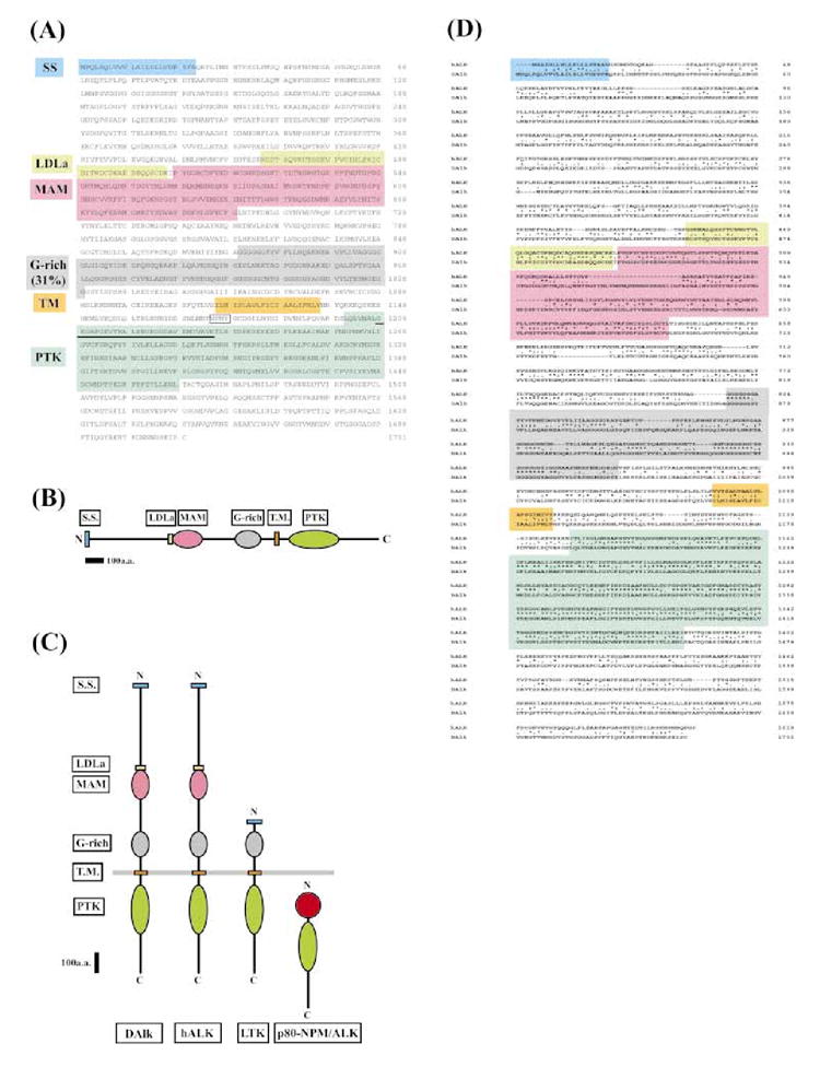
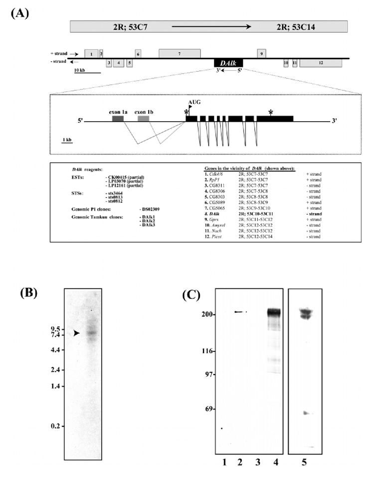
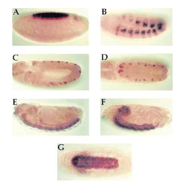
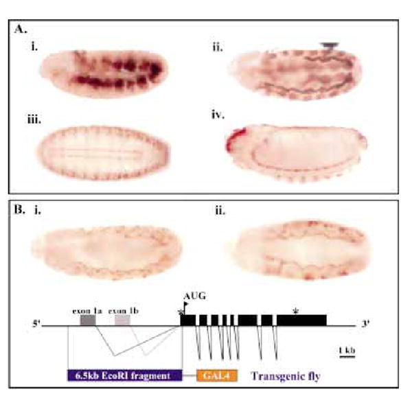
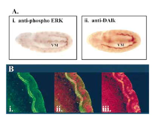
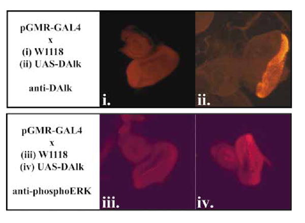
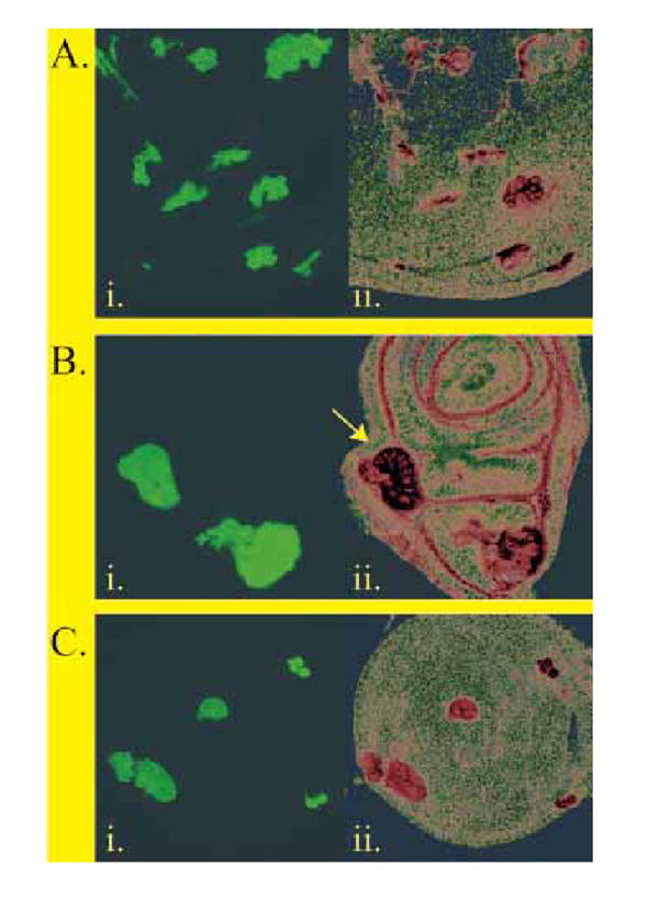
Results: We describe here a novel Drosophila melanogaster RTK, DAlk, which we have mapped to band 53 on the right arm of the second chromosome. Full-length DAlk cDNA encodes a phosphoprotein of 200 kDa, which shares homology not only with mammalian ALK but also with the orphan RTK LTK. Analysis of both mammalian and Drosophila ALK reveals that the ALK family of RTKs contains a newly identified MAM domain within their extracellular domains. Like its mammalian counterpart, DAlk appears to be expressed in the developing CNS by in situ analysis. However, in addition to expression of DAlk in the Drosophila brain, careful analysis reveals an additional early role for DAlk in the developing visceral mesoderm where its expression is coincident with activated ERK.

Conclusion: In this paper we describe a Drosophila melanogaster Alk RTK which is expressed in the developing embryonic mesoderm and CNS. Our data provide evidence for the existence of a DAlk RTK pathway in Drosophila. We show that ERK participates in this pathway, and that it is activated by DAlk in vivo. Expression patterns of dALK, together with activated ERK, suggest that DAlk fulfils the criteria of the missing RTK pathway, leading to ERK activation in the developing visceral mesoderm.


Figures

Figure 1
Figure 1
Molecular characterization of DAlk. (A) Protein sequence of DAlk. The protein sequence for DAlk is shown. Though not shown here, an in-frame upstream STOP codon precedes the initial methionine residue. The termination STOP codon is indicated by an asterisk (*). The signal sequence, LDLa, MAM, G-rich, transmembrane and PTK domains are shaded, and the consensus GxGxxG and AxK in the ATP binding site is underlined. The originally identified 64 amino acid PCR clone is double underlined. The putative IRS/SHC SH2 binding site (NPNY1170) is boxed. (B) DAlk schematic outline. (C) Schematic comparison of DAlk with the mammalian LTK/ALK family RTKs, ALK. Also shown is the p80-NPM/ALK protein product of the t(2;5) chromosomal translocation found in non-Hodgkins lymphoma. Structural conservation between DAlk, hALK and LTK include the presence of an intracellular PTK domain, an amino-terminal signal sequence, and an extracellular glycine-rich domain. Only DAlk and hALK share extracellular LDLa and MAM domains. (D) Alignment of the deduced amino acid sequences of Drosophila and human ALK. The signal sequence, LDLa, MAM, G-rich, transmembrane and PTK domains are shaded. The putative IRS/SHC SH2 binding site (NPNY1170) is boxed. The amino acid residues which are identical between the two species are marked by asterisks.
Figure 2
Figure 2
(A) Genomic characterization of DAlk. (Top panel) A map corresponding to the genomic region 53C7–53C14 on the second right chromosome comprising approximately 100 kb of genomic DNA containing the DAlk locus is shown. DAlk (centre) is depicted by the solid black box, and the surrounding genes, numbered 1 through 12, are indicated as grey boxes. (Middle panel) The deduced structure of the ≈15 kb DAlk transcription unit is shown below in expanded form. The 10 exons are represented by solid black boxes, and thin lines represent intron regions. The stop codon preceding the first AUG initiation codon (marked with a flag), is indicated by an asterisk (*), as is the TAA termination codon at the 3′ end of the DAlk transcript. Three EST sequences, CK00415, LP03070 and LP12161, encode partial DAlk sequences. (Bottom panel) ESTs, STSs, Genomic clones and genes in the vicinity of DAlk are indicated. (B) Expression of DAlk mRNA. Northern blot analysis using total RNA from embryos. 20 μg total RNA was loaded. (C) Over-expression of DAlk in 293 cells. 293 cells were cDNA encoding full length DAlk (lanes 2 and 4). Vector DNA alone was used as a control (lanes 1 and 3). DAlk was analysed by SDS-PAGE followed by anti-DAlk immunoblot (lanes 1 and 2) and antiphosphotyrosine immunoblot (lanes 3 and 4). In addition, anti-DAlk antibodies recognize a doublet at 200 kDa in embryo extracts (lane 5).
Figure 3
Figure 3
Expression of DAlk during embyrogenesis: in situ hybridization. In situ hybridization of DAlk to whole-mount wild-type embryos showing the expression of DAlk at: (A) stage 6 in the presumptive amnioserosa, (B) stage 10 in a subset of mesodermal cells, (C and D) stage 11 in a subset of ectodermal cells, and (E) stage 14 and (F and G) stage 17 in the developing ventral nerve cord (VNC). Anterior is to the left and dorsal is up, except for (D) which is a dorsal view and (G) which is a ventral view.
Figure 4
Figure 4
DAlk expression. (A) DAlk expression during embryogenesis. DAlk immunostaining of developing embryos is shown in brown. Embryo orientation is anterior to the left, dorsal up (i, ii, iv), dorsal facing (iii). (i) At stage 10, DAlk is highly expressed within a subset of mesodermal cells. (ii) Stage 11. DAlk protein is found in the visceral mesoderm. Staining can also be observed at the invagination of the tracheal pits. (iii and iv) Strong expression of DAlk is restricted to the brain and the ventral nerve cord. (B) DAlk enhancer promoter region analysis. The DAlk transcription unit is depicted as a schematic diagram. A 6.5 kb EcoRI fragment 5′ of the ORF of DAlk (black box) was placed upstream of Gal4 as shown. Transgenic flies carrying DAlkEI6.5-GAL4 crossed to UAS-nuclearGFP reporter flies displayed a visceral mesoderm expression pattern (i) lateral view, stage 11 embryo (ii) ventral view, stage 11 embryo.
Figure 5
Figure 5
DAlk and phospho-MAPK co-localize in the visceral mesoderm. (A) DAlk and phospho-ERK co-localize in the visceral mesoderm. (i) At stage 11 phospho-ERK is detected in the visceral mesoderm (VM), prior to and after the generation of a continuous visceral stripe. (ii) anti-DAlk staining of a stage 11 embryo, showing DAlk protein in the visceral mesoderm (VM), also prior to and after the generation of a continuous visceral stripe. (B) Stage 11 embryo stained with (i) anti-phosphoERK (red) and (iii) anti-DAlk (green). (iii) Expression of both phospho-ERK and DAlk co-localize and are specifically detected in the visceral mesoderm at this stage (merged image).
Figure 6
Figure 6
DAlk drives ERK activation in the developing eye disc. Wild-type DAlk is capable of driving ERK activation in vivo. (i) Third instar eye disc from a control animal carrying pGMR-GAL4; anti-DAlk. (ii) Third instar eye disc from an animal carrying pGMR-GAL4 driving UAS-DAlk displaying DAlk overexpression: anti-DAlk. (iii) Third instar eye disc from a control animal carrying pGMR-GAL4; anti-phosphoERK. (iv) Third instar eye disc from an animal carrying pGMR-GAL4 driving UAS-DAlk displaying DAlk over-expression: anti-phosphoERK.
Figure 7
Figure 7
DAlk clonal over-expression in developing imaginal discs. (A) Heat-shock induced clones over-expressing wild-type DAlk under the Actin-5C promoter. (i) Imaginal discs displaying heat-shock induced over-expression clones marked by the overexpression of UAS-GFP. (ii) The DAlk immunostaining of imaginal discs with anti-DAlk antibodies is shown in red. (B) Heat-shock induced clones over-expressing wild-type DAlk under the Actin-5C promoter contain increased levels of phosphotyrosine. (i) Third instar wing disc displaying heat-shock induced over-expression clones marked by the over-expression of UAS-GFP. (ii) Anti-phosphotyrosine immunostaining of the same tissues with 4G10 anti-phosphotyrosine antibodies shown in red. Arrow indicates tissue perturbation observed during analysis of larger DAlk clones. (C) Heat-shock induced clonal overexpression of wild-type DAlk under the Actin-5C promoter results in ERK activation. (i) Third instar wing disc displaying heat-shock induced DAlk over-expression clones marked by the over-expression of UAS-GFP. (ii) Anti-phosphoERK immunostaining of the same tissues antibodies is shown in red.

Similar articles

Cited by

References

    1. Adams MD, Celniker SE, Holt RA, et al. The genome sequence of Drosophila melanogaster. Science. 2000;287:2185–2195. - PubMed
    1. Altschul SF, Gish W, Miller W, Myers EW, Lipman DJ. Basic local alignment search tool. J Mol Biol. 1990;215:403–410. - PubMed
    1. Beckmann G, Bork P. An adhesive domain detected in functionally diverse receptors. Trends Biochem Sci. 1993;18:40– 41. - PubMed
    1. Beiman M, Shilo BZ, Volk T. Heartless, a Drosophila FGF receptor homolog, is essential for cell migration and establishment of several mesodermal lineages. Genes Dev. 1996;10:2993–3002. - PubMed
    1. Bourgouin C, Lundgren SE, Thomas JB. Apterous is a Drosophila LIM domain gene required for the development of a subset of embryonic muscles. Neuron. 1992;9:549–561. - PubMed

Publication types

MeSH terms

Substances