. 2001 Aug;183(15):4421-34.
doi: 10.1128/JB.183.15.4421-4434.2001.

Methionine regeneration and aspartate aminotransferase in parasitic protozoa

Affiliations

Methionine regeneration and aspartate aminotransferase in parasitic protozoa

L C Berger et al. J Bacteriol. 2001 Aug.

Abstract

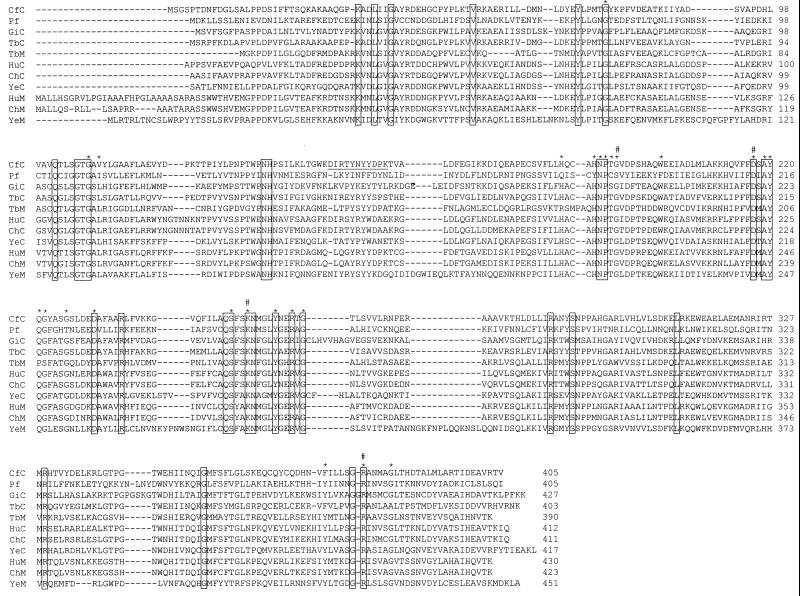
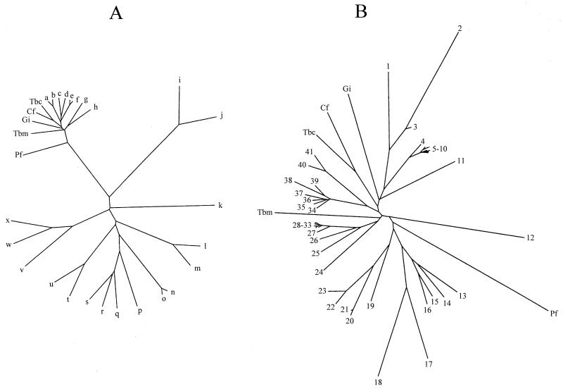
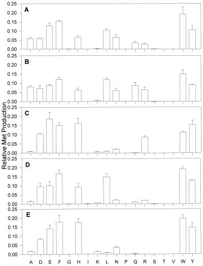
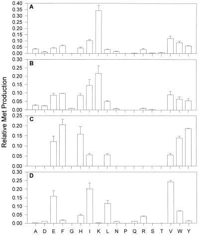
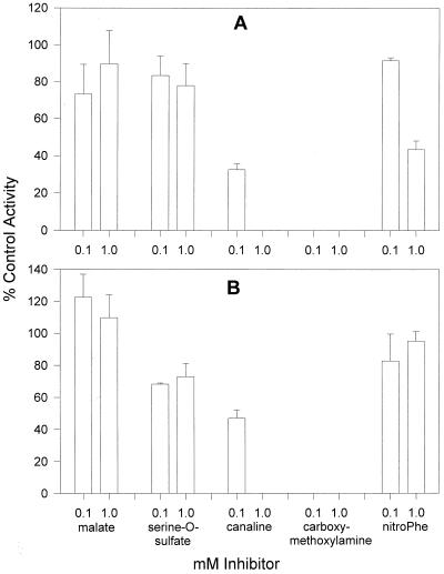
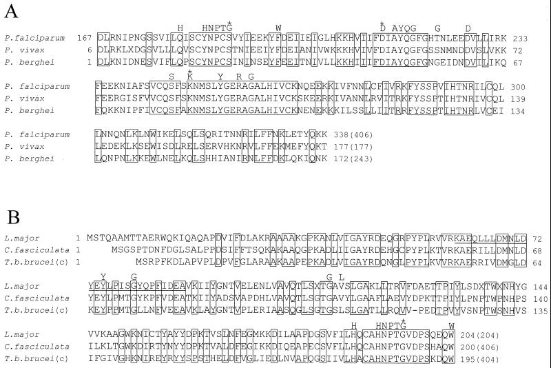
Aspartate aminotransferases have been cloned and expressed from Crithidia fasciculata, Trypanosoma brucei brucei, Giardia intestinalis, and Plasmodium falciparum and have been found to play a role in the final step of methionine regeneration from methylthioadenosine. All five enzymes contain sequence motifs consistent with membership in the Ia subfamily of aminotransferases; the crithidial and giardial enzymes and one trypanosomal enzyme were identified as cytoplasmic aspartate aminotransferases, and the second trypanosomal enzyme was identified as a mitochondrial aspartate aminotransferase. The plasmodial enzyme contained unique sequence substitutions and appears to be highly divergent from the existing members of the Ia subfamily. In addition, the P. falciparum enzyme is the first aminotransferase found to lack the invariant residue G197 (P. K. Mehta, T. I. Hale, and P. Christen, Eur. J. Biochem. 214:549-561, 1993), a feature shared by sequences discovered in P. vivax and P. berghei. All five enzymes were able to catalyze aspartate-ketoglutarate, tyrosine-ketoglutarate, and amino acid-ketomethiobutyrate aminotransfer reactions. In the latter, glutamate, phenylalanine, tyrosine, tryptophan, and histidine were all found to be effective amino donors. The crithidial and trypanosomal cytosolic aminotransferases were also able to catalyze alanine-ketoglutarate and glutamine-ketoglutarate aminotransfer reactions and, in common with the giardial aminotransferase, were able to catalyze the leucine-ketomethiobutyrate aminotransfer reaction. In all cases, the kinetic constants were broadly similar, with the exception of that of the plasmodial enzyme, which catalyzed the transamination of ketomethiobutyrate significantly more slowly than aspartate-ketoglutarate aminotransfer. This result obtained with the recombinant P. falciparum aminotransferase parallels the results seen for total ketomethiobutyrate transamination in malarial homogenates; activity in the latter was much lower than that in homogenates from other organisms. Total ketomethiobutyrate transamination in Trichomonas vaginalis and G. intestinalis homogenates was extensive and involved lysine-ketomethiobutyrate enzyme activity in addition to the aspartate aminotransferase activity. The methionine production in these two species could be inhibited by the amino-oxy compounds canaline and carboxymethoxylamine. Canaline was also found to be an uncompetitive inhibitor of the plasmodial aspartate aminotransferase, with a K(i) of 27 microm.


Figures

FIG. 1
FIG. 1
Alignment of parasite AspATs with selected eukaryotic enzymes. The enzymes are as follows: CfC, C. fasciculata cytoplasmic AspAT; Pf, P. falciparum AspAT; GiC, G. intestinalis cytoplasmic AspAT; TbC, T. brucei brucei cytoplasmic AspAT; TbM, T. brucei brucei mitochondrial AspAT, HuC, Homo sapiens cytoplasmic AspAT; ChC, Gallus gallus cytoplasmic AspAT; YeC, S. cerevisiae cytoplasmic AspAT; HuM, H. sapiens mitochondrial AspAT; ChM, G. gallus mitochondrial AspAT; and YeM, S. cerevisiae mitochondrial AspAT. Boxes surround residues which are conserved across all 11 sequences, while the underlined residues in the C. fasciculata enzyme represent the sequence determined previously by amino acid sequencing of a purified aminotransferase (7). The residues marked with asterisks are those reported by Jensen and Gu (23) as being conserved in all members of the Ia subfamily of aminotransferases, while those marked with number signs are those reported by Mehta et al. (33) as being conserved in all aminotransferase families.
FIG. 2
FIG. 2
Phylogenetic analysis of parasite AspATs. In both trees, constructed by neighbor joining with the Phylip package, parasite AspATs are abbreviated as follows: Cf, C. fasciculata cytoplasmic AspAT; Tbc, T. brucei brucei cytoplasmic AspAT; Tbm, T. brucei brucei mitochondrial AspAT; Gi, G. intestinalis cytoplasmic AspAT; and Pf, P. falciparum AspAT. Tree A was formed using the following additional sequences: a, H. sapiens cytoplasmic AspAT; b, G. gallus cytoplasmic AspAT; c, Lupinus angustifolicus mitochondrial AspAT; d, Oryza sativa cytoplasmic AspAT; e, H. sapiens mitochondrial AspAT; f, G. gallus mitochondrial AspAT; g, E. coli AspAT; h, E. coli TyrAT; i, Schizosaccharomyces pombe TyrAT; j, S. cerevisiae TyrAT; k, E. coli alanine-valine aminotransferase; l, H. sapiens AlaAT; m, S. cerevisiae AlaAT; n, H. sapiens kynurenine aminotransferase; o, Rattus norvegicus kynurenine aminotransferase; p, B. subtilis YHDR gene product; q, B. subtilis AspAT; r, B. subtilis PATA gene product; s, B. subtilis YUGH gene product; t, H. sapiens TyrAT; u, Trypanosoma cruzi TyrAT; v, Halobacter sp. histidinol-phosphate aminotransferase; w, E. coli histidinol-phosphate aminotransferase; and x, S. pombe histidinol-phosphate aminotransferase. Tree B was formed using the following additional sequences from members of the aminotransferase Ia subfamily: 1, C. elegans CE07462 gene product; 2, C. elegans CE06829 gene product; 3, C. elegans CE07461 gene product; 4, G. gallus cytoplasmic AspAT; 5, Mus muris cytoplasmic AspAT; 6, R. norvegicus cytoplasmic AspAT; 7, H. sapiens cytoplasmic AspAT; 8, Equus caballus cytoplasmic AspAT; 9, Bos taurus cytoplasmic AspAT; 10, Sus scrofus cytoplasmic AspAT; 11, S. cerevisiae cytoplasmic AspAT; 12, S. cerevisiae mitochondrial AspAT; 13, Vibrio cholerae AspAT; 14, E. coli AspAT; 15, Haemophilus influenzae AspAT; 16, Neisseria gonorrhoeae AspAT; 17, Paracoccus denitrificans TyrAT; 18, Rhizobium meliloti TyrAT; 19, Pseudomonas aeruginosa TyrAT; 20, N. gonorrhoeae TyrAT; 21, N. meningitidis TyrAT; 22, E. coli TyrAT; 23, K. pneumoniae TyrAT; 24, A. thaliana AspAT1; 25, Drosophila melanogaster CT10757 gene product; 26, C. elegans CE02477 gene product; 27, G. gallus mitochondrial AspAT; 28, H. sapiens mitochondrial AspAT; 29, E. caballus mitochondrial AspAT; 30, B. taurus mitochondrial AspAT; 31, S. scrofus mitochondrial AspAT; 32, R. norvegicus mitochondrial AspAT; 33, M. muris mitochondrial AspAT; 34, Medicago sativa cytoplasmic AspAT; 35, Daucus caroti cytoplasmic AspAT; 36, O. sativa cytoplasmic AspAT; 37, A. thaliana AspAT3; 38, A. thaliana AspAT4; 39, A. thaliana AspAT2; 40, L. angustifolicus mitochondrial AspAT; and 41, A. thaliana mitochondrial AspAT.
FIG. 3
FIG. 3
Amino donor spectrum for recombinant parasite AspATs. Purified recombinant aminotransferases were incubated with a 2 mM concentration of a single amino acid and 1 mM KMTB as described in Materials and Methods and analyzed for Met production by HPLC. Met production for each amino acid is shown as the fraction of total Met production. (A) C. fasciculata cytoplasmic AspAT. (B) T. brucei brucei cytoplasmic AspAT. (C) T. brucei brucei mitochondrial AspAT. (D) G. intestinalis cytoplasmic AspAT. (E) P. falciparum AspAT.
FIG. 4
FIG. 4
Amino donor spectrum for parasite or tissue homogenates. Aliquots of subcellular homogenates were incubated with a 2 mM concentration of a single amino acid and 1 mM KMTB as described in Materials and Methods and analyzed for Met production by HPLC. Met produced by each amino acid is shown as the fraction of total Met production. (A) T. vaginalis homogenates. (B) G. intestinalis homogenates. (C) P. falciparum homogenates. (D) Pig kidney homogenates.
FIG. 5
FIG. 5
Inhibition of Met production in homogenates of T. vaginalis (A) and G. intestinalis (B). Homogenates of each parasite were incubated with 2 mM ADEFGHIKLNQRSTWY, 1 mM KMTB, and 0.1 or 1.0 mM individual inhibitors as described in Materials and Methods and analyzed for Met production. Inhibition of Met production relative to the results for control incubations is shown. nitroPhe, nitrophenylalanine.
FIG. 6
FIG. 6
Alignment of plasmodial and trypanosomatid AspATs. (A) Clustal alignment of a portion of the P. falciparum AspAT with the deduced amino acid sequences of gene fragments obtained from the P. vivax and P. berghei sequence tag projects (9). (B) Clustal alignment of portions of the C. fasciculata cytoplasmic AspAT and T. brucei brucei cytoplasmic (c) AspAT with the deduced amino acid sequence of a gene fragment obtained from the L. major genome project (35). For both sets of sequences, the boxed residues were conserved by all three sequences, the residues above the sequences are those reported to be conserved in all aminotransferases in the Ia subfamily (23), and the residues marked with asterisks are those reported to be conserved in all aminotransferase families (33). The residues underlined in the L. major fragment represent the amino acid sequence determined by Vernal et al. (45) from a purified L. mexicana enzyme. The numbers in parentheses represent the total length of the amino acid sequence obtained for each enzyme.
FIG. 7
FIG. 7
Reverse transcription-PCR of P. falciparum AspAT. RNA isolated from asynchronous P. falciparum was incubated in the presence (lanes E) or absence (lanes C) of reverse transcriptase and then subjected to PCR with primers specific for the full-length P. falciparum AspAT gene (PfASAT) or the P. falciparum lactate dehydrogenase gene (PfLDH) as a positive control. The products were then analyzed on an agarose gel together with DNA markers (lane M). The lengths of the markers are (from the gel bottom) 0.5, 0.75, 1.0, 1.5, 2.0, 2.5, 3.0, 4.0, 5.0, 6.0, 8.0, and 10.0 kbp. The expected length of AspAT was 1,218 bp, and that of lactate dehydrogenase was 951 bp.

Similar articles

Cited by

References

    1. Altschul S F, Madden T L, Schaffer A A, Zhang J, Zhang Z, Miller W, Lipman D J. Gapped BLAST and PSI-BLAST: a new generation of protein database search programs. Nucleic Acids Res. 1997;25:3389–3402. - PMC - PubMed
    1. Ayala F J, Escalante A A, Lal A A, Rich S M. Evolutionary relationships of human malaria parasites. In: Sherman I, editor. Malaria: parasite biology, pathogenesis, and protection. Washington, D.C.: ASM Press; 1998. pp. 285–300.
    1. Bacchi C J, Sufrin J R, Nathan H C, Spiess A J, Hannon T, Garafolo J, Alecia K, Katz L, Yarlett N. 5′-Alkyl-substituted analogs of 5′-methylthioadenosine as trypanocides. Antimicrob Agents Chemother. 1991;35:1904–1906. - PMC - PubMed
    1. Backlund P S, Chang C P, Smith R A. Identification of 2-keto-4-methylthiobutyrate as an intermediate compound in methionine synthesis from 5′-methylthioadenosine. J Biol Chem. 1982;257:4196–4202. - PubMed
    1. Backlund P S, Smith R A. Methionine synthesis from 5′-methylthioadenosine in rat liver. J Biol Chem. 1980;256:1533–1535. - PubMed

Publication types

MeSH terms

Associated data