

doi: 10.1128/JB.183.15.4659-4663.2001.
Activation by gene amplification of pitB, encoding a third phosphate transporter of Escherichia coli K-12
Affiliations
- PMID: 11443103
- PMCID: PMC95363
- DOI: 10.1128/JB.183.15.4659-4663.2001
Item in Clipboard
Activation by gene amplification of pitB, encoding a third phosphate transporter of Escherichia coli K-12
J Bacteriol.
2001 Aug.
Abstract
Two systems for the uptake of inorganic phosphate (P(i)) in Escherichia coli, PitA and Pst, have been described. A revertant of a pitA pstS double mutant that could grow on P(i) was isolated. We demonstrate that the expression of a new P(i) transporter, PitB, is activated in this strain by a gene amplification event.
Figures
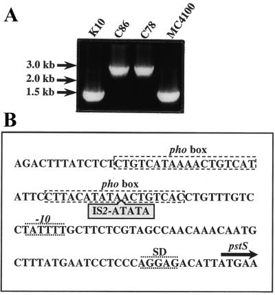
Characterization of the pstS mutation in C86. (A) Amplification of the pstS gene and its promoter region by PCR using chromosomal DNA of strains K10, C86, C78, and MC4100 (5) and the primers pst4 (5′-GAGTAATAAATGGATGCCC-3′) and pst5 (5′-CGGTGGGTTAAAAGCAGGC-3′). Strains K10 (pitA10), C86 (pstS21 derivative of K10), and C78 (pstS28 derivative of K10) were kindly provided by the E. coli Genetic Stock Center (Department of Biology, Yale University, New Haven, Conn.). The positions of the molecular size markers are indicated at the left. (B) Position of the IS2 element in the promoter region of the pstS gene in strain C86. The two pho boxes, the −10 region and the Shine-Dalgarno sequence (SD), are indicated. The start of the coding region of pstS is indicated by an arrow.

Expression of the PstS and the PhoU proteins in pitA mutant strain K10 (lane 1), pitA pstS mutant strain CE1485 (lane 2), and the pseudorevertant strain CE1487 (lane 3). Cells were grown in a peptone-based, phosphate-poor medium (11) supplemented with 0.5% glucose, 660 μM K2HPO4, and 1 mM G3P (HPi medium) (A) or with no K2HPO4 or G3P added (LPi medium) (B). The alternative phosphate source G3P was omitted from the LPi medium, since it may be degraded by alkaline phosphatase, thereby generating Pi. Although growth of CE1485 in the LPi medium was very poor, enough cells could be collected for this analysis. Proteins were separated by sodium dodecyl sulfate-polyacrylamide gel electrophoresis (12) and, for Western blotting (1), transferred to a nitrocellulose membrane (Schleicher & Schuell). Immunodetection was performed with polyclonal antisera directed against the Pi-binding protein (PstS) and PhoU. The positions of molecular size standard proteins are indicated on the left in kilodaltons.
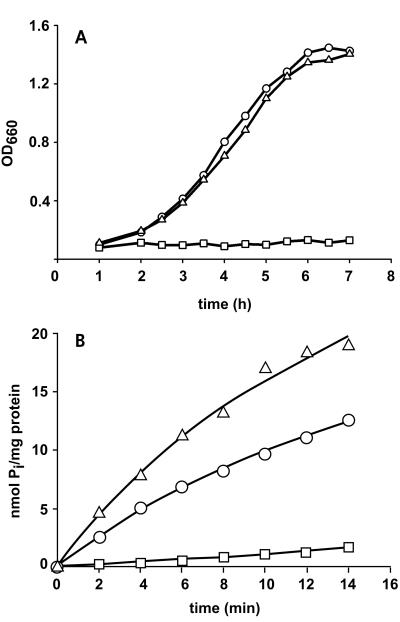
Growth and 33Pi uptake. (A) Growth curve of pitA mutant strain K10 (○), pitA pstS double-mutant CE1485 (□), and the pseudorevertant CE1487 (▵). Cells were grown overnight in Luria broth supplemented with G3P, pelleted, and resuspended in HEPES-buffered synthetic medium (25) supplemented with 0.5% glucose and 660 μM K2HPO4. Growth was monitored for 7 h. (B) Uptake of 33Pi by cells of strains K10 (○), CE1485 (□), and CE1487 (▵). Cells were grown in Luria broth supplemented with 20 mM glucose and 1 mM G3P to an optical density at 660 nm (OD660) of approximately 0.9, washed, and resuspended in a solution of 20 mM potassium piperazine-N,N′-bis(2-ethanesulfonate) (PIPES) (pH 7.0)–10 mM MgSO4. These cells were stored on ice, and, within 2 h, transport assays were performed at 30°C with 50 μM 33P-labeled potassium phosphate as described previously (27). The experiments were repeated three times with essentially the same results, and data from a representative experiment are shown.
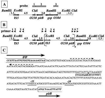
(A and B) Maps of the pitB chromosomal region of strains CE1485 (A) and CE1487 (B). Only the relevant BamHI, ClaI, and EcoRI sites are depicted. At the top of panel A, the probes used for Southern hybridization are indicated. At the top of panel B, the arrowheads indicate PCR primers with the following sequences: pr1, 5′-GGAAGATCGATGCGCTGG-3′; pr2, 5′-CCATTACCAGCCTTGGGG-3′; pr3, 5′-GGGGAAATTCTTCTCGGC-3′; pr4, 5′-GGATATCGTCAGCGGCGC-3′; pr5, 5′-CCTGTGTATATATCAAGGCC-3′; pr6, 5′-CAGGTAACGATGGTGCGG-3′; and pr7, 5′-CCTGCTCGGCACTCTCGG-3′. The numbers between the restriction sites indicate the lengths of the fragments in kilobases. (C) Nucleotide sequence of the pitB-gsp intercistronic region. Coding sequences for the glutathionylspermidine synthetase (Gsp), including the stop codon, and PitB proteins are indicated in bold italics and boxed. Dashed arrows indicate inverted repeats, which may function as the transcriptional terminator of the gsp gene. Putative −35 and −10 sequences of the pitB promoter are indicated. The insertion in strain CE1487 of the amplified DNA fragment containing IS5-pitB is indicated.
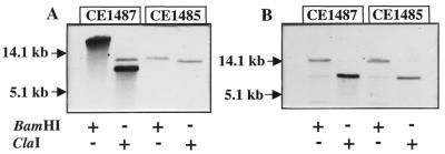
Southern blot analysis of chromosomal DNA of CE1485 and CE1487. Blots were hybridized with the pitB probe (A) and probe 3 (B) (see Fig. 4). DNA was digested with BamHI or ClaI, and fragments were separated by electrophoresis on a 0.8% agarose gel. The DNA was transferred from the gel to Hybond-N+ membranes (Amersham) with a vacuum blotter (Bio-Rad model 785). After transfer, the filter was washed in 2× SSC (1× SSC is 0.15 M NaCl plus 0.015 M sodium citrate [pH 7.0]) for 5 min, and DNA was cross-linked by UV irradiation for 2 min. Labeling of the probes, hybridization, and detection were done with digoxigenin labeling and detection kits (Boehringer Mannheim). Hybridization and stringency washes were carried out at 68°C. The positions of molecular size standard DNA fragments are indicated in kilobases.
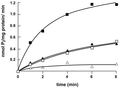
Uptake of 33Pi in right-side-out membrane vesicles of strain CE1491 expressing pitB from plasmid pSL41. Membrane vesicles were diluted to a final protein concentration of 0.1 to 0.5 mg of protein/ml in air-saturated 50 mM potassium PIPES (pH 7.0)–10 mM MgSO4. The membrane vesicles were preincubated for 3 min at 30°C with 2 μM pyrroloquinoline quinone in the absence (▵) or presence (■) of 20 mM glucose to generate the PMF and in the presence of glucose in combination with 2.5 μM valinomycin (□) or 2.5 μM nigericin (▴). Transport was initiated on addition of 50 μM 33P-labeled potassium phosphate and analyzed by rapid filtration (27). The experiments were repeated twice with essentially the same results, and data from a representative experiment are shown.
Similar articles
-
The phosphate-binding protein of Escherichia coli is not essential for P(i)-regulated expression of the pho regulon.J Bacteriol. 2001 Oct;183(19):5768-71. doi: 10.1128/JB.183.19.5768-5771.2001. J Bacteriol. 2001. PMID: 11544243 Free PMC article.
-
Characterization of PitA and PitB from Escherichia coli.J Bacteriol. 2001 Sep;183(17):5008-14. doi: 10.1128/JB.183.17.5008-5014.2001. J Bacteriol. 2001. PMID: 11489853 Free PMC article.
-
The pst operon of Bacillus subtilis has a phosphate-regulated promoter and is involved in phosphate transport but not in regulation of the pho regulon.J Bacteriol. 1997 Apr;179(8):2534-9. doi: 10.1128/jb.179.8.2534-2539.1997. J Bacteriol. 1997. PMID: 9098050 Free PMC article.
-
Molybdate transport.Res Microbiol. 2001 Apr-May;152(3-4):311-21. doi: 10.1016/s0923-2508(01)01202-5. Res Microbiol. 2001. PMID: 11421278 Review.
-
More than just "histone-like" proteins.Cell. 1990 Nov 2;63(3):451-3. doi: 10.1016/0092-8674(90)90438-k. Cell. 1990. PMID: 2121364 Review. No abstract available.
Cited by
-
Isolation, Characterization, and Tea Growth-Promoting Analysis of JW-CZ2, a Bacterium With 1-Aminocyclopropane-1-Carboxylic Acid Deaminase Activity Isolated From the Rhizosphere Soils of Tea Plants.Front Microbiol. 2022 Feb 28;13:792876. doi: 10.3389/fmicb.2022.792876. eCollection 2022. Front Microbiol. 2022. PMID: 35295310 Free PMC article.
-
Induction of the Pho regulon suppresses the growth defect of an Escherichia coli sgrS mutant, connecting phosphate metabolism to the glucose-phosphate stress response.J Bacteriol. 2012 May;194(10):2520-30. doi: 10.1128/JB.00009-12. Epub 2012 Mar 16. J Bacteriol. 2012. PMID: 22427626 Free PMC article.
-
The phosphate starvation stimulon of Corynebacterium glutamicum determined by DNA microarray analyses.J Bacteriol. 2003 Aug;185(15):4519-29. doi: 10.1128/JB.185.15.4519-4529.2003. J Bacteriol. 2003. PMID: 12867461 Free PMC article.
-
Autoamplification of a two-component regulatory system results in "learning" behavior.J Bacteriol. 2001 Aug;183(16):4914-7. doi: 10.1128/JB.183.16.4914-4917.2001. J Bacteriol. 2001. PMID: 11466297 Free PMC article.
-
Strategies of organic phosphorus recycling by soil bacteria: acquisition, metabolism, and regulation.Environ Microbiol Rep. 2022 Feb;14(1):3-24. doi: 10.1111/1758-2229.13040. Epub 2022 Jan 10. Environ Microbiol Rep. 2022. PMID: 35001516 Free PMC article. Review.
References
-
- Agterberg M, Fransen R, Tommassen J. Expression of Escherichia coli PhoE protein in avirulent Salmonella typhimurium aroA and galE strains. FEMS Microbiol Lett. 1988;50:295–299.
-
- Alexeyev M F, Shokolenko I N, Croughan T P. Improved antibiotic-resistance gene cassettes and omega elements for Escherichia coli vector construction and in vitro deletion/insertion mutagenesis. Gene. 1995;160:63–67. - PubMed
-
- Bennett R L, Malamy M H. Arsenate resistant mutants of Escherichia coli and phosphate transport. Biochem Biophys Res Commun. 1970;40:496–503. - PubMed
-
- Blattner F R, Plunkett III G, Block C A, Perna N T, Burland V, Riley M, et al. The complete genome sequence of Escherichia coli K-12. Science. 1997;277:1453–1474. - PubMed
-
- Casadaban M J. Transposition and fusion of the lac genes to selected promoters in Escherichia coli using bacteriophage lambda and Mu. J Mol Biol. 1976;104:541–544. - PubMed
Publication types
MeSH terms
Substances
LinkOut - more resources
Full Text Sources
Other Literature Sources
Molecular Biology Databases
Research Materials
Miscellaneous
