

The velocity of ultrasound in human primary melanoma tissue - implications for the clinical use of high resolution sonography
- PMID: 11445001
- PMCID: PMC34515
- DOI: 10.1186/1471-5945-1-1
The velocity of ultrasound in human primary melanoma tissue - implications for the clinical use of high resolution sonography
Abstract
Background: Ultrasonography with 20 MHz frequency can be used to estimate tumour thickness preoperatively in malignant melanoma (MM) of the skin. The vertical invasion depth is the single most important prognostic factor for localised MM, and its preoperative knowledge would be very useful for the planning of surgical procedures. Since ultrasonographic distance measurements directly depend upon the tissue specific ultrasound velocity, we determined the ultrasound velocity in primary melanoma.
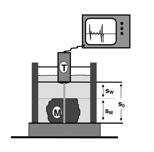
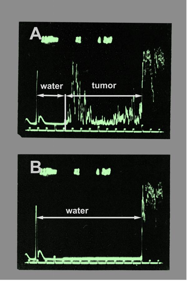
Results: Ultrasound velocity was calculated from runtime differences of a 20 MHz ultrasound signal along a known distance either through water alone or through thick specimens of primary MM. The ultrasound velocities varied between 1553 m/s and 1588 m/s with a mean of 1564 m/s in four different MM specimens. The analysis of different parts of the specimens showed that the variation of the calculated velocities was larger between different specimens than within one individual specimen.
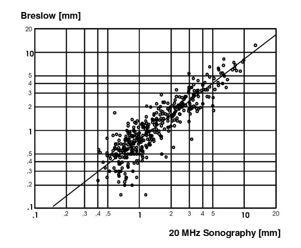
Conclusions: The ultrasound velocity in MM tissue may be slightly lower than normally assumed, thereby explaining a part of the overestimation usually found in sonographic measurement of melanoma invasion depth. Additionally, the variation of ultrasound velocity between individual tumours may contribute to the impairment of the correlation found between sonometry and Breslow's measurement of MM invasion depth. For practical reasons, a setting of 1580 m/s will be appropriate for ultrasonography of primary malignant melanoma.
Figures

References
-
- Balch CM. Cutaneous melanoma: prognosis and treatment results worldwide. Semin Surg Oncol. 1992;8:400–14. - PubMed
-
- Breitbart EW, Rehpenning W. Potentials and limits of ultrasonic diagnosis for the in vivo determination of the depth of invasion of malignant melanoma. Hautkr. 1983;58:975–87. - PubMed
-
- Gassenmaier G, Kiesewetter F, Schell H, Zinner M. Value of high resolution ultrasound in determination of vertical tumor thickness in malignant melanoma of the skin. Hautarzt. 1990; 41:360–4. - PubMed
-
- Tacke J, Haagen G, Hornstein OP, Huettinger G, Kiesewetter F, Schell H, Diepgen TL. Clinical relevance of sonometry-derived tumour thickness in malignant melanoma-a statistical analysis. Br J Dermatol. 1995;132:209–14. - PubMed
Publication types
MeSH terms
LinkOut - more resources
Full Text Sources
Medical
