. 2001 Aug;127(1):23-38.
doi: 10.1016/s0034-5687(01)00230-4.

Genioglossal inspiratory activation: central respiratory vs mechanoreceptive influences

Affiliations

Genioglossal inspiratory activation: central respiratory vs mechanoreceptive influences

G Pillar et al. Respir Physiol. 2001 Aug.

Abstract

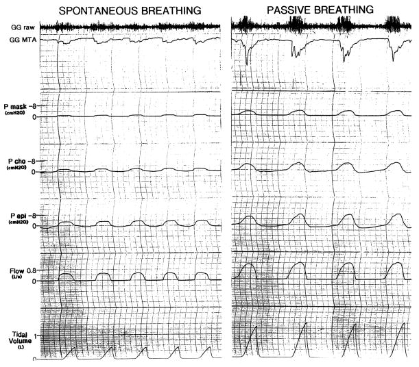
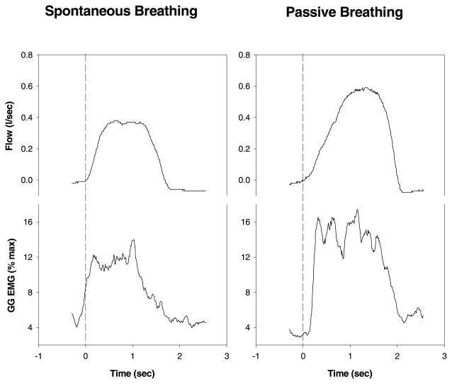
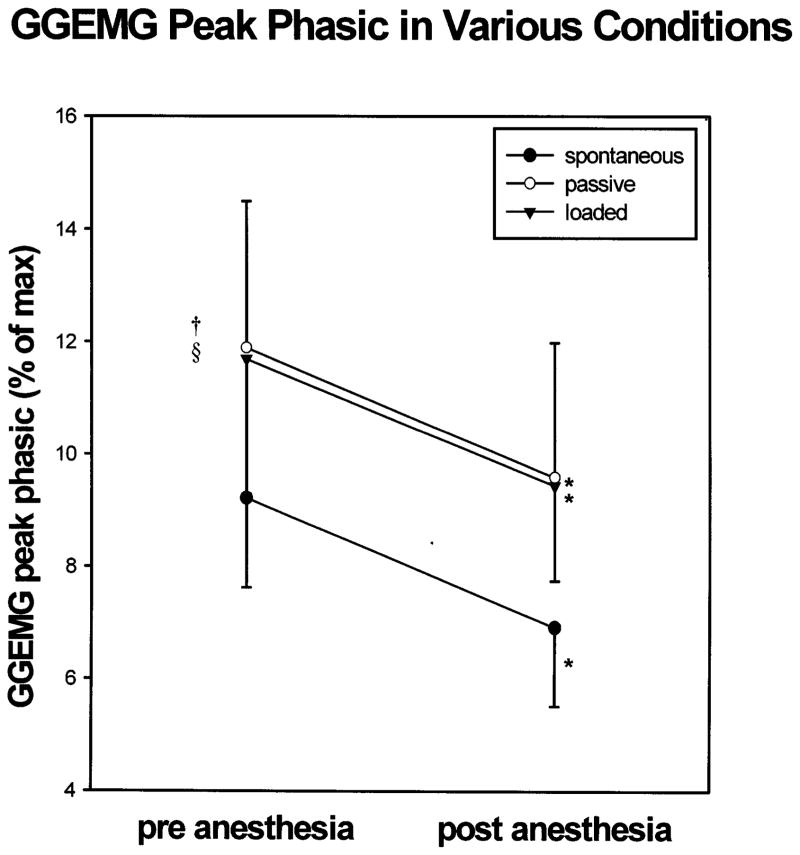
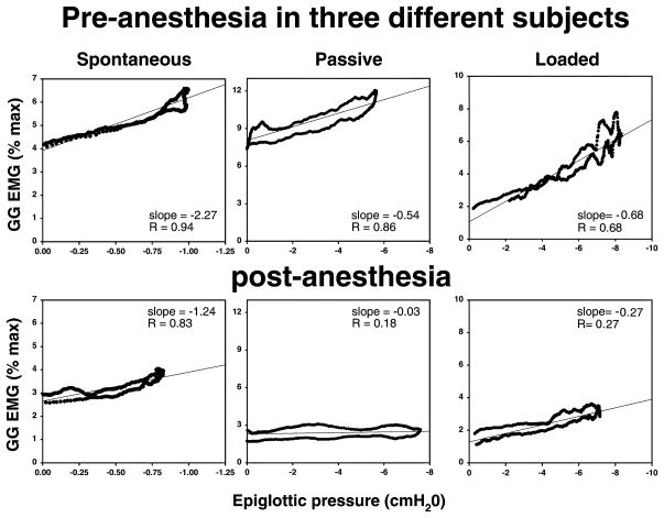
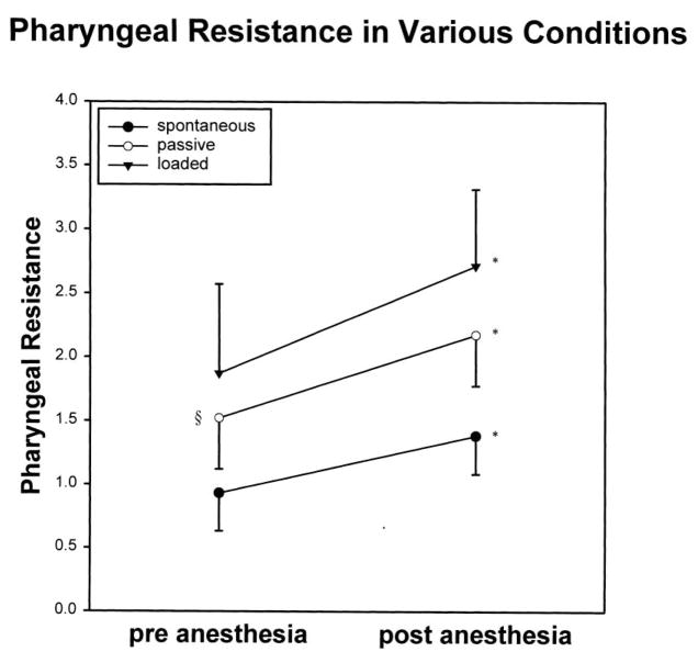
Upper airway dilator muscles are phasically activated during respiration. We assessed the interaction between central respiratory drive and local (mechanoreceptive) influences upon genioglossal (GG) activity throughout inspiration. GG(EMG) and airway mechanics were measured in 16 awake subjects during baseline spontaneous breathing, increased central respiratory drive (inspiratory resistive loading; IRL), and decreased respiratory drive (hypocapnic negative pressure ventilation), both prior to and following dense upper airway topical anesthesia. Negative epiglottic pressure (P(epi)) was significantly correlated with GG(EMG) across inspiration (i.e. within breaths). Both passive ventilation and IRL led to significant decreases in the sensitivity of the relationship between GG(EMG) and P(epi) (slope GG(EMG) vs P(epi)), but yielded no change in the relationship (correlation) between GG(EMG) and P(epi). During negative pressure ventilation, pharyngeal resistance increased modestly, but significantly. Anesthesia in all conditions led to decrements in phasic GG(EMG), increases in pharyngeal resistance, and decrease in the relationship between P(epi) and GG(EMG). We conclude that both central output to the GG and local reflex mediated activation are important in maintaining upper airway patency.


Figures

Fig. 1
Fig. 1
Depicted are raw data from the polygraphic recording. The passive breathing condition (on the right) is remarkable for the more negative epiglottic pressures, the greater genioglossus EMG activity and the loss of pre-activation when compared with spontaneous (on the left). The loss of pre-activation is more easily appreciated in Fig. 2. [raw electromyogram in arbitary units, GGMTA: moving time average genioglossus electromyogram in %maximum unit (an increase in activity is in a downward direction on this tracing), Pmask: mask pressure in cmH2O, Pcho: choanal pressure in cmH2O, Pepi: epiglottic pressure in cmH2O]. Only inspiratory values can be seen due to the one way valve used in the breathing apparatus.
Fig. 2
Fig. 2
Raw data example of the loss of pre-activation in changing from spontaneous breathing to iron lung passive breathing. Note the clear increase in GG activity prior to the onset of inspiratory flow in the spontaneous but not the passive breathing example. The peak GGEMG is higher in the passive example due to the more negative pharyngeal pressure generated by the ventilator as compared with spontaneous breathing.
Fig. 3
Fig. 3
Peak phasic GGEMG increased significantly with both mechanical ventilation and IRL as compared with spontaneous breathing. In all three conditions, anesthesia was associated with a significant reduction in peak phasic GGEMG. * = P < 0.05 pre- vs post-anesthesia; † = P < 0.05 spontaneous vs negative pressure ventilation; § = P < 0.05 spontaneous vs IRL.
Fig. 4
Fig. 4
During spontaneous breathing, this individual showed a decline in both the slope and the R value for the GG/Pepi relationship following anesthesia. During passive ventilation, this example illustrates a loss of the GG/Pepi relationship (based on both R value and slope) following anesthesia. During loaded breathing, dense topical anesthesia led to a substantial decline in the slope of the GG/Pepi relationship although the R value was preserved in this individual.
Fig. 5
Fig. 5
In all three conditions, (spontaneous breathing, negative pressure ventilation and IRL), there was an increase in pharyngeal resistance following dense topical anesthesia. * = P < 0.05 pre- vs post-anesthesia; § = P < 0.05 spontaneous vs negative pressure (passive) ventilation.

References

    1. Akahoshi T, White DP, Edwards JK, Beauregard J, Shea SA. Phasic mechanoreceptor stimuli can induce phasic activation of upper airway muscles in humans. J Physiol London. 2001;531:677–691. - PMC - PubMed
    1. Berry R, McNellis M, Kouchi K, Light R. Upper airway anesthesia reduces phasic genioglossus activity during sleep apnea. Am J Respir Crit Care Med. 1997;156:127–132. - PubMed
    1. Bianchi A, Denavit-Saubie M, Champagnat J. Central control of breathing in mammals: neuronal circuitry, membrane properties, and neurotransmitters. Physiol Rev. 1995;75:1–31. - PubMed
    1. Corfield D, Murphy K, Guz A. Does the motor cortical control of the diaphragm ‘bypass’ the brain stem respiratory centres in man? Respir Physiol. 1998;114:109–117. - PubMed
    1. Fogel R, Malhotra A, Edwards JK, Shea SA, White DP. Reduced genioglossal activity with upper airway anesthesia in awake patients with OSA. J Appl Physiol. 2000;88:1346–1354. - PubMed

Publication types

LinkOut - more resources