. 2001 Jul 1;15(13):1674-87.
doi: 10.1101/gad.902601.

Nuclear reorganization and homologous chromosome pairing during meiotic prophase require C. elegans chk-2

Affiliations

Nuclear reorganization and homologous chromosome pairing during meiotic prophase require C. elegans chk-2

A J MacQueen et al. Genes Dev. .

Abstract

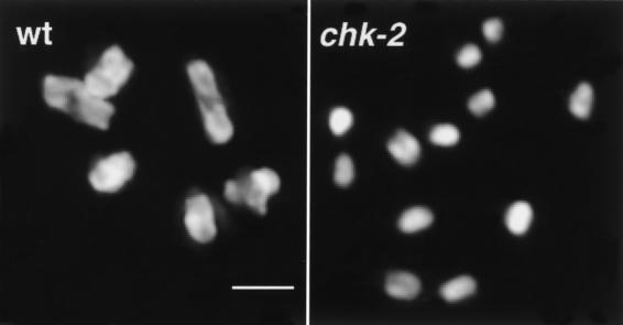
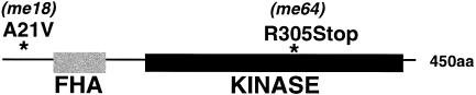
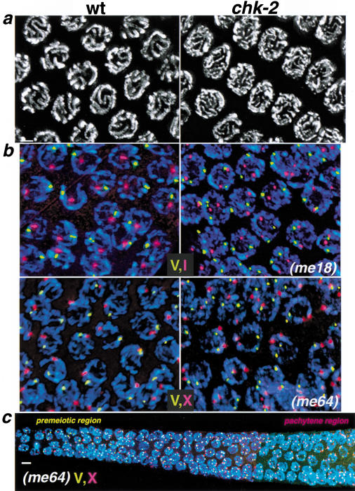
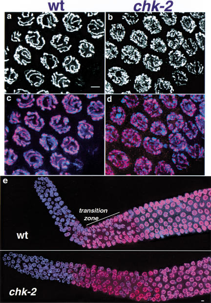
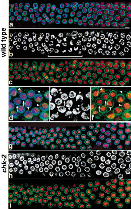
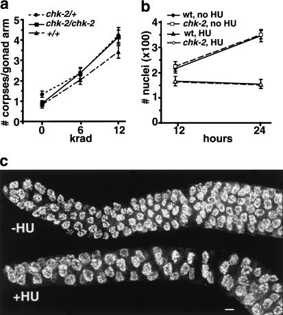
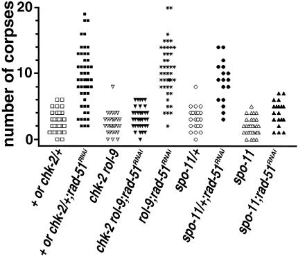
Analysis of mutants defective in meiotic chromosome pairing has uncovered a role for Caenorhabditis elegans chk-2 in initial establishment of pairing between homologous chromosomes during early meiotic prophase. chk-2 is also required for the major spatial reorganization of nuclei that normally accompanies the onset of pairing, suggesting a mechanistic coupling of these two events. Despite failures in pairing, nuclear reorganization, and crossover recombination, chk-2 mutants undergo many other aspects of meiotic chromosome morphogenesis and complete gametogenesis. Although chk-2 encodes a C. elegans ortholog of the Cds1/Chk2 checkpoint protein kinases, germ-line nuclei in chk-2 mutants are competent to arrest proliferation in response to replication inhibition and to trigger DNA damage checkpoint responses to ionizing radiation. However, chk-2 mutants are defective in triggering the pachytene DNA damage checkpoint in response to an intermediate block in the meiotic recombination pathway, suggesting that chk-2 is required either for initiation of meiotic recombination or for monitoring a specific subset of DNA damage lesions. We propose that chk-2 functions during premeiotic S phase to enable chromosomes to become competent for subsequent meiotic prophase events and/or to coordinate replication with entry into prophase.


Figures

Figure 1
Figure 1
Absence of chiasmata in chk-2 mutants. Each panel shows DAPI-stained chromosomes in a single oocyte nucleus at diakinesis, the last stage of meiotic prophase. (Left) The wild-type nucleus (wt) contains six bivalents, each corresponding to a pair of homologous chromosomes linked by a chiasma. (Right) The chk-2 oocyte nucleus contains 12 univalent chromosomes, indicating an absence of chiasmata. Bar, 2 μm.
Figure 2
Figure 2
Changes in CHK-2 protein caused by chk-2 mutations. The positions of the conserved forkhead-associated (FHA) and serine/threonine protein kinase domains are indicated by a gray and black box, respectively. Changes to the predicted protein caused by the me18 and me64 mutations are shown above asterisks indicating the locations of these changes. (aa) amino acids.
Figure 3
Figure 3
Failure to establish homolog pairing and alignment in chk-2 mutants. (a) DAPI-stained nuclei from the pachytene region of wild-type (wt) and chk-2 mutant germ lines. In wild-type pachytene nuclei, parallel pairs of DAPI-stained tracks correspond to pairs of homologous chromosomes intimately aligned along their entire lengths. In chk-2 mutants, nuclei from this region of the germ line exhibit disorganized DAPI-stained tracks that are not aligned in parallel pairs. The images are projections approximately halfway through three-dimensional data stacks of whole nuclei to allow visual resolution of individual chromosomal stretches. (b,c) Two-color FISH analysis, indicating failure of homolog pairing in chk-2 mutants. (b) Nuclei shown are from the pachytene regions of the germ lines; images are projections through three-dimensional data stacks encompassing whole nuclei. DAPI signal is in blue; FISH signals corresponding to different chromosomal regions are shown in red and yellow: (I) Y13H5; (X) Y51E2; (V) 5srDNA locus. In control nuclei, either a single hybridization signal or a closely spaced doublet is observed for both probes, indicating close juxtaposition of homologous sequences. In chk-2 mutants, two separate hybridization signals are seen for each probe. (c) A composite of images taken at three overlapping positions along the distal–proximal axis of a chk-2 mutant germ line, from the premeiotic region to a region that corresponds to late pachytene stage in wild-type germ lines. For many (but not all) of the nuclei shown, the projection encompasses the full nuclear volume. Unpaired FISH signals for chromosome V and X probes are seen throughout the germ line. Bars, (a,b) 2 μm, (c) 4 μm.
Figure 4
Figure 4
Localization of HIM-3 on meiotic chromosomes. HIM-3, a meiosis-specific component of chromosome axes, was visualized by immunofluorescence. Anti-HIM-3 is shown in white (a,b) or red (c–e); DAPI is shown in blue (c–e). (a–d) High-resolution images of pachytene region nuclei; images represent projections approximately halfway through three-dimensional data stacks of whole nuclei. In wild-type (wt) pachytene nuclei (a,c), HIM-3 localizes at the interface between aligned chromosome pairs. Nuclei from the same region of the germ line in chk-2 mutants (b,d) show extensive, continuous HIM-3 localization along chromosomes, but these HIM-3 lines are more numerous and often appear thinner than those in wild-type pachytene nuclei, reflecting the fact that chromosomes are not aligned lengthwise with a homologous partner (see Fig. 3). (e) Low magnification images of distal tip through mid-pachytene region of control and chk-2 mutant germ lines; images shown are projections approximately halfway through three-dimensional data stacks of the entire germ line, encompassing whole nuclei. In wild-type germ lines, HIM-3 begins to localize to nuclei and chromosomes in the transition zone region (marked by the presence of several nuclei with crescent-shaped chromatin configurations), which corresponds to the beginning of meiotic prophase (Zetka et al. 1999). In the chk-2 mutant germ line, HIM-3 begins to localize onto chromosomes in nuclei at the same position, relative to the distal tip, as in the wild-type germ line. Bars, 2 μm.
Figure 5
Figure 5
Failure in spatial reorganization of early prophase nuclei in chk-2 mutants. Germ-line nuclei were triple-labeled with DAPI (blue in a,d,g, white in b,e,h) to label chromosomes, mAbD77 (red) to label nucleoli, and anti-Ce-lamin (green) to indicate nuclear outline. (a–c) Composite image of three overlapping regions along the distal–proximal axis of a wild-type germ line, extending from the premeiotic region (left) through the pachytene stage. In premeiotic nuclei, chromatin is dispersed about the periphery of most nuclei, imparting a round appearance to the DAPI signals. The transition zone, indicated by a bracket in b, contains nuclei that have recently entered meiotic prophase; the DAPI signals in many nuclei in this region have a distinct crescent-shaped appearance, reflecting a highly polarized nuclear organization (see below). DAPI signals again appear more round in pachytene nuclei (right), in which chromosomes are once more dispersed about the nuclear periphery and surround the nucleolus. For the DAPI and lamin signals, images are projections through three-dimensional data stacks encompassing 0.7- to 1.2-μm-thick sections centered slightly above or below the equatorial planes of most nuclei; for nucleolar signals, projections encompass the whole nuclei. (d–f) Detail of nuclear organization in the wild-type transition zone. These images are projections of three-dimensional data stacks that encompass whole nuclei for the DAPI and nucleolar signals; for the lamin signals, 0.7-μm-thick sections centered approximately at the nuclear equator are shown to indicate the outline of the nucleus without obstructing the other signals. Arrows point to nuclei with premeiotic morphology, in which chromatin completely surrounds a prominent, central nucleolus. Arrowheads point to nuclei with a highly polarized nuclear organization: the chromatin is concentrated toward one side of the nucleus and no longer surrounds the nucleolus, which has adopted a reciprocal position adjacent to the nuclear periphery on the opposite side of the nucleus (note that DAPI-dark regions in b, e, and h correspond to the nucleolus). (g–i) Composite image of three overlapping regions along the distal–proximal axis of a chk-2 mutant germ line. Images are projections through three-dimensional data stacks encompassing a 0.7-μm-thick equatorial section of most nuclei. In all nuclei in both premeiotic (roughly the leftmost third of the image) and meiotic regions of the germ line, the chromatin completely surrounds the nucleolus; no nuclei with polarized organization are observed. Bars, (a) 4 μm, (d) 2 μm.
Figure 6
Figure 6
Functional pachytene DNA damage checkpoint and hydroxyurea (HU)-induced proliferation arrest in chk-2 mutant germ lines. (a) chk-2 and control germ lines were tested for the ability to trigger checkpoint-induced apoptosis of late pachytene meiocytes in response to ionizing radiation. The number of germ cell corpses was scored at 22–24 h after exposure of late-stage L4 hermaphrodites to the indicated doses of γ irradiation. The Y-axis indicates the mean number of germ-line corpses scored per gonad arm; 33–47 gonad arms were scored for each data point. Error bars indicate standard error of the mean. (b) Quantitation of numbers of germ cell nuclei in wild-type and chk-2 mutant worms chronically exposed to HU and in untreated (age-matched) controls. HU treatment began at the late L4 larval stage, and germ-line nuclei in optically bisected gonad arms (see Materials and Methods) were counted 12 or 24 h later. Each data point indicates the average value from 4–10 germ lines; error bars indicate standard deviations. (c) Morphological changes in chk-2 premeiotic nuclei after HU exposure; wild-type germ lines exhibited an identical response. Images shown are projections encompassing whole DAPI-stained nuclei, from an untreated germ line (top), and from a germ line after a 12-h exposure to HU (bottom); in the HU-treated germ line, nuclei are substantially enlarged and reduced in number, and DAPI signals appear more diffuse. Bar, 4 μm.
Figure 7
Figure 7
chk-2 mutants are defective in triggering the pachytene checkpoint in response to rad-51 RNAi. Scatterplot depicting number of apoptotic germ cell corpses detected in individual gonad arms of control ([chk-2 or +]/+, rol-9, spo-11/+), chk-2, or spo-11 worms with or without rad-51 RNAi treatment. For these experiments, all worms were homozygous for the ced-1(e1735) mutation, which inhibits removal of corpses (Ellis et al. 1991).

References

    1. Albertson DG, Rose AM, Villeneuve AM. Chromsome organization, mitosis, and meiosis. In: Riddle DL, Blumenthal T, Meyer BJ, Priess JR, editors. C. elegans II. Cold Spring Harbor, NY: Cold Spring Harbor Laboratory Press; 1997. pp. 47–78. - PubMed
    1. Aris JP, Blobel G. Identification and characterization of a yeast nucleolar protein that is similar to a rat liver nucleolar protein. J Cell Biol. 1988;107:17–31. - PMC - PubMed
    1. Bailis JM, Roeder GS. Synaptonemal complex morphogenesis and sister-chromatid cohesion require Mek1-dependent phosphorylation of a meiotic chromosomal protein. Genes & Dev. 1998;12:3551–3563. - PMC - PubMed
    1. Bailis JM, Roeder GS. Pachytene exit controlled by reversal of Mek1-dependent phosphorylation. Cell. 2000;101:211–221. - PubMed
    1. Bass HW, Marshall WF, Sedat JW, Agard DA, Cande WZ. Telomeres cluster de novo before the initiation of synapsis: A three-dimensional spatial analysis of telomere positions before and during meiotic prophase. J Cell Biol. 1997;137:5–18. - PMC - PubMed

Publication types

MeSH terms