

The interaction of insulin-like growth factor-I with the N-terminal domain of IGFBP-5
- PMID: 11447105
- PMCID: PMC125553
- DOI: 10.1093/emboj/20.14.3638
The interaction of insulin-like growth factor-I with the N-terminal domain of IGFBP-5
Abstract
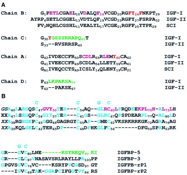
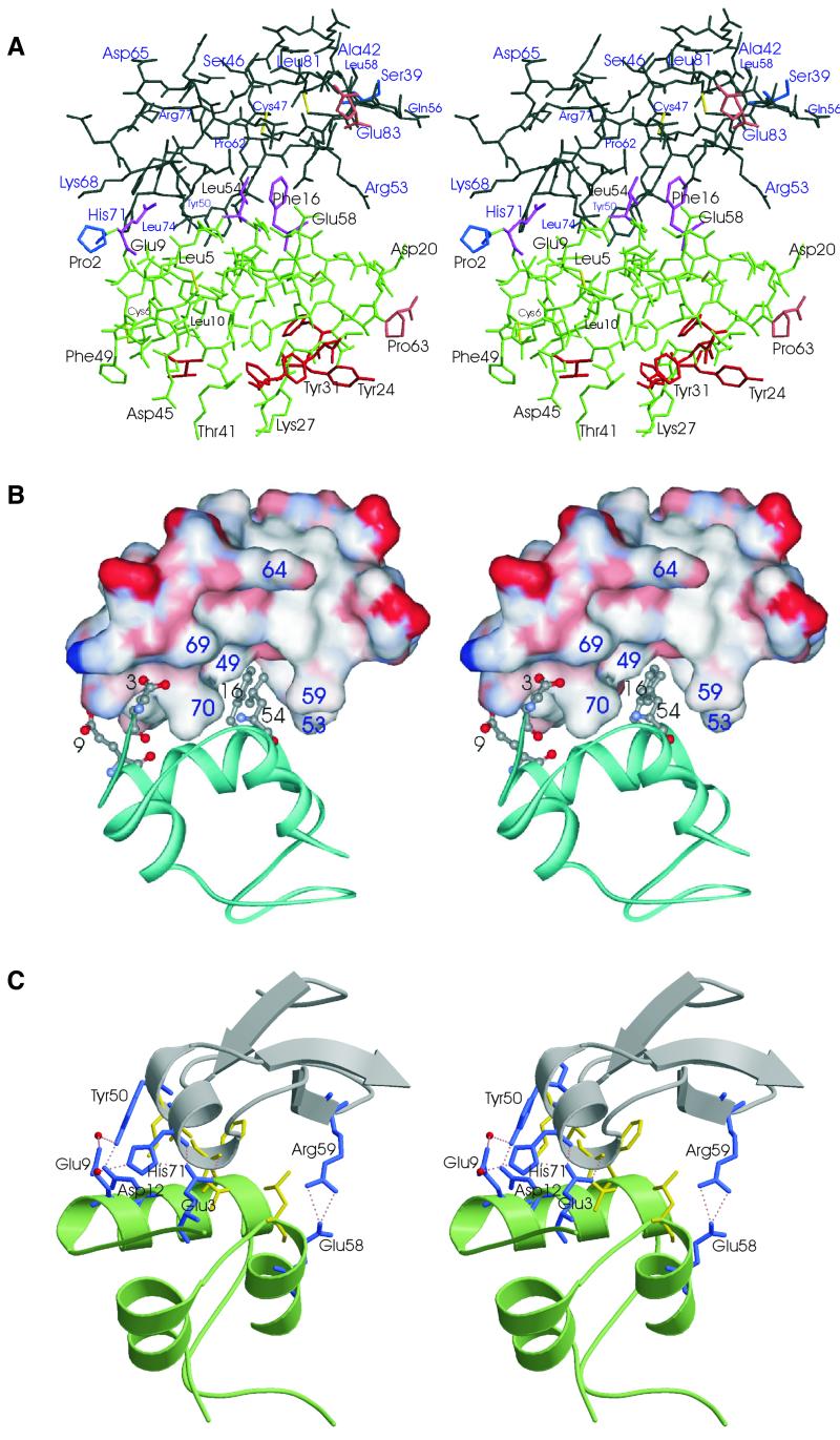
Insulin-like growth factors (IGFs) are key regulators of cell proliferation, differentiation and transformation, and are thus pivotal in cancer, especially breast, prostate and colon neoplasms. They are also important in many neurological and bone disorders. Their potent mitogenic and anti-apoptotic actions depend primarily on their availability to bind to the cell surface IGF-I receptor. In circulation and interstitial fluids, IGFs are largely unavailable as they are tightly associated with IGF-binding proteins (IGFBPs) and are released after IGFBP proteolysis. Here we report the 2.1 A crystal structure of the complex of IGF-I bound to the N-terminal IGF-binding domain of IGFBP-5 (mini-IGFBP-5), a prototype interaction for all N-terminal domains of the IGFBP family. The principal interactions in the complex comprise interlaced hydrophobic side chains that protrude from both IGF-I and the IGFBP-5 fragment and a surrounding network of polar interactions. A solvent-exposed hydrophobic patch is located on the IGF-I pole opposite to the mini-IGFBP-5 binding region and marks the IGF-I receptor binding site.
Figures
Similar articles
-
Structure of the IGF-binding domain of the insulin-like growth factor-binding protein-5 (IGFBP-5): implications for IGF and IGF-I receptor interactions.EMBO J. 1998 Nov 16;17(22):6558-72. doi: 10.1093/emboj/17.22.6558. EMBO J. 1998. PMID: 9822601 Free PMC article.
-
Heparin-binding, highly basic regions within the thyroglobulin type-1 repeat of insulin-like growth factor (IGF)-binding proteins (IGFBPs) -3, -5, and -6 inhibit IGFBP-4 degradation.Endocrinology. 1997 Jun;138(6):2280-5. doi: 10.1210/endo.138.6.5182. Endocrinology. 1997. PMID: 9165012
-
Partial IGF affinity of circulating N- and C-terminal fragments of human insulin-like growth factor binding protein-4 (IGFBP-4) and the disulfide bonding pattern of the C-terminal IGFBP-4 domain.Biochemistry. 2000 May 2;39(17):5082-8. doi: 10.1021/bi992513s. Biochemistry. 2000. PMID: 10819974
-
IGF binding proteins and their functions.Mol Reprod Dev. 1993 Aug;35(4):368-74; discussion 374-5. doi: 10.1002/mrd.1080350409. Mol Reprod Dev. 1993. PMID: 7691098 Review.
-
The insulin-like growth factor-binding protein (IGFBP) superfamily.Endocr Rev. 1999 Dec;20(6):761-87. doi: 10.1210/edrv.20.6.0382. Endocr Rev. 1999. PMID: 10605625 Review.
Cited by
-
Characterization and Expression Analysis of Insulin Growth Factor Binding Proteins (IGFBPs) in Pacific White Shrimp Litopenaeus vannamei.Int J Mol Sci. 2021 Jan 21;22(3):1056. doi: 10.3390/ijms22031056. Int J Mol Sci. 2021. PMID: 33494370 Free PMC article.
-
Biological effects and regulation of IGFBP5 in breast cancer.Front Endocrinol (Lausanne). 2022 Aug 25;13:983793. doi: 10.3389/fendo.2022.983793. eCollection 2022. Front Endocrinol (Lausanne). 2022. PMID: 36093095 Free PMC article. Review.
-
Defining the pathway to insulin-like growth factor system targeting in cancer.Biochem Pharmacol. 2010 Oct 15;80(8):1115-24. doi: 10.1016/j.bcp.2010.06.013. Epub 2010 Jun 23. Biochem Pharmacol. 2010. PMID: 20599789 Free PMC article. Review.
-
Structural basis for assembly and disassembly of the IGF/IGFBP/ALS ternary complex.Nat Commun. 2022 Jul 30;13(1):4434. doi: 10.1038/s41467-022-32214-2. Nat Commun. 2022. PMID: 35907924 Free PMC article.
-
Customized biomaterials to augment chondrocyte gene therapy.Acta Biomater. 2017 Apr 15;53:260-267. doi: 10.1016/j.actbio.2017.02.008. Epub 2017 Feb 7. Acta Biomater. 2017. PMID: 28185909 Free PMC article.
References
-
- Abrahams J.P. and Leslie,A.G.W. (1996) Methods used in the structure determination of bovine mitochondrial F1 ATPase. Acta Crystallogr. D, 52, 30–42. - PubMed
-
- Adams M.J., Blundell,T.L., Dodson,E.J., Vijazan,M., Baker,E.N., Hodgkin,D.C., Rimm,B. and Sheat,S. (1969) Structure of rhombohedral 2 zinc insulin crystals. Nature, 224, 491–495.
-
- Bach L.A., Hsieh,S., Sakano,K., Fujiwara,H., Perdue,J.F. and Rechler,M.M. (1993) Binding of mutants of human insulin-like growth factor II to insulin-like growth factor binding proteins 1–6. J. Biol. Chem., 268, 9246–9254. - PubMed
-
- Baxter R.C., Bayne,M.L. and Cascieri,M.A. (1992) Structural determinants for binary and ternary complex formation between insulin-like growth factor-I (IGF-I) and IGF binding protein-3. J. Biol. Chem., 267, 60–65. - PubMed
-
- Bayne M.L., Applebaum,J., Chicchi,G.G., Miller,R.E. and Cascieri,M.A. (1990) The roles of tyrosine 24, 31, and 60 in the high affinity binding of insulin-like growth factor-I and the type 1 insulin-like growth factor receptor. J. Biol. Chem., 265, 15648–15652. - PubMed
Publication types
MeSH terms
Substances
Associated data
- Actions
LinkOut - more resources
Full Text Sources
Other Literature Sources
