. 2001 Jul 16;20(14):3645-56.
doi: 10.1093/emboj/20.14.3645.

Crystal structure of the M-fragment of alpha-catenin: implications for modulation of cell adhesion

Affiliations

Crystal structure of the M-fragment of alpha-catenin: implications for modulation of cell adhesion

J Yang et al. EMBO J. .

Abstract

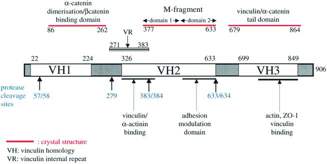
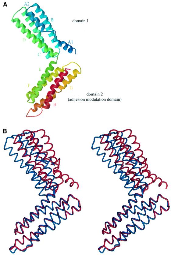
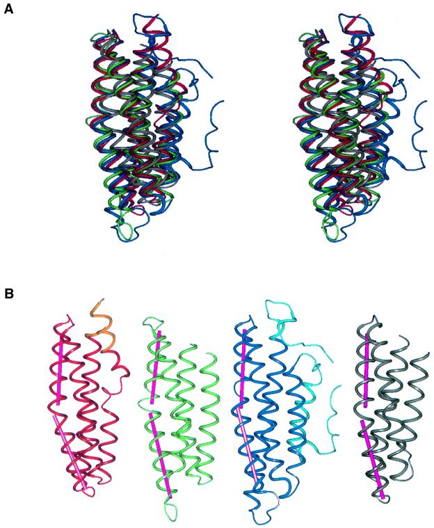
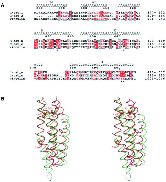
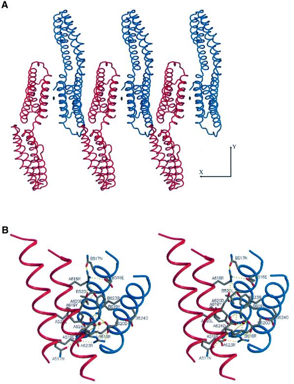
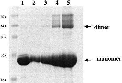
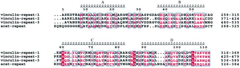
The cytoskeletal protein alpha-catenin, which shares structural similarity with vinculin, is required for cadherin-mediated cell adhesion, and functions to modulate cell adhesive strength and to link the cadherins to the actin-based cytoskeleton. Here we describe the crystal structure of a region of alpha-catenin (residues 377-633) termed the M-fragment. The M-fragment is composed of a tandem repeat of two antiparallel four-helix bundles of virtually identical architectures that are related in structure to the dimerization domain of alpha-catenin and the tail region of vinculin. These results suggest that alpha-catenin is composed of repeating antiparallel helical domains. The region of alpha-catenin previously defined as an adhesion modulation domain corresponds to the C-terminal four-helix bundle of the M-fragment, and in the crystal lattice these domains exist as dimers. Evidence for dimerization of the M-fragment of alpha-catenin in solution was detected by chemical cross-linking experiments. The tendency of the adhesion modulation domain to form dimers may explain its biological activity of promoting cell-cell adhesiveness by inducing lateral dimerization of the associated cadherin molecule.


Figures

None
Fig. 1. Schematic of α-catenin indicating that the protein is composed of tandem repeats of antiparallel α-helical bundles with defined biological functions. The positions of the VH1, VH2, VH3 and the α-catenin VR regions, together with sites of proteolysis are indicated. The domains of α-catenin for which structural data are available (β-catenin/dimerization domain; M-fragment of α-catenin; α-catenin tail) and the position of the predicted VR are shown.
None
Fig. 2. Detection of protease-resistant domains of α-catenin using limited proteolysis. Incubation of α-catenin with increasing concentra tions of trypsin for 20 min reveals the formation of a partially resistant 63 kDa fragment.
None
Fig. 3. Structure of α-catenin M-fragment. (A) Overall view of the structure with two four-helix domains, coloured from the N-terminus (blue) to the C-terminus (red). (B) Stereo-view showing the two crystallographically independent molecules (A and B) superimposed using equivalent Cα atoms of domain 2. Red denotes molecule A and blue denotes molecule B. The figure was prepared using BOBSCRIPT (Esnouf, 1997), Raster3D (Merrit and Murphy, 1994) and PREPI (S.Islam and M.Sternberg).
None
Fig. 4. Similarities between α-catenin M-fragment domains 1 and 2 and domains of other structurally related proteins. (A) Superimposition of α-catenin M-fragment domains 1 (red) and 2 (green), the vinculin tail domain (blue) and the α-catenin dimerization domain (grey). (B) A comparison of the α-catenin M-fragment domains 1 (red/orange) and 2 (green) with the vinculin tail domain (dark/light blue) and α-catenin dimerization domain (grey). Darker colours denote the structures used for alignment onto domain 2 of the α-catenin M-fragment. The figures were prepared using PREPI (S.Islam and M.Sternberg).
None
Fig. 5. Similarities between α-catenin M-fragment domains 1 and 2. (A) Sequence alignment of α-catenin M-fragment domains 1 and 2 and the vinculin tail domain. (B) Superimposition of α-catenin M-fragment domains 1 and 2 showing residues Val433, Pro462, Ala501 and Val502 of domain 1 and the structurally equivalent residues of domain 2 [indicated with an asterisk in (A)].
None
Fig. 6. View of the dimer interface of the α-catenin M-fragment domain 2. (A) Crystal packing of dimers of molecules A and B of the α-catenin M-fragment viewed perpendicular to the 2-fold non-crystallographic symmetry axis. (B) Stereo-view showing the interactions between the monomers at the dimer interface formed from domain 2. Colouring scheme as in Figure 3B.
None
Fig. 7. Evidence that the M-fragment of α-catenin exists as a dimer in solution. Lane 1: 0.25 mg/ml M-fragment of α-catenin in the absence of the cross-linking reagent dimethyl suberimidate (DMS). Lanes 2–5: 0.05, 0.1, 0.25 and 0.5 mg/ml of the α-catenin M-fragment incubated with a 30-fold molar access of DMS for 1.5 h at 20°C. The formation of dimers of the α-catenin M-fragment at concentrations of >0.1 mg/ml (3 µM) (lanes 3–5) is detected by the formation of dimers cross-linked with DMS visualized on an SDS–polyacrylamide gel.
None
Fig. 8. Sequence alignment of three internal repeats of human meta-vinculin (vinculin repeats) and the related sequence in human α(E)-catenin. The mean sequence identity between the α-catenin VR and the three VRs of vinculin is ∼10%. The positions of the four predicted α-helices are shown. The figure was produced using ESPript (Gouet et al., 1999).

References

    1. Aberle H., Butz,S., Stappert,J., Weissig,H., Kemler,R. and Hoschuetzky,H. (1994) Assembly of the cadherin–catenin complex in vitro with recombinant proteins. J. Cell Sci., 107, 3655–3663. - PubMed
    1. Abrahams J.P. and Leslie,A.G.W. (1996) Methods used in the structure determination of the bovine mitochondrial F1 ATPase. Acta Crystallogr. D, 52, 30–42. - PubMed
    1. Bakolitsa C., de Pereda,J.M., Bagshaw,C.R., Critchley,D.R. and Liddington,R.C. (1999) Crystal structure of the vinculin tail suggests a pathway for activation. Cell, 99, 603–613. - PubMed
    1. Berman H.M., Westbrook,J., Feng,Z., Gillialnd,G., Bhat,T.N., Weissig,H., Shindyalov,I.N. and Bourne,P.E. (2000) The Protein Data Bank. Nucleic Acids Res., 28, 235–242. - PMC - PubMed
    1. Birchmeier W. and Behrens,J. (1994) Cadherin expression in carcinomas: role in the formation of cell junctions and the prevention of invasiveness. Biochim. Biophys. Acta, 1198, 11–26. - PubMed

Publication types

MeSH terms

Associated data