. 2001 Jul 16;20(14):3667-75.
doi: 10.1093/emboj/20.14.3667.

Adenylyl cyclase Rv1625c of Mycobacterium tuberculosis: a progenitor of mammalian adenylyl cyclases

Affiliations

Adenylyl cyclase Rv1625c of Mycobacterium tuberculosis: a progenitor of mammalian adenylyl cyclases

Y L Guo et al. EMBO J. .

Abstract

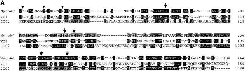
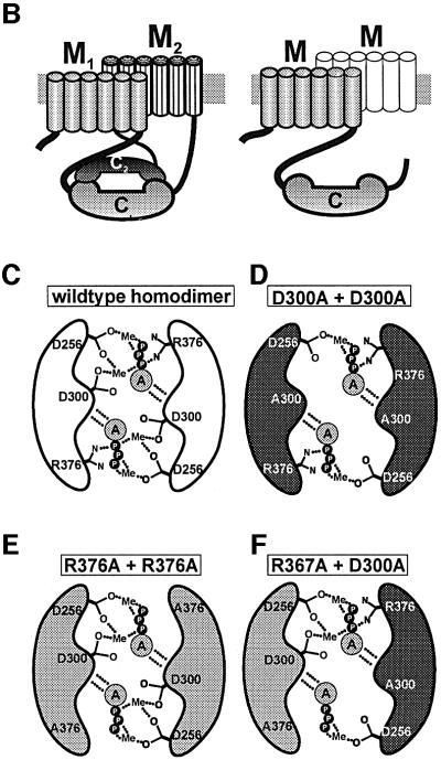
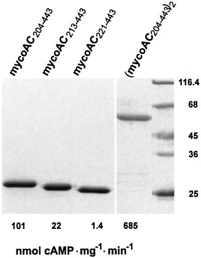
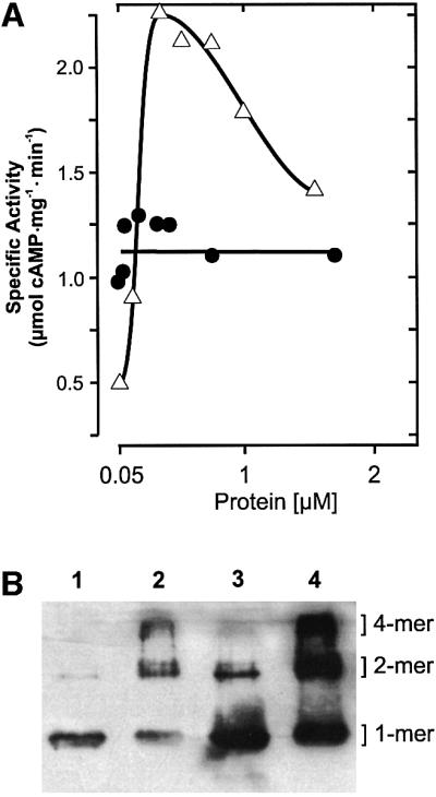
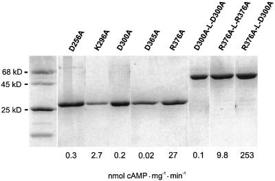
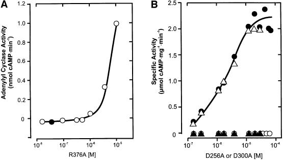
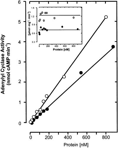
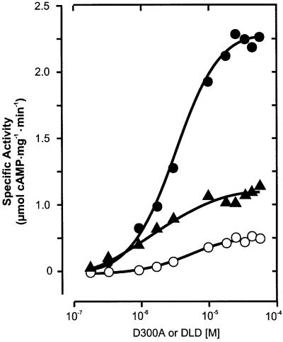
The gene Rv1625c from Mycobacterium tuberculosis encodes a membrane-anchored adenylyl cyclase corresponding to exactly one-half of a mammalian adenylyl cyclase. An engineered, soluble form of Rv1625c was expressed in Escherichia coli. It formed a homodimeric cyclase with two catalytic centers. Amino acid mutations predicted to affect catalysis resulted in inactive monomers. A single catalytic center with wild-type activity could be reconstituted from mutated monomers in stringent analogy to the mammalian heterodimeric cyclase structure. The proposed existence of supramolecular adenylyl cyclase complexes was established by reconstitution from peptide-linked, mutation-inactivated homodimers resulting in pseudo-trimeric and -tetrameric complexes. The mycobacterial holoenzyme was expressed successfully in E.coli and mammalian HEK293 cells, i.e. its membrane targeting sequence was compatible with the bacterial and eukaryotic machinery for processing and membrane insertion. The membrane-anchored mycobacterial cyclase expressed in E.coli was purified to homogeneity as a first step toward the complete structural elucidation of this important protein. As the closest progenitor of the mammalian adenylyl cyclase family to date, the mycobacterial cyclase probably was spread by horizontal gene transfer.


Figures

None
Fig. 1. (A) Alignment of the catalytic domains of the mycobacterial adenylyl cyclase with C1 from canine type V (VC1) and C2 from rat type II (IIC2) adenylyl cyclases (residues shared with either mammalian sequence are inverted). The triangles indicate D204, E213 and A221 as starting points of the cytosolic constructs. The arrows mark the mutated amino acids (to alanine) that are involved in substrate definition (K296 and D365), coordination of metal ions (D256 and D300) and transition state stabilization (R376). Note that in the mammalian domains, the equivalents of D256 and D300 are contributed by C1 whereas D365 and R376 are contributed by C2. (B) Predicted topology of the pseudoheterodimeric mammalian adenylyl cyclases (left) and the monomeric mycobacterial AC. M designates a membrane cassette of six transmembrane spans. In the mycobacterial enzyme, the homodimerization is intimated by a sketchy second M domain. (C) Symbolized homodimeric catalytic center of the mycobacterial adenylyl cyclase capable of forming two catalytic sites. D256, D300 and R376 are outlined; binding of the adenine ring A is indicated by dotted lines. (D and E) Proposed homodimeric structure of the (D) D300A and (E) R376A mutants. (F) Symbolized heterodimer with a single catalytic site reconstituted from the D300A and R376A mutant monomers. The same model may be applied to the D256A mutation (not depicted). P = phosphate; Me = divalent metal cation.
None
Fig. 2. Purification and activity of recombinant mycobacterial adenylyl cyclase catalytic domains (SDS–PAGE analysis, Coomassie blue staining). The soluble cytoplasmic domains started at D204 (mycoAC204–443), E213 (mycoAC213–443) and A221 (mycoAC221–443) as indicated in Figure 1A. The specific activities indicated below each lane were determined with 75 µM ATP at a protein concentration of 2 µg/assay (750 nM). The dimer (mycoAC204–443)2 was assayed at 0.6 µg protein/assay (110 nM).
None
Fig. 3. (A) Protein dependence of mycoAC204–443 (open triangles) and linked (mycoAC204–443)2 (filled circles) activity (850 µM Mn-ATP, 4 min). (B) Western blot analysis of mycoAC204–443 oligomers cross-linked by 20 mM glutaraldehyde. Lane 1, 300 nM mycoAC204–443 (0.24 µg protein), no glutaraldehyde; lane 2, + 20 mM glutaraldehyde; lane 3, 2 µM mycoAC204–443 (1.6 µg protein), no glutaraldehyde; lane 4, + 20 mM glutaraldehyde. Densitometric evaluation (lanes 1–4): the monomers accounted for 96, 31, 80 and 30%, the dimers for 4, 44, 18 and 33%, and the tetramers for 0, 25, 2 and 37%, respectively.
None
Fig. 4. SDS–PAGE analysis of purified mycoAC204–443 mutant constructs (Coomassie blue staining). Molecular mass standards are on the left. The following amounts of protein were applied (left to right): D256A, 4 µg; K296A, 1.5 µg; D300A, 4 µg; D365A, 2 µg; R376A, 2.3 µg; D300A-L-D300A, R376A-L-R376A and R376A-L-D300A, 4 µg each. The specific AC activities determined at 75 µM Mn-ATP and 4 µM protein are indicated below each lane (114 nM for R376A-L-D300A).
None
Fig. 5. Reconstitution of mycobacterial adenylyl cyclase activity from the D256A, D300A and R376A mutants. (A) Protein dependency of R376A activity. The protein concentration marked by the filled circle (45 nM) was used in (B) for reconstitution with increasing amounts of D256A (open triangles) and D300A (filled circles); D256A (filled triangles) and D300A (open circles) alone. Note the different scales (assays with 850 µM Mn-ATP, 4 min).
None
Fig. 7. Intermolecular formation of adenylyl cyclase catalysts from mutated, inactive monomers and homodimers. Filled circles: 45 nM R376A (inactive) titrated with increasing amounts of D300A-L-D300A (DLD). Filled triangles: 36 nM R376A-L-R376A titrated with D300A. Open circles: 36 nM R376A-L-R376A titrated with D300A-L-D300A. Assays were carried out with 850 µM Mn-ATP as a substrate for 4 min. The specific activity was calculated on the basis of the fixed concentration of the minor constituent.
None
Fig. 6. One and two catalytic centers formed by (mycoAC204–443)2 (open circles) and R376A-L-D300A (filled circles), respectively, demonstrated by the linearity of the protein dependences and the unchanging specific activities. Inset: specific activity (same symbols; 850 µM Mn-ATP, 4 min).
None
Fig. 8. Electrophoretic analysis (SDS–PAGE and western blot) of the mycobacterial adenylyl cyclase holoenzyme. Lane 1, Coomassie blue-stained SDS–PAGE of purified enzyme expressed in E.coli; lanes 2 and 3, western blot of purified protein probed with antibodies against the N-terminal His tag. Note the distinct 92 kDa band in lane 3 demonstrating a homodimer. Escherichia coli (lane 4) and HEK293 homogenates (lane 5) expressing the holoenzyme were probed with an antibody directed against mycoAC204–443.

Similar articles

Cited by

References

    1. Baltimore D. (2001) Our genome unveiled. Nature, 409, 814–816. - PubMed
    1. Bhatnagar N.B., Bhatnagar,R. and Venkitasubramanian,T.A. (1984) Characterization and metabolism of cyclic guanosine 3′,5′-monophosphate in Mycobacterium smegmatis. Biochem. Biophys. Res. Commun., 121, 634–640. - PubMed
    1. Chang G., Spencer,R.H., Lee,A.T., Barclay,M.T. and Rees,D.C. (1998) Structure of the MscL homolog from Mycobacterium tuberculosis: a gated mechanosensitive ion channel. Science, 282, 2220–2226. - PubMed
    1. Chen G.Q., Cui,C., Mayer,M.L. and Gouaux,E. (1999) Functional characterization of a potassium-selective prokaryotic glutamate receptor. Nature, 402, 817–821. - PubMed
    1. Chen J. et al. (1995) A region of adenylyl cyclase 2 critical for regulation by G protein βγ subunits. Science, 268, 1166–1169. - PubMed

Publication types

MeSH terms