. 2001 Jul 16;20(14):3781-8.
doi: 10.1093/emboj/20.14.3781.

Acf1, the largest subunit of CHRAC, regulates ISWI-induced nucleosome remodelling

Affiliations

Acf1, the largest subunit of CHRAC, regulates ISWI-induced nucleosome remodelling

A Eberharter et al. EMBO J. .

Abstract

The chromatin accessibility complex (CHRAC) was originally defined biochemically as an ATP-dependent 'nucleosome remodelling' activity. Central to its activity is the ATPase ISWI, which catalyses the transfer of histone octamers between DNA segments in cis. In addition to ISWI, four other potential subunits were observed consistently in active CHRAC fractions. We have now identified the p175 subunit of CHRAC as Acf1, a protein known to associate with ISWI in the ACF complex. Interaction of Acf1 with ISWI enhances the efficiency of nucleosome sliding by an order of magnitude. Remarkably, it also modulates the nucleosome remodelling activity of ISWI qualitatively by altering the directionality of nucleosome movements and the histone 'tail' requirements of the reaction. The Acf1-ISWI heteromer tightly interacts with the two recently identified small histone fold proteins CHRAC-14 and CHRAC-16. Whether topoisomerase II is an integral subunit has been controversial. Refined analyses now suggest that topoisomerase II should not be considered a stable subunit of CHRAC. Accordingly, CHRAC can be molecularly defined as a complex consisting of ISWI, Acf1, CHRAC-14 and CHRAC-16.


Figures
None
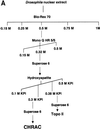
Fig. 1. CHRAC consists of ISWI, Acf1, CHRAC-16 and CHRAC-14. (A) Purification scheme for the isolation of native CHRAC from Drosophila embryos. (B) Bio-gel HT hydroxyapatite chromatography separates CHRAC from topoisomerase II. CHRAC-containing Superose 6 fractions were pooled and applied onto a hydroxyapatite column. Western blot analyses with the indicated antisera show that co-eluting ISWI, Acf1 and CHRAC-14/16 were separated from topo II.
None
Fig. 2. Size exclusion chromatography of CHRAC, native topo II and recombinant topo II. Size standards are depicted by arrows. (A) Pooled CHRAC hydroxyapatite fractions (21–24) were separated on a Superose 6 column. Western blot analyses showed that CHRAC eluted with a mol. wt of ∼600 kDa. (B) Superose 6 chromatography of topo II-containing hydroxyapatite fractions (28–32). (C) Superose 6 chromatography of purified recombinant Drosophila topo II. (D) Analysis of recombinant topo II by MALS coupled to a Superdex S-200 sizing column.
None
Fig. 3. Immunoprecipitation of the CHRAC components from Drosophila nuclear extract. Protein from crude extract was immunoprecipitated with anti-ISWI, anti-CHRAC-14 and anti-CHRAC-14/16 antibodies as well as pre-immune serum. Aliquots of the immunoprecipitated material were resolved by SDS–PAGE and detected by western blotting using the indicated antisera.
None
Fig. 4. Purification of recombinant ISWI, Acf1 and ISWI/Acf1. The indicated factors (Flag-ISWI, Acf1-Flag or Flag-ISWI + Acf1) were expressed via a baculovirus system and purified by α-Flag immuno affinity chromatography. Two different concentrations for each protein were resolved by 6% SDS–PAGE and stained with Coomassie blue. Arrows depict the positions of Acf1 and ISWI.
None
Fig. 5. Nucleosome sliding by recombinant ISWI/Acf1 (= ACF) and ISWI. (A) Nucleosomes positioned at the centre of the 248 rDNA fragment were incubated with the indicated amounts of affinity-purified Acf1-Flag, ACF or Flag-ISWI. Nucleosomes were then analysed on a native 4.5% polyacrylamide gel. Terminal or central nucleosome positions are indicated to the left. Lane (–) indicates the untreated centre nucleosome. (B) End-positioned nucleosomes were incubated with the indicated amounts of affinity-purified Flag-ISWI, ACF or Acf1-Flag. Nucleosome analysis and figure labelling are described in (A).
None
Fig. 6. Functional comparison of ISWI and ACF. (A) Nucleosome sliding was monitored during a time course of reactions containing either 2 fmol of ACF and end-positioned nucleosome, or 25 fmol of F-ISWI with a centrally positioned nucleosome substrate. The reactions were analysed by native gel electrophoresis as before. Signals corresponding to the moved and unmoved nucleosome fraction were quantified by a PhosphoImager (FujiFilm BAS-1500) and the fraction of moved nucleosomes plotted as a function of time. (B) ATPase assays: 400 fmol each of F-ISWI or ACF were tested for ATPase in the presence of 100 ng of naked DNA (open bars) or 100 ng of nucleosomal DNA reconstituted by salt gradient dialysis (black bars). A low background of hydrolysed ATP in the absence of enzyme was subtracted. The results have been reproduced qualitatively under various circumstances, although the absolute numbers vary as a function of ATP batch, age and enzyme preparation.
None
Fig. 7. Histone tail requirement for nucleosome mobilization by ACF. Recombinant ACF complex was incubated with end-positioned nucleosomes reconstituted with either wild-type histones (intact) or, alternatively, a histone mixture consisting of three wild-type histones and one tailless variant (Δ) as indicated. The reactions were analysed by native polyacrylamide gel electrophoresis. Positions of nucleosomes and free DNA are indicated. Reactions contained 0.1, 0.2, 0.5, 0.75, 1 and 2 fmol of ACF (the 2 fmol sample was omitted in the panel ‘intact’).

Similar articles

Cited by

References

    1. Alexiadis V., Varga-Weisz,P.D., Bonte,E., Becker,P.B. and Gruss,C. (1998) In vitro chromatin remodelling by chromatin accessibility complex (CHRAC) at the SV40 origin of DNA replication. EMBO J., 17, 3428–3438. - PMC - PubMed
    1. Arents G., Burlingame,R.W., Wang,B.C., Love,W.E. and Moudrianakis,E.N. (1991) The nucleosomal core histone octamer at 3.1 Å resolution: a tripartite protein assembly and a left-handed superhelix. Proc. Natl Acad. Sci. USA, 88, 10148–10152. - PMC - PubMed
    1. Bochar D.A., Savard,J., Wang,W., Lafleur,D.W., Moore,P., Cote,J. and Shiekhattar,R. (2000) A family of chromatin remodeling factors related to Williams syndrome transcription factor. Proc. Natl Acad. Sci. USA, 97, 1038–1043. - PMC - PubMed
    1. Boyer L.A., Logie,C., Bonte,E., Becker,P.B., Wade,P.A., Wolffe,A.P., Wu,C., Imbalzano,A.N. and Peterson,C.L. (2000) Functional delineation of three groups of the ATP-dependent family of chromatin remodeling enzymes. J. Biol. Chem., 275, 18864–18870. - PubMed
    1. Brehm A., Längst,G., Kehle,J., Clapier,C.R., Imhof,A., Eberharter,A., Müller,J. and Becker,P.B. (2000) dMi-2 and ISWI chromatin remodeling factors have distinct nucleosome binding and mobilization properties. EMBO J., 19, 4332–4341. - PMC - PubMed

Publication types