. 2001 Jul 16;20(14):3821-30.
doi: 10.1093/emboj/20.14.3821.

The RNA binding protein YB-1 binds A/C-rich exon enhancers and stimulates splicing of the CD44 alternative exon v4

Affiliations

The RNA binding protein YB-1 binds A/C-rich exon enhancers and stimulates splicing of the CD44 alternative exon v4

E Stickeler et al. EMBO J. .

Abstract

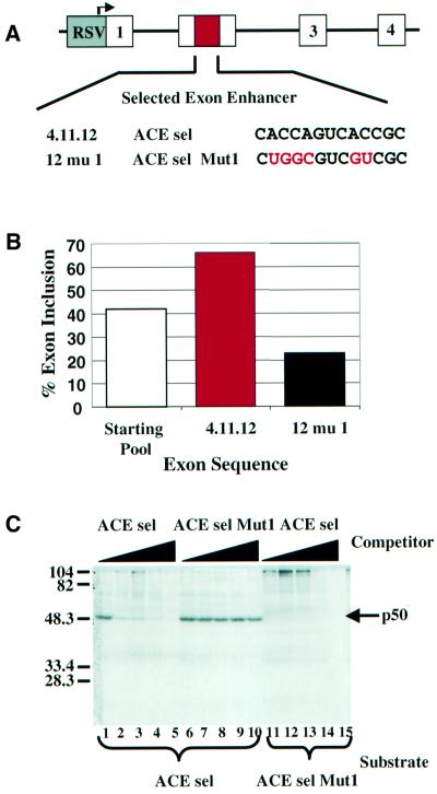
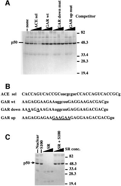
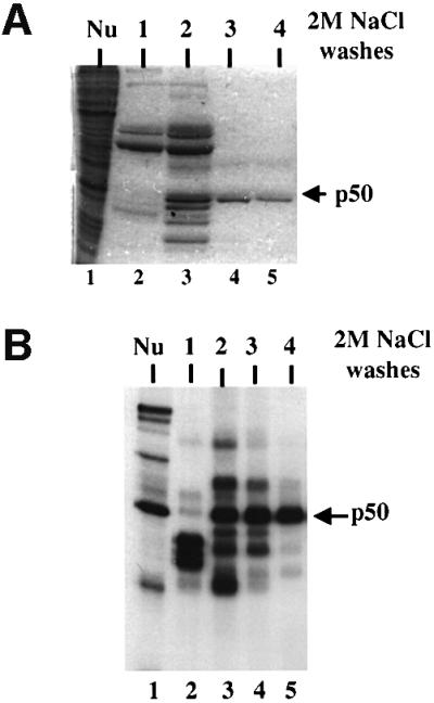
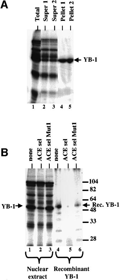
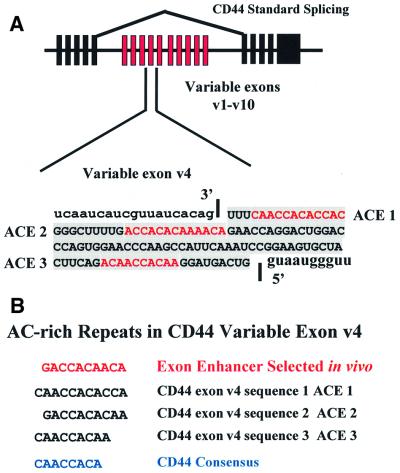
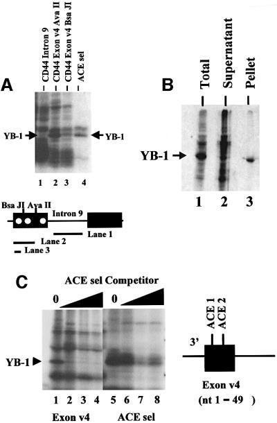
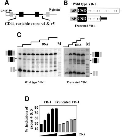
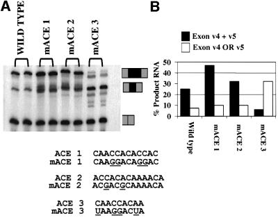
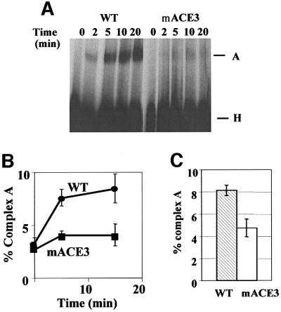
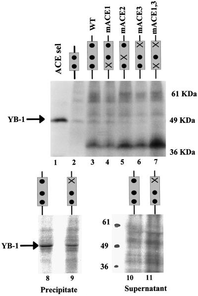
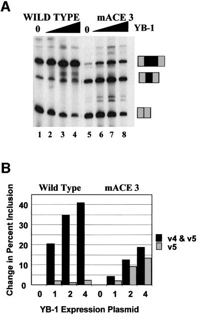
Exon enhancers are accessory pre-mRNA splicing signals that stimulate exon splicing. One class of proteins, the serine-arginine-rich (SR) proteins, have been demonstrated to bind enhancers and activate splicing. Here we report that A/C-rich exon enhancers (ACE elements) are recognized by the human YB-1 protein, a non-SR protein. Sequence-specific binding of YB-1 was observed both to an ACE derived from an in vivo iterative selection protocol and to ACE elements in an alternative exon (v4) from the human CD44 gene. The ACE element that was the predominant YB-1 binding site in CD44 exon v4 was required for maximal in vivo splicing and in vitro spliceosome assembly. Expression of wild-type YB-1 increased inclusion of exon v4, whereas a truncated form of YB-1 did not. Stimulation of exon v4 inclusion by wild-type YB-1 required the ACE necessary for YB-1 binding in vitro, suggesting that YB-1 stimulated exon inclusion in vivo by binding to an exonic ACE element. These observations identify a protein in addition to SR proteins that participates in the recognition of exon enhancers.


Figures

None
Fig. 1. A/C-rich exon enhancers bind a 50 kDa nuclear protein. (A) Sequence of one of the 13 nucleotide ACE exon enhancers identified by iterative in vivo selection from 13 random nucleotides using the diagrammed mini-gene (Coulter et al., 1997). The 4.11.12 isolate, referred to here as ACE sel, is shown along with a mutant version. (BIn vivo exon inclusion activity of the sequences shown in (A). Activity was tested in a heterologous exon context (Coulter et al., 1997). (CIn vitro UV cross-linking of a 51 nucleotide RNA including two copies of the ACE sel sequence from (A) (see Materials and methods). Radiolabeled RNA substrates as indicated beneath the gel were incubated for 10 min under standard in vitro splicing conditions using HeLa nuclear extract in the presence of increasing concentrations (0, 1, 5, 10 and 25 pmol) of competitor RNAs containing the sequences shown above the gel. Following cross-linking and RNase digestion, cross-linked proteins were resolved by 11% SDS–PAGE. A prominent 50 kDa band that was cross-linked to wild-type but not mutant RNA is indicated by an arrow.
None
Fig. 2. p50 is distinct from the SR proteins that recognize purine-rich enhancers. (A) RNAs containing purine-rich enhancers do not compete UV cross-linking of p50 to ACE elements. The ACE sel RNA described in Figure 1 was subjected to UV cross-linking in the presence of competitor RNAs containing the sequences indicated above the gel. The positions of p50 and molecular weight markers are indicated. (B) Sequences of the competitor RNAs which are derived from exon 5 of the chicken cardiac troponin T gene [Ramchatesingh et al., 1995, where they are referred to as D2WT (GAR wt), D2A2 (GAR down mutant), D2EY1 (GAR up mutant)]. Not shown are the first 17 nucleotides (ACE sel; see Materials and methods) or 15 nucleotides (GAR wt, GAR down mutant and GAR up mutant) derived from the transcription vector. (C) Preparations of HeLa SR proteins do not contain p50, and SR proteins do not stimulate the binding of p50. UV cross-linking was performed using in vitro splicing conditions (Materials and methods) in either HeLa nuclear (lane 1) or S100 (lanes 2, 6, 7 and 8) extract or with increasing amounts (50, 100 or 200 ng) of SR proteins (Zahler, 1999) in the absence (lanes 3, 4 and 5) or presence (lanes 6, 7 and 8) of S100 extract. The positions of p50 and marker proteins are indicated.
None
Fig. 3. Purification of p50. (A) HeLa nuclear extract was fractionated on poly(U)–agarose (Materials and methods). The starting material (lane 1) and four successive elution washes using 2 M NaCl are shown (lanes 2–5) in Coomassie Blue-stained 10% SDS–polyacrylamide gel. The position of a 50 kDa protein in fractions 2, 3 and 4 is denoted with an arrow. (B) UV cross-linking of the column fractions shown in (A). Column fractions were dialyzed against Roeder D (Dignam et al., 1983) and used in a standard UV cross-linking assay with the ACE sel RNA. The cross-linked p50 in nuclear extract (lane 1) and fractions 2–4 is indicated.
None
Fig. 4. Antibodies to YB-1 peptides immunoprecipitate p50. Polyclonal antibodies raised against a C-terminal peptide of human YB-1 were used to immunoprecipitate proteins cross-linked to the ACE sel RNA. (A) Immunoprecipitation of cross-linking proteins in HeLa nuclear extract. Duplicate immunoprecipitations are shown. Lane 1, total cross-linking; lanes 2 and 3, supernatants for immunoprecipitations 1 and 2; lanes 4 and 5, pellets from immunoprecipitations 1 and 2. (B) Competition of UV cross-linking of nuclear proteins (lanes 1–3) or gel-purified and renatured recombinant human YB-1 (lanes 4–6). Cross-linking reactions were performed without competitor (lanes 1 and 4) or with 10 pmol of the ACE sel competitor (lanes 2 and 5) or the ACE sel Mut 1 competitor (lanes 3 and 6). The positions of HeLa YB-1 and recombinant YB-1 are indicated; the latter is slightly larger as a result of the presence of a His tag.
None
Fig. 5. Human CD44 alternative splicing. (A) The exon–intron architecture of the human CD44 gene is drawn at the top. The 10 alternative cassette exons are depicted in red. The exon studied in this report is the fourth variable exon v4. The sequence of this exon is shown at the bottom of the figure. Sequences within the exon are indicated with a gray background. The red sequence indicates A/C-rich elements ACE 1, ACE 2 and ACE 3 within exon v4 that are potential binding sites for YB-1. (B) A/C-rich repeats within CD44 variable exons 4 and 5. The A/C-rich sequences from the two exons are aligned and a consensus repeat sequence (blue) is derived. At the top is the derived repeat consensus ACE from the exon selection experiments.
None
Fig. 6. CD44 exon v4 can be UV cross-linked to YB-1. (A) The substrate RNAs diagrammed below the gel were UV cross-linked in a standard splicing assay using HeLa nuclear extract. A diagram of the region of the human CD44 gene containing the fourth and fifth alternative exons is indicated below the gel. Intron 9 is the naturally occurring intron separating exons v4 and v5. ACE elements within exon v4 are indicated by white circles. The partial exon v4 RNAs shown in lanes 2 and 3 were generated by transcript termination at the indicated restriction sites. The ACE sel RNA used for lane 4 is longer than the RNA used for previous figures (see Materials and methods) resulting in the cross-linking of a protein slightly larger thanYB-1 in addition to YB-1. The position of YB-1 is indicated. (B) Immunoprecipitation of cross-linked proteins with an antibody specific for YB-1. A substrate RNA containing the first half of exon 4 and including the ACE 1 and ACE 2 elements (diagrammed in C) was subjected to UV cross-linking in a standard HeLa in vitro splicing assay. Cross-linked proteins were immunoprecipitated using the anti-YB-1 antibody and displayed by SDS–PAGE. Lane 1, total proteins; lane 2, supernatant; lane 3, precipitated protein (3-fold more of the reaction was loaded in lanes 2 and 3 than 1; lanes 2 and 3 came from the same reaction). (C) Competition of YB-1 cross-linking to CD44 exon v4 with an RNA containing ACE sel. Two substrate RNAs were employed: the exon 4 substrate described in (B) (lanes 1–4) and the ACE sel RNA itself (lanes 5–8). Competitor concentrations were 0, 0.3, 3.0 and 10 pmol. The position of cross-linked YB-1 is indicated.
None
Fig. 7. Transfection of human YB-1 increased the inclusion of CD44 exons v4 and v5. (A) The reporter mini-gene containing CD44 variable exons v4 and v5. CD44 sequences are in black, human β-globin sequences are in gray, and the CMV promoter is in white. The CD44 insert is a contiguous genomic fragment mapping from 792 nucleotides upstream of exon v4 to 515 nucleotides downstream of exon v5. (B) Diagram of the wild-type and mutant YB-1 proteins made from co-expressing plasmids. The three indicated domains include an N-terminal alanine- and proline-rich region (AP), the single-stranded nucleic acid binding CSD and the C-terminal highly charged region. (C) RT–PCR analysis of RNAs produced upon transfection of HeLa cells with the reporter shown in (A) and increasing amounts (0, 1, 2, 3 or 4 µg) of either wild-type or mutant YB-1 expression plasmids. Duplicate lanes are shown for each concentration of the wild-type YB-1. PCR oligonucleotides were complementary to sequences in the flanking β-globin exons. Product RNAs resulting from inclusion of neither CD44 exon, either exon v4 or v5, or both exons v4 and v5 are indicated. Note that exons v4 and v5 have such similar sizes that inclusion bands resulting from either of the exons will be in the same region of the gel. Increasing amounts of the YB-1 expression plasmid resulted in a linear increase in the amount of YB-1 mRNA (data not shown). (D) Quantification of the effect of YB-1 on inclusion of CD44 exons v4 and v5. Results of four independent experiments are shown with 1 SD indicated.
None
Fig. 8. Mutation of CD44 exon v4 ACE 1 or 3 alters in vivo recognition of exon v4. (A) The diagrammed mutations were introduced into exon v4 of the CD44 mini-gene described in Figure 7. Wild-type and ACE 1, 2 or 3 mini-genes were transfected in duplicate into HeLa cells and the splicing phenotype was determined by RT–PCR as described in Figure 7. Note that because exons v4 and v5 are almost the same size, products containing exons v4 or v5 alone are indistinguishable in size. Sequencing of the band produced with the ACE 3 mutant indicates that this RNA product contains exon v5 but not v4. Product RNAs including no, one or two CD44 alternative exons are indicated. (B) Quantification of the results in (A).
None
Fig. 9. Mutation of ACE 3 depressed spliceosome assembly on CD44 exon v4. The wild-type or ACE3 mutant substrate RNAs including all of variable exon v4 and its flanking 3′ and 5′ splicing signals were incubated under in vitro splicing conditions for 0, 2, 5, 10 or 20 min. Mutations are identical to those described in Figure 8. (A) Formed complexes were analyzed by native gel electrophoresis (Carlo et al., 2000). The non-specific H complex and initial ATP-dependent spliceosomal A complex are indicated. (B) Quantification of the reaction in (A). The gel was scanned in the phosphoimager and the percentage of RNA in complex A was plotted. (C) Quantification of the amount of complex A formed at 20 min from multiple experiments using mutant and wild-type substrate RNA.
None
Fig. 10. Mutation of CD44 ACE sequences depresses UV cross-linking of YB-1. Top: the binding of YB-1 to wild-type and mutant CD44 exon 4 sequences was compared by UV cross-linking. The substrates are diagrammed above the gel. The position of YB-1 is indicated. Bottom: immunoprecipitation of YB-1 UV cross-linked to wild-type or mutant substrates. Cross-linked reactions were immunoprecipitated using the YB-1-specific antibody. Proteins from both the precipitate and the supernatant were displayed by SDS–PAGE.
None
Fig. 11. Mutation of CD44 ACE sequences depresses the ability to respond to YB-1 in vivo. (A) The mini-gene containing CD44 variable exons v4 and v5 was co-transfected with an expression vector coding for YB-1 as in Figure 9. The mini-gene contained a wild-type exon v4 (lanes 1–4) or an exon v4 containing the ACE 3 mutation (lanes 5–8). Increasing amounts of YB-1 vector were used (0, 1, 2 or 4 µg). The positions of RNA species resulting from the inclusion of no, one or two CD44 exons are indicated. (B) Quantification of the results in (A).

References

    1. Arning S., Gruter,P., Bilbe,G. and Kramer,A. (1996) Mammalian splicing factor SF1 is encoded by variant cDNAs and binds to RNA. RNA, 2, 794–810. - PMC - PubMed
    1. Bargou R.C. et al. (1997) Nuclear localization and increased levels of transcription factor YB-1 in primary human breast cancers are associated with intrinsic MDR1 gene expression. Nature Med., 3, 447–478. - PubMed
    1. Bell M.V., Cowper,A.E., Lefranc,M.P., Bell,J.I. and Screaton,G.R. (1998) Influence of intron length on alternative splicing of CD44. Mol. Cell. Biol., 18, 5930–5941. - PMC - PubMed
    1. Berglund J.A., Abovich,N. and Rosbash,M. (1998) A cooperative interaction between U2AF65 and mBBP/SF1 facilitates branchpoint region recognition. Genes Dev., 12, 858–867. - PMC - PubMed
    1. Black D. (1995) Finding splice sites within a wilderness of RNA. RNA, 1, 763–771. - PMC - PubMed

Publication types