. 2001 Jul 16;20(14):3840-8.
doi: 10.1093/emboj/20.14.3840.

Synthesis of a novel hepatitis C virus protein by ribosomal frameshift

Affiliations

Synthesis of a novel hepatitis C virus protein by ribosomal frameshift

Z Xu et al. EMBO J. .

Abstract

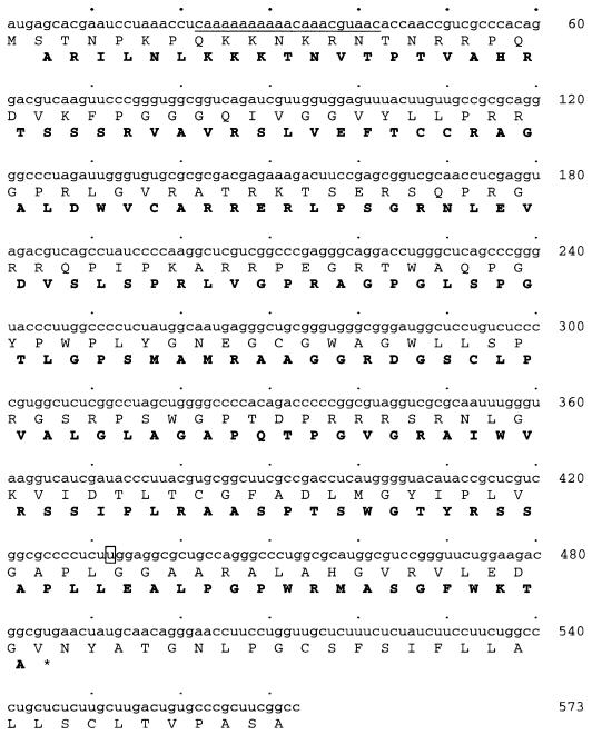
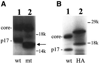
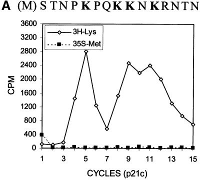
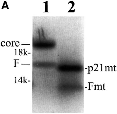
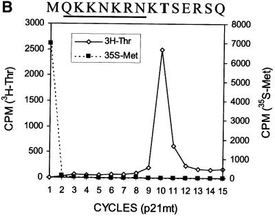
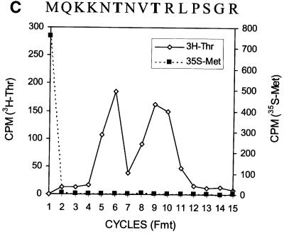
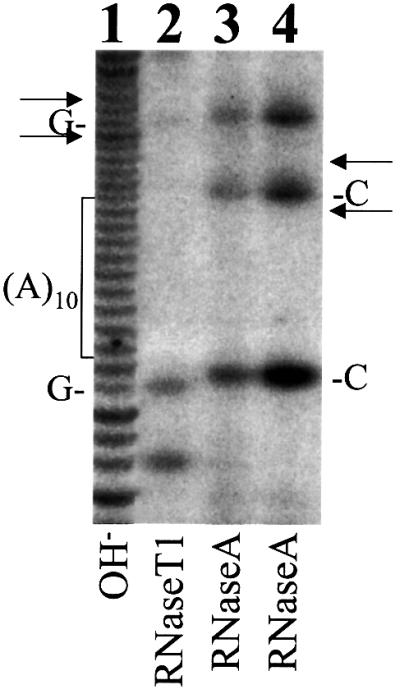
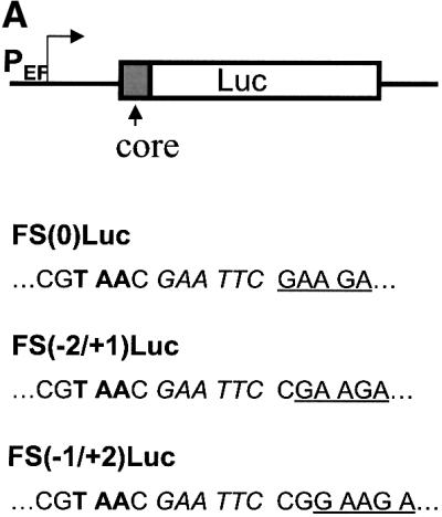
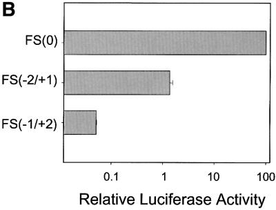
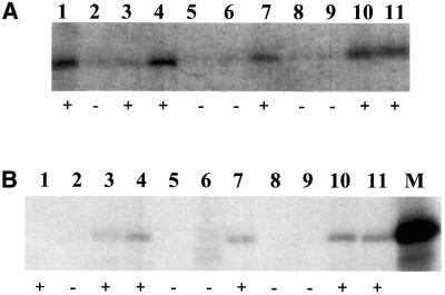
Hepatitis C virus (HCV) is an important human pathogen that affects approximately 100 million people worldwide. Its RNA genome codes for a polyprotein, which is cleaved by viral and cellular proteases to produce at least 10 mature viral protein products. We report here the discovery of a novel HCV protein synthesized by ribosomal frameshift. This protein, which we named the F protein, is synthesized from the initiation codon of the polyprotein sequence followed by ribosomal frameshift into the -2/+1 reading frame. This ribosomal frameshift requires only codons 8-14 of the core protein-coding sequence, and the shift junction is located at or near codon 11. An F protein analog synthesized in vitro reacted with the sera of HCV patients but not with the sera of hepatitis B patients, indicating the expression of the F protein during natural HCV infection. This unexpected finding may open new avenues for the development of anti-HCV drugs.


Figures

None
Fig. 1. The HCV core protein sequence and its overlapping coding sequence. The amino acid sequences, shown by one-letter code, were deduced from the sequence of the HCV-1 isolate (Choo et al., 1991). The sequence shown ends at the C-terminus of the core protein-coding sequence. The amino acid sequence encoded by the overlapping coding sequence is shown in bold. Codons 8–14 of the core protein sequence are underlined and nucleotide 432 is boxed.
None
Fig. 2. Analysis of translational termination and initiation sites of the 17 kDa protein. (A) Termination of the 17 kDa protein sequence in the overlapping coding sequence. pCMV-CC contained the wild-type core protein-coding sequence (lane 1), and pCMV-CCmt contained the core protein-coding sequence with a premature termination codon in the alternative ORF (lane 2). The HCV sequences in these two DNA constructs were under the control of the T7 promoter and the immediate early promoter of cytomegalovirus. The RNA was synthesized using the T7 RNA polymerase and translated using the rabbit reticulocyte lysates or wheat germ extracts. The locations of p21c (core) and the 17 kDa (p17) protein bands are marked. The arrow denotes the location of the truncated 17 kDa protein. The locations of the molecular weight markers are also shown. (B) Translation initiation of the 17 kDa protein from the core protein initiation codon. Lane 1, translation of the wild-type HCV core protein-coding sequence; lane 2, translation of the core protein sequence that had been fused to the HA tag.
None
Fig. 3. Radiosequencing of p21c (A) and the F protein (B). A comparison of the [3H]lysine sequencing results of p21c and the F protein is shown in (C). The amino acid sequences of p21c and a portion of the F protein are aligned at the top of the sequencing cycles. The initiating methionine residue is shown in parentheses and the lysine residues are shown in bold letters. The arrow in (C) indicates the disappearance of the peak at cycle 11 when the F protein was sequenced. p21c and the F protein were synthesized using RNA derived from pCMV-CC and radiolabeled with [35S]methionine and [3H]lysine. These two proteins were then gel purified and subjected to radiosequencing as described in Materials and methods.
None
Fig. 4. Expression and radiosequencing of p21c and the F protein deletion mutants. (A) Expression of the deletion mutants of p21c and the F protein. Lane 1, the translation of the wild-type core protein sequence; lane 2, the translation of the core protein sequence with the deletions of codons 2–7 and 15–50. (B) Radiosequencing of the p21c deletion mutant that had been labeled with [3H]threonine and [35S]methionine. The predicted sequence of the p21c mutant is aligned at the top of the sequencing cycles. Codons 8–14 are underlined and the sequence following these codons represents the p21c sequence starting from codon 51. (C) Radiosequencing of the F deletion mutant that had been labeled with [3H]threonine and [35S]methionine. The translation and the radiosequencing were conducted as described in Materials and methods. The sequence of the F protein mutant, predicted based on a –2 ribosomal frameshift, is also aligned at the top of the sequencing cycles.
None
Fig. 5. Enzymatic sequencing of the T7 transcript containing codons 8–14 of the HCV sequence. Lane 1, partial alkaline (OH) hydrolysis of the HCV RNA; lane 2, partial digestion with RNase T1, which cuts after G; lanes 3 and 4, partial digestion with RNase A, which cuts after U and C. The locations of the 10 A stretch and its two flanking C residues and G residues are indicated. Arrows indicate the locations of RNase T1 and RNase A bands that would be produced if there was a one nucleotide deletion or a two nucleotide insertion in the 10 A stretch. Note that there was no apparent sequence heterogeneity flanking the 10 A stretch.
None
Fig. 6. Analysis of the ribosomal frameshift efficiency in Huh7 cells. (A) Schematic illustration of the luciferase reporter constructs. PEF indicates the EF1α promoter, Luc indicates the luciferase reporter, and core indicates codons 1–14 of the core protein sequence. Sequences located at the fusion junction of the core protein and the luciferase reporter (underlined) are also shown. Italic letters indicate the EcoRI site that was used to fuse the two sequences. Bold letters denote the termination codon located in the –1/+2 reading frame. (B) The luciferase reporter assay. Huh7 cells were transfected with various luciferase reporter constructs, and the relative frameshift efficiencies were determined by the procedures described in Materials and methods. The experiments were carried out in triplicate and repeated at least twice. Data represent the average of the results.
None
Fig. 7. Analysis of F protein-reactive antibodies in patients. (A) Immunoprecipitation of the F protein analog. The F RNA was synthesized from pGEM-core9a with T7 RNA polymerase and translated using rabbit reticulocyte lysates. The protein was radiolabeled with [35S]methionine. A 10 µl aliquot of the translational mixture was then incubated with 10 µl of the sera isolated from HCV or HBV patients for radioimmunoprecipitation analysis. ‘+’ and ‘–’ indicate HCV and HBV sera, respectively. (B) Immunoprecipitation of the truncated F protein. The truncated F protein was radiolabeled with [35S]methionine and radioimmunoprecipitated as in (A). The protein without immunoprecipitation was run in parallel in lane M to serve as a marker.

References

    1. Beard M.R. et al. (1999) An infectious molecular clone of a Japanese genotype 1b hepatitis C virus. Hepatology, 30, 316–324. - PubMed
    1. Blight K.J., Kolykhalov,A.A. and Rice,C.M. (2000) Efficient initiation of HCV RNA replication in cell culture. Science, 290, 1972–1974. - PubMed
    1. Brierley I., Digard,P. and Inglis,S.C. (1989) Characterization of an efficient coronavirus ribosomal frameshifting signal: requirement for an RNA pseudoknot. Cell, 57, 537–547. - PMC - PubMed
    1. Bukh J., Apgar,C.L. and Yanagi,M. (1999) Toward a surrogate model for hepatitis C virus: an infectious molecular clone of the GB virus-B hepatitis agent. Virology, 262, 470–478. - PubMed
    1. Choo Q.-L., Kuo,G., Weiner,A.J., Overby,L.R., Bradley,D.W. and Houghton,M. (1989) Isolation of a cDNA clone derived from a blood-borne non-A, non-B viral hepatitis genome. Science, 244, 359–362. - PubMed

Publication types