. 2001 Jul 16;20(14):3849-60.
doi: 10.1093/emboj/20.14.3849.

Two overlapping reading frames in a single exon encode interacting proteins--a novel way of gene usage

Affiliations

Two overlapping reading frames in a single exon encode interacting proteins--a novel way of gene usage

M Klemke et al. EMBO J. .

Abstract

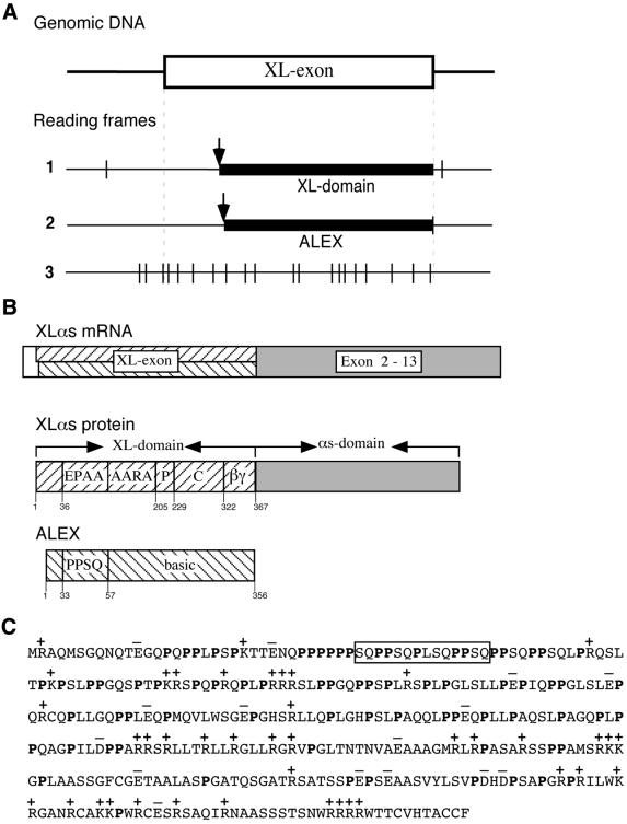
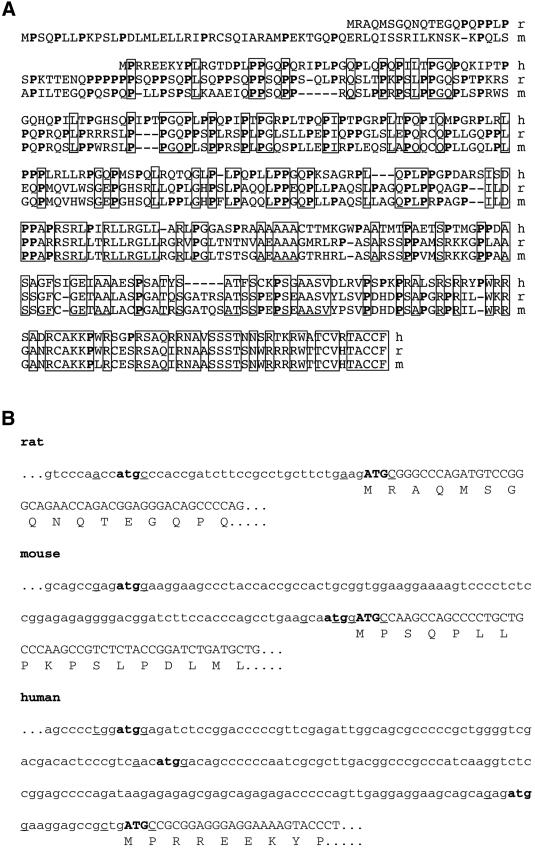
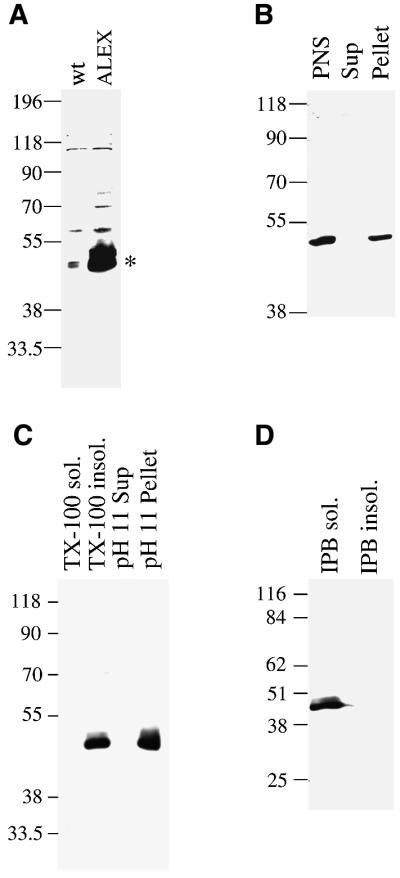
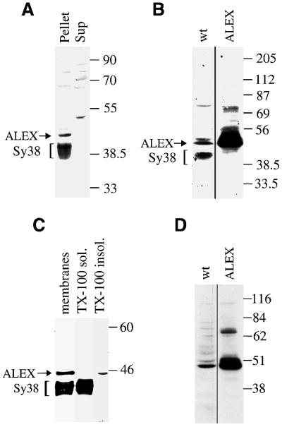
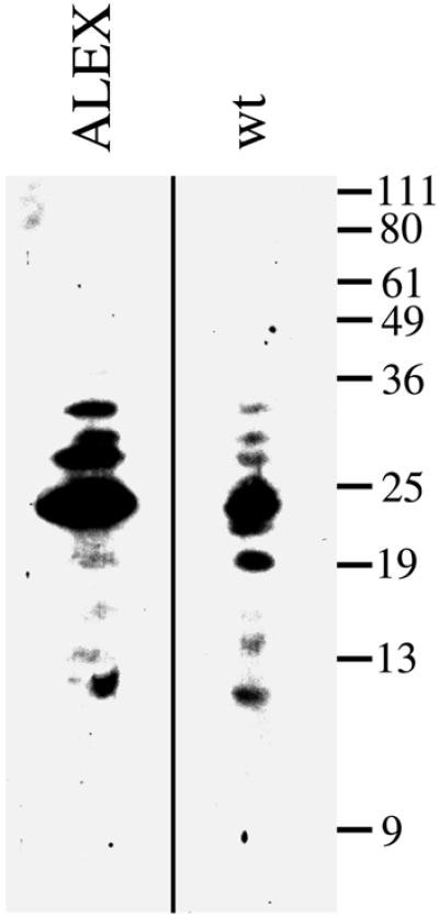
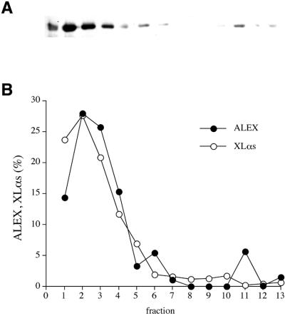
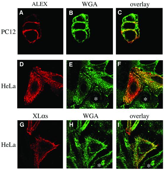
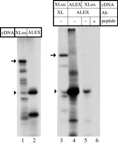
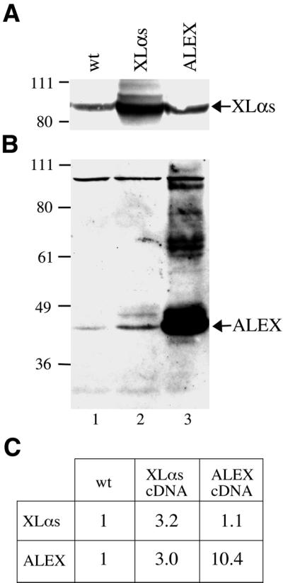
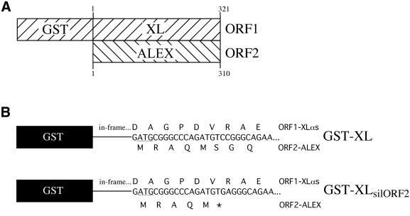
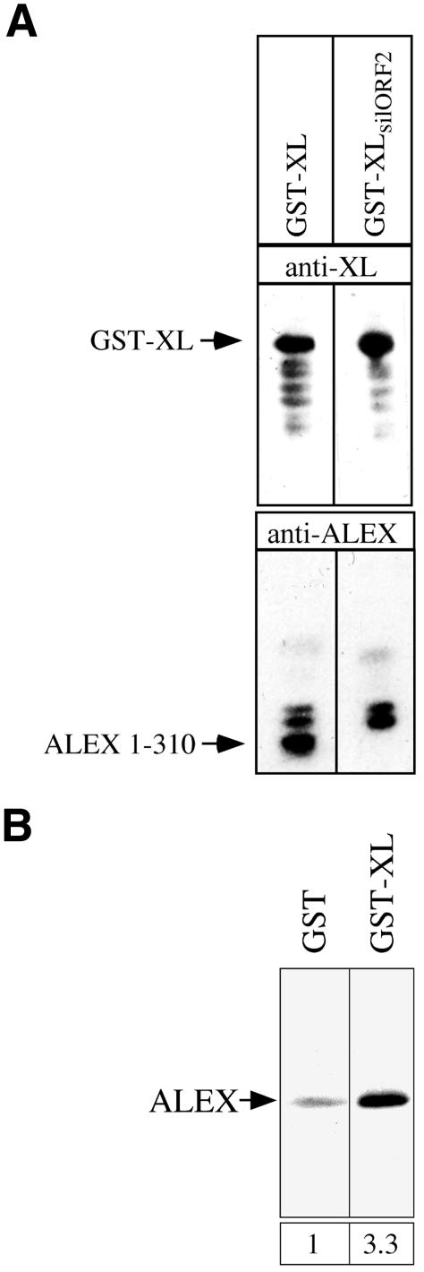
The >1 kb XL-exon of the rat XLalphas/Galphas gene encodes the 37 kDa XL-domain, the N-terminal half of the 78 kDa neuroendocrine-specific G-protein alpha-subunit XLalphas. Here, we describe a novel feature of the XL-exon, the presence of an alternative >1 kb open reading frame (ORF) that completely overlaps with the ORF encoding the XL-domain. The alternative ORF starts 32 nucleotides downstream of the start codon for the XL-domain and is terminated by a stop codon exactly at the end of the XL-exon. The alternative ORF encodes ALEX, a very basic (pI 11.8), proline-rich protein of 356 amino acids. Both XLalphas and ALEX are translated from the same mRNA. Like XLalphas, ALEX is expressed in neuroendocrine cells and tightly associated with the cytoplasmic leaflet of the plasma membrane. Remarkably, ALEX binds to the XL-domain of XLalphas. Our results reveal a mechanism of gene usage that is without precedent in mammalian genomes.


Figures

None
Fig. 1. Existence of two overlapping open reading frames in the XL-exon. (A) The XL-exon of the rat XLαs/Gαs gene (DDBJ/EMBL/GenBank accession No. AF093569) contains two ORFs. ORF1 (1) encodes the XL-domain of XLαs, ORF2 (2) the ALEX protein. The third reading frame (3) does not encode a protein due to the presence of multiple stop codons. The protein-encoding region of the XL-exon is indicated by the thick line, start codons by arrows, and stop codons by vertical lines. (B) Structure of the XLαs mRNA and the two proteins, XLαs and ALEX, derived from it. XLαs: EPAA, ARAA, alanine-rich repeats; P, proline-rich region; C, cysteine-rich region; βγ, βγ-binding region; numbers refer to the corrected translational start (Kehlenbach et al., 1995). ALEX: PPSQ, proline-rich repeats. (C) Deduced amino acid sequence of the rat ALEX protein. The N-terminal methionine corresponds to the 5′ most ATG in the second reading frame of the XL-exon, whose 5′ end was determined by primer extension analysis of PC12 cell mRNA (Y.Wang and W.B.Huttner, unpublished data). Basic amino acids are indicated by (+), acidic amino acids by (–); proline residues are shown in bold. The box indicates the peptide used as antigen to raise the anti-ALEX antibody.
None
Fig. 2. Comparison of the human, mouse and rat amino acid sequence of ALEX and the 5′ nucleotide sequence of the XL-exon. (A) Deduced amino acid sequence of ALEX. As for rat (r) ALEX, the N-terminal methionine in human (h) and mouse (m) ALEX corresponds to the 5′ most ATG in the second reading frame of the human (Hayward et al., 1998a) and mouse (AF116268) XL-exon, respectively. Proline residues are shown in bold; boxes indicate residues conserved between all three species. (B) Nucleotide sequence at the 5′ end of the XL-exon. Potential translational start codons are shown in bold; lower case letters, XLαs-ORF; upper case letters, ALEX-ORF. Nucleotides at the –3 and +4 position around the putative start codons, known to be crucial for initiation of translation (Kozak, 1991), are underlined. DDBJ/EMBL/GenBank accession Nos for the nucleotide sequence of the XL-exon are: AF093569 (rat), AJ245739 (mouse) and AJ224868 (human).
None
Fig. 3. Membrane association of cDNA-expressed ALEX. (A) PNS from PC12 cells either untransfected (wt) or transfected with the ALEX cDNA (ALEX) was analyzed by SDS–PAGE followed by immunoblotting using the anti-ALEX antibody. ALEX is indicated by an asterisk. (B) A PNS from PC12 cells transfected with the ALEX cDNA, as well as a supernatant (Sup) and pellet derived from it by ultracentrifugation, were analyzed by SDS–PAGE followed by immunoblotting using the anti-ALEX antibody. (C) Total membranes from PC12 cells transfected with the ALEX cDNA were treated with Triton X-100 (TX-100) or carbonate (pH 11). Soluble (TX-100 sol., pH 11 Sup) and insoluble (TX-100 insol., pH 11 Pellet) material obtained by ultracentrifugation was analyzed by SDS–PAGE followed by immunoblotting using the anti-ALEX antibody. (D) Total membranes from PC12 cells transfected with the ALEX cDNA were solubilized in immunoprecipitation buffer (IPB) containing ionic detergents, and subjected to ultracentrifugation. Supernatant (IPB sol.) and pellet (IPB insol.) were analyzed by SDS–PAGE followed by immunoblotting using the anti-ALEX antibody.
None
Fig. 4. Membrane association of endogenous ALEX. (A) PNS from untransfected PC12 cells was subjected to ultracentrifugation, and supernatant (Sup) and pellet were analyzed by SDS–PAGE followed by immunoblotting using the anti-ALEX antibody. (B) Immunoblot analysis, using the anti-ALEX antibody, of a total membrane fraction of PC12 cells either untransfected (wt) or transfected with the ALEX cDNA (ALEX). (C) Total membranes from PC12 cells were treated with Triton X-100 (TX-100). Soluble (TX-100 sol.) and insoluble (TX-100 insol.) material obtained by ultracentrifugation, as well as an aliquot of the total membranes, were analyzed by SDS–PAGE followed by immunoblotting using the anti-ALEX antibody. (D) Immunoblot analysis, using the anti-ALEX antibody, of the Triton X-100-insoluble material of total membranes of PC12 cells either untransfected (wt) or transfected with the ALEX cDNA (ALEX). (A–C) The positions of ALEX and of synaptophysin (Sy38), which is also detected by the anti-ALEX antibody, are indicated by arrows and brackets, respectively. (B and D) Exposures of the immunoblot lanes ‘ALEX’ were shorter than those of the lanes ‘wt’.
None
Fig. 5. Comparison of cDNA-expressed and endogenous ALEX by peptide mapping. The Triton X-100-insoluble material obtained from total membranes of PC12 cells, either transfected with the ALEX cDNA (ALEX) or untransfected (wt), was subjected to SDS–PAGE. The region of the gel containing ALEX was subjected to limited proteolysis by the S.aureus V8 protease during a second SDS–PAGE, and ALEX-derived peptides were detected by immunoblotting using the anti-ALEX antibody. Exposure of the immunoblot lane ‘ALEX’ was shorter than that of the lane ‘wt’.
None
Fig. 6. Subcellular fractionation of cDNA-expressed ALEX. PNS from PC12 cells either untransfected (B, XLαs) or transfected with the ALEX cDNA (A and B, ALEX) was subjected to velocity sucrose gradient centrifugation. (A) Fractions were analyzed by SDS–PAGE followed by immunoblotting with either the anti-ALEX antibody, as shown, or an antibody specific for the XL-domain of XLαs (not shown). (B) Immunoreactive ALEX (filled circles) and XLαs (open circles) in the fractions were quantitated and expressed as a percentage of total recovered per gradient (1 = top of gradient).
None
Fig. 7. Immunofluorescence analysis of PC12 and HeLa cells transfected with the ALEX cDNA or the XLαs cDNA. PC12 cells (AC) transfected with the ALEX cDNA and HeLa cells (D–I) transfected with either the ALEX cDNA (DF) or the XLαs cDNA (GI) were double-stained with the anti-ALEX antibody (ALEX, red) or the anti-XL antibody (XLαs, red) and FITC-conjugated wheat germ agglutinin (WGA, green); (A–C) double-staining after fixation and permeabilization; (D–I) cell surface staining with WGA followed by fixation, permeabilization and immunostaining. Analysis using confocal microscopy; single optical X–Y sections through the middle of the cells (A–C) or at the level of the coverslip (D–I) are shown.
None
Fig. 8. Generation of ALEX upon in vitro translation of the XLαs cDNA. The original PC12 cell full-length XLαs cDNA (Kehlenbach et al., 1994) or the ALEX cDNA fragment derived from it were subjected to in vitro transcription/translation in the presence of [35S]methionine/cysteine. The 35S-labeled translation products were analyzed by SDS–PAGE, either before (lanes 1 and 2) or after (lanes 3–6) immunoprecipitation, and visualized by phosphoimaging. Immunoprecipitation was performed with antibodies (Ab) directed against the C-terminus of XLαs (lane 3) or against ALEX (lanes 4–6), in the absence (–) or presence (+) of 10 µg/ml of the peptide used as antigen to raise the anti-ALEX antibody. Arrows, full-length XLαs; arrowheads, ALEX.
None
Fig. 9. Generation of ALEX upon transfection of the XLαs cDNA in PC12 cells. PC12 cells were either not transfected (wt) or transfected with the full-length XLαs cDNA (Kehlenbach et al., 1994) (XLαs) or the ALEX cDNA fragment derived from it (ALEX). (A) Total cell membranes (280 µg protein) were analyzed by immunoblotting using the anti-XL antibody. (B) The Triton X-100-insoluble material obtained from these membranes was analyzed by immunoblotting using the anti-ALEX antibody. (C) Quantification of the XLαs band of (A) and the ALEX band of (B) by densitometric scanning. The values obtained for the untransfected cells were set arbitrarily to 1 and the other values expressed relative to this.
None
Fig. 10. Fusion protein constructs between GST and the XL-domain of XLαs that allow or prevent translation of ALEX. (A) Structure of the GST–XL cDNA construct, which allows translation of (i) a fusion protein comprising GST linked to amino acid residues 1–321 of the XL-domain of XLαs (ORF1) and (ii) a C-terminally truncated ALEX, ALEX1–310 (ORF2, see also Figure 1B). (B) The GST–XL cDNA construct (top) was mutated to the GST–XLsilORF2 construct (bottom) by altering a nucleotide triplet [TCC (top) to TGA (bottom)] such that the ORF1-encoded protein sequence, which is in-frame with GST, remains unaffected but a stop codon (asterisk) is introduced into ORF2.
None
Fig. 11. ALEX binds specifically to the XL-domain of XLαs. (A) Generation of ALEX, and its binding to the XL-domain of XLαs, upon transformation of bacteria with the GST–XL cDNA construct. Escherichia coli were transformed with either the GST–XL or the GST–XLsilORF2 cDNA construct. The GST–XL fusion protein was purified via glutathione–Sepharose 4B and eluted by an excess of reduced glutathione. Eluates (15 µg of protein) were analyzed by SDS–PAGE and immunoblotting using either the anti-XL antibody (top) or the anti-ALEX antibody (bottom). (BIn vitro translated ALEX binds specifically to the XL-domain of a GST–XL fusion protein. Escherichia coli were transformed with pGEX-2T or the GST–XLsilORF2 cDNA construct, and GST and the GST–XL fusion protein, respectively, were purified by binding to glutathione– Sepharose 4B. 35S-Labeled ALEX, obtained by in vitro transcription/translation of the ALEX cDNA, was incubated with 50 µg of either GST or GST–XL bound to glutathione–Sepharose 4B. GST and GST–XL, as well as the 35S-labeled ALEX bound to either protein, were eluted by an excess of reduced glutathione, and eluates analyzed by SDS–PAGE and fluorography. The ALEX band was quantified by densitometric scanning and the amount of ALEX bound to the GST–XL fusion protein is expressed relative to that bound to GST only, which was set arbitrarily to 1. Both lanes of the fluorogram are the same exposure.

References

    1. Ausubel F.M., Brent,R., Kingston,R.E., Moore,D., Seidman,J.G., Smith,J.A. and Struhl,K. (eds.) (1997) Current Protocols in Molecular Biology. John Wiley & Sons, New York, NY.
    1. Barrell B.G., Air,G.M. and Hutchinson,C.A. (1976) Overlapping genes in bacteriophage φX174. Nature, 264, 34–41. - PubMed
    1. Barrell B.G., Shaw,D.C., Walker,J.E., Northop,F.D., Godson,G.N. and Fiddes,J.C. (1978) Overlapping genes in bacteriophages φX174 and G4. Biochem. Soc. Trans., 6, 63–67. - PubMed
    1. Bartsch D., Casadio,A., Karl,K.A., Serodio,P. and Kandel,E.R. (1998) CREB1 encodes a nuclear activator, a repressor and a cytoplasmatic modulator that form a regulatory unit critical for long-term facilitation. Cell, 95, 211–223. - PubMed
    1. Cleveland, D.W., Fischer S.G., Kirschner M.W. and Laemmli U.K. (1977) Peptide mapping by limited proteolysis in sodium dodecyl sulfate and analysis by gel electrophoresis. J. Biol. Chem., 252, 1102–1106. - PubMed

Publication types

MeSH terms