

In situ activation of helper T cells in the lung
- PMID: 11447152
- PMCID: PMC98566
- DOI: 10.1128/IAI.69.8.4790-4798.2001
In situ activation of helper T cells in the lung
Abstract
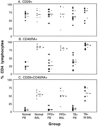
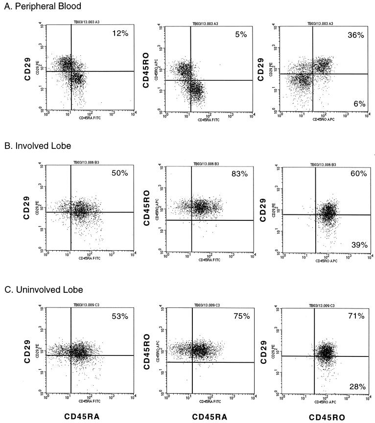
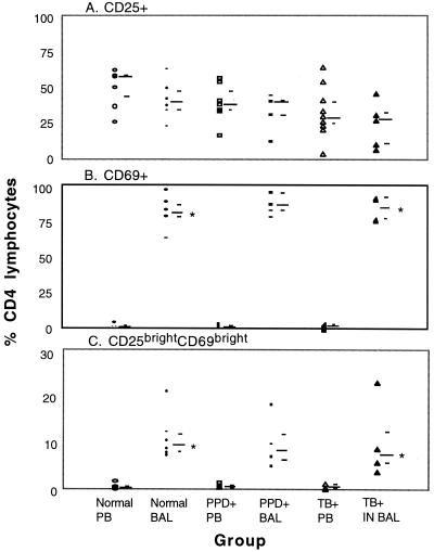
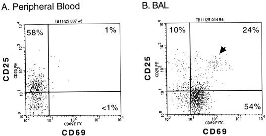
To better understand the lung and systemic responses of helper T cells mediating memory immunity to Mycobacterium tuberculosis, we used three- and four-color flow cytometry to study the surface phenotype of CD4(+) lymphocytes. Bronchoalveolar lavage (BAL) fluid and peripheral blood (PB) samples were obtained from a total of 25 subjects, including 10 tuberculosis (TB)-infected subjects, 8 purified-protein-derivative-negative subjects, and 7 purified-protein-derivative-positive subjects. In marked contrast to CD4(+) lymphocytes from PB (9% +/- 5% expressing CD45RA and CD29), the majority (55% +/- 16%) of CD4(+) lymphocytes in BAL (ALs) simultaneously expressed CD45RA, a naïve T-cell marker, and CD29, members of the very late activation family. Further evaluation revealed that CD4(+) ALs expressed both CD45RA and CD45RO, a memory T-cell marker. In addition, the proportion of CD4(+) lymphocytes expressing CD69, an early activation marker, was drastically increased in BAL fluid (83% +/- 9%) compared to PB (1% +/- 1%), whereas no significant difference was seen in the expression of CD25, the low-affinity interleukin 2 receptor (34% +/- 15% versus 40% +/- 16%). More importantly, we identified a minor population of CD69(bright) CD25(bright) CD4(+) lymphocytes in BAL (10% +/- 6%) that were consistently absent from PB (1% +/- 1%). Thus, CD4(+) lymphocytes in the lung paradoxically coexpress surface molecules characteristic of naïve and memory helper T cells as well as surface molecules commonly associated with early and late stages of activation. No difference was observed for ALs obtained from TB-infected and uninfected lung segments in this regard. It remains to be determined if these surface molecules are induced by the alveolar environment or if CD4(+) lymphocytes coexpressing this unusual combination of surface molecules are selectively recruited from the circulation. Our data suggest that ex vivo experiments on helper T-cell subsets that display distinctive phenotypes may be pivotal to studies on the human immune response to potential TB vaccines.
Figures
Similar articles
-
Flow cytometric characterisation of the "false naive" (CD45RA+, CD45RO-, CD29 bright+) peripheral blood T-lymphocytes in health and in rheumatoid arthritis.Rheumatol Int. 1996;16(2):77-87. doi: 10.1007/BF01816439. Rheumatol Int. 1996. PMID: 8853229
-
Phenotypic analysis of lymphocytes and monocytes/macrophages in peripheral blood and bronchoalveolar lavage fluid from patients with pulmonary sarcoidosis.Thorax. 1999 Apr;54(4):339-46. doi: 10.1136/thx.54.4.339. Thorax. 1999. PMID: 10092696 Free PMC article.
-
Immunophenotypic characterization of peripheral T lymphocytes in Mycobacterium tuberculosis infection and disease.Clin Exp Immunol. 2002 Apr;128(1):149-54. doi: 10.1046/j.1365-2249.2002.01809.x. Clin Exp Immunol. 2002. PMID: 11982602 Free PMC article.
-
Th22 response induced by Mycobacterium tuberculosis strains is closely related to severity of pulmonary lesions and bacillary load in patients with multi-drug-resistant tuberculosis.Clin Exp Immunol. 2021 Feb;203(2):267-280. doi: 10.1111/cei.13544. Epub 2020 Nov 18. Clin Exp Immunol. 2021. PMID: 33128773 Free PMC article. Review.
-
P. Rambotti Lecture. Human naive and memory T cells revisited: new markers (CD31 and CD27) that help define CD4+ T cell subsets.Clin Exp Rheumatol. 1993 May-Jun;11(3):241-7. Clin Exp Rheumatol. 1993. PMID: 8394793 Review.
Cited by
-
CD4 T cell depletion exacerbates acute Mycobacterium tuberculosis while reactivation of latent infection is dependent on severity of tissue depletion in cynomolgus macaques.AIDS Res Hum Retroviruses. 2012 Dec;28(12):1693-702. doi: 10.1089/AID.2012.0028. Epub 2012 May 4. AIDS Res Hum Retroviruses. 2012. PMID: 22480184 Free PMC article.
-
Relationship between chemokine receptor expression, chemokine levels and HIV-1 replication in the lungs of persons exposed to Mycobacterium tuberculosis.Eur J Immunol. 2013 Feb;43(2):540-9. doi: 10.1002/eji.201242804. Epub 2012 Dec 13. Eur J Immunol. 2013. PMID: 23147374 Free PMC article.
-
Aerosolized gamma interferon (IFN-gamma) induces expression of the genes encoding the IFN-gamma-inducible 10-kilodalton protein but not inducible nitric oxide synthase in the lung during tuberculosis.Infect Immun. 2004 Mar;72(3):1275-83. doi: 10.1128/IAI.72.3.1275-1283.2004. Infect Immun. 2004. PMID: 14977928 Free PMC article.
-
Identification of M. tuberculosis-specific Th1 cells expressing CD69 generated in vivo in pleural fluid cells from patients with tuberculous pleurisy.PLoS One. 2011;6(8):e23700. doi: 10.1371/journal.pone.0023700. Epub 2011 Aug 22. PLoS One. 2011. PMID: 21887301 Free PMC article.
-
Early events in Mycobacterium tuberculosis infection in cynomolgus macaques.Infect Immun. 2006 Jul;74(7):3790-803. doi: 10.1128/IAI.00064-06. Infect Immun. 2006. PMID: 16790751 Free PMC article.
References
-
- Adema G J, Hartgers F, Verstraten R, de Vries E, Marland G, Menon S, Foster J, Xu Y, Nooyen P, McClanahan T, Bacon K B, Figdor C G. A dendritic-cell-derived C-C chemokine that preferentially attracts naive T cells. Nature. 1997;387:713–717. - PubMed
-
- Ancochea J, Gonzalez A, Sanchez M J, Aspa J, Lopez-Botet M. Expression of lymphocyte activation surface antigens in bronchoalveolar lavage and peripheral blood cells from young healthy subjects. Chest. 1993;104:32–37. - PubMed
-
- Becker S, Harris D T, Koren H S. Characterization of normal human lung lymphocytes and interleukin-2-induced lung T cell lines. Am J Respir Cell Mol Biol. 1990;3:441–448. - PubMed
-
- Bellocq A, Lecossier D, Pierre-Audigier C, Tazi A, Valeyre D, Hance A J. T cell receptor repertoire of T lymphocytes recovered from the lung and blood of patients with sarcoidosis. Am J Respir Crit Care Med. 1994;149:646–654. - PubMed
Publication types
MeSH terms
Substances
Grants and funding
LinkOut - more resources
Full Text Sources
Research Materials
Miscellaneous
