

In situ activation of helper T cells in the lung
- PMID: 11447152
- PMCID: PMC98566
- DOI: 10.1128/IAI.69.8.4790-4798.2001
In situ activation of helper T cells in the lung
Abstract
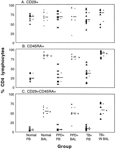
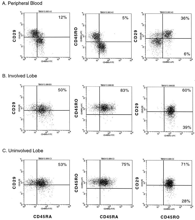
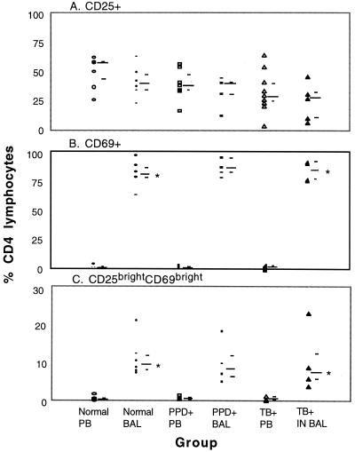
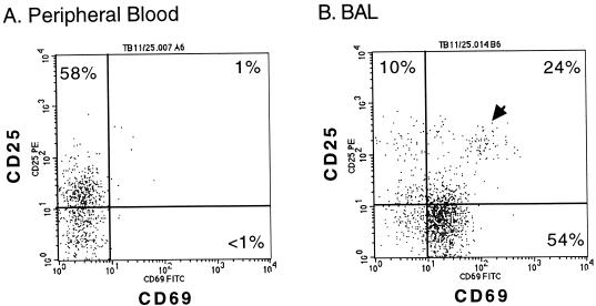
To better understand the lung and systemic responses of helper T cells mediating memory immunity to Mycobacterium tuberculosis, we used three- and four-color flow cytometry to study the surface phenotype of CD4(+) lymphocytes. Bronchoalveolar lavage (BAL) fluid and peripheral blood (PB) samples were obtained from a total of 25 subjects, including 10 tuberculosis (TB)-infected subjects, 8 purified-protein-derivative-negative subjects, and 7 purified-protein-derivative-positive subjects. In marked contrast to CD4(+) lymphocytes from PB (9% +/- 5% expressing CD45RA and CD29), the majority (55% +/- 16%) of CD4(+) lymphocytes in BAL (ALs) simultaneously expressed CD45RA, a naïve T-cell marker, and CD29, members of the very late activation family. Further evaluation revealed that CD4(+) ALs expressed both CD45RA and CD45RO, a memory T-cell marker. In addition, the proportion of CD4(+) lymphocytes expressing CD69, an early activation marker, was drastically increased in BAL fluid (83% +/- 9%) compared to PB (1% +/- 1%), whereas no significant difference was seen in the expression of CD25, the low-affinity interleukin 2 receptor (34% +/- 15% versus 40% +/- 16%). More importantly, we identified a minor population of CD69(bright) CD25(bright) CD4(+) lymphocytes in BAL (10% +/- 6%) that were consistently absent from PB (1% +/- 1%). Thus, CD4(+) lymphocytes in the lung paradoxically coexpress surface molecules characteristic of naïve and memory helper T cells as well as surface molecules commonly associated with early and late stages of activation. No difference was observed for ALs obtained from TB-infected and uninfected lung segments in this regard. It remains to be determined if these surface molecules are induced by the alveolar environment or if CD4(+) lymphocytes coexpressing this unusual combination of surface molecules are selectively recruited from the circulation. Our data suggest that ex vivo experiments on helper T-cell subsets that display distinctive phenotypes may be pivotal to studies on the human immune response to potential TB vaccines.
Figures
References
-
- Adema G J, Hartgers F, Verstraten R, de Vries E, Marland G, Menon S, Foster J, Xu Y, Nooyen P, McClanahan T, Bacon K B, Figdor C G. A dendritic-cell-derived C-C chemokine that preferentially attracts naive T cells. Nature. 1997;387:713–717. - PubMed
-
- Ancochea J, Gonzalez A, Sanchez M J, Aspa J, Lopez-Botet M. Expression of lymphocyte activation surface antigens in bronchoalveolar lavage and peripheral blood cells from young healthy subjects. Chest. 1993;104:32–37. - PubMed
-
- Becker S, Harris D T, Koren H S. Characterization of normal human lung lymphocytes and interleukin-2-induced lung T cell lines. Am J Respir Cell Mol Biol. 1990;3:441–448. - PubMed
-
- Bellocq A, Lecossier D, Pierre-Audigier C, Tazi A, Valeyre D, Hance A J. T cell receptor repertoire of T lymphocytes recovered from the lung and blood of patients with sarcoidosis. Am J Respir Crit Care Med. 1994;149:646–654. - PubMed
Publication types
MeSH terms
Substances
Grants and funding
LinkOut - more resources
Full Text Sources
Research Materials
Miscellaneous
