. 2001 Aug;69(8):4898-905.
doi: 10.1128/IAI.69.8.4898-4905.2001.

Neutrophil depletion during Toxoplasma gondii infection leads to impaired immunity and lethal systemic pathology

Affiliations

Neutrophil depletion during Toxoplasma gondii infection leads to impaired immunity and lethal systemic pathology

S K Bliss et al. Infect Immun. 2001 Aug.

Abstract

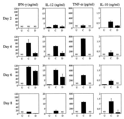
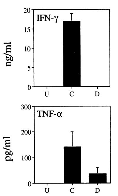
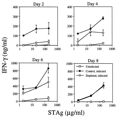
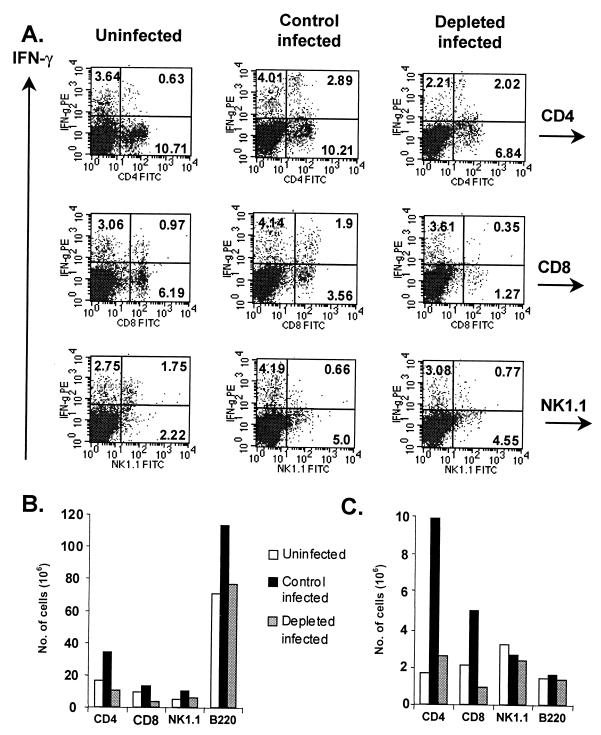
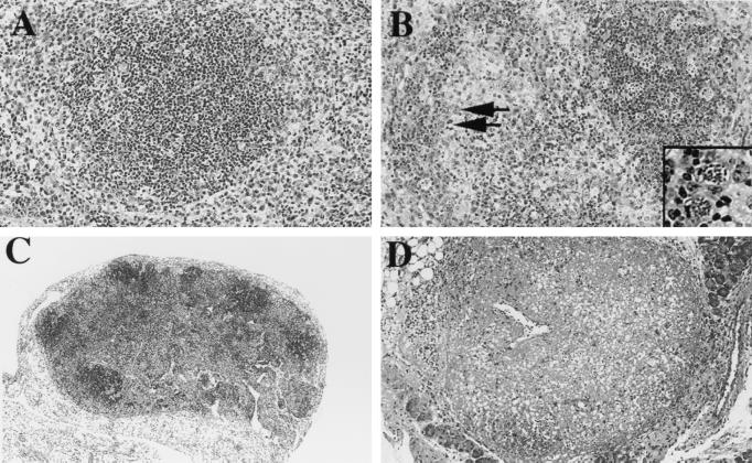
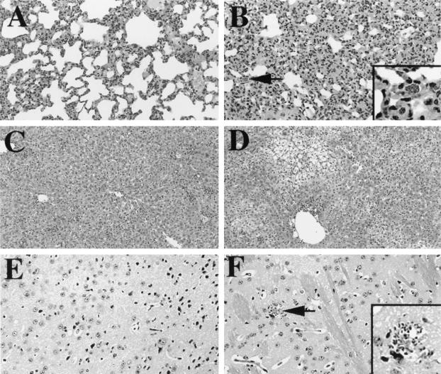
The immunomodulatory role of neutrophils during infection with Toxoplasma gondii was investigated. Monoclonal antibody-mediated depletion revealed that neutrophils are essential for survival during the first few days of infection. Moreover, neutrophil depletion was associated with a weaker type 1 immune response as measured by decreased levels of gamma interferon, interleukin-12 (IL-12) and tumor necrosis factor alpha. IL-10 was also decreased in depleted animals. Additionally, splenic populations of CD4(+) T cells, CD8(+) T cells, and NK1.1(+) cells were decreased in depleted mice. Neutrophil-depleted mice exhibited lesions of greater severity in tissues examined and a greater parasite burden as determined by histopathology and reverse transcription-PCR. We conclude that neutrophils are critical near the time of infection because they influence the character of the immune response and control tachyzoite replication.


Figures

FIG. 1
FIG. 1
Granulocytes are necessary for survival of T. gondii infection only during the first few days of infection. C57BL/6 mice were injected i.p. either with 200 μg of the granulocyte-depleting Ab RB6C6.8C5 on days −2, 0, and +2, days +2, +4, and +6, or days +6, +8, and +10 or with a control rat IgG on days −2, 0, and +2. Mice (four per group) were infected i.p. with 100 ME49 cysts on day 0 (four mice per group). Survival was monitored daily. Results represent three experiments.
FIG. 2
FIG. 2
Constitutive expression of cytokines from cultured splenocytes of neutrophil-depleted mice is impaired. C57BL/6 mice were administered either the anti-Gr-1 MAb or a control rat IgG on days −2, 0, +2, and +4. Mice (four per group) were infected i.p. with 100 ME49 cysts on day 0 as described in Materials and Methods (three mice per group). On days +2, +4, +6, and +8 postinfection, mice were euthanatized and spleens were collected for culture. Additionally, spleens from uninfected mice were cultured. Splenocytes were cultured in medium for 3 days, and cell-free supernatants were harvested for cytokine level determination by ELISA. U, cells from uninfected mice; C, cells from animals administered control Ab; D, mice depleted of neutrophils by anti-Gr-1 Ab injection; ND, not detected. Results are expressed as means + standard deviations and represent three experiments.
FIG. 3
FIG. 3
Levels of IFN-γ and TNF-α in plasma of neutrophil-depleted mice are decreased at 2 days postinfection. Plasma was obtained from mice (four per group) treated as described in the legend to Fig. 2 at 2 days postinfection. Cytokine levels were determined by ELISA. U, cells from uninfected animals; C, cells from mice injected with control Ab; D, animals depleted of neutrophils by anti-Gr-1 MAb administration. Results are expressed as means + standard deviations and represent three experiments.
FIG. 4
FIG. 4
Ability of splenocytes from depleted infected mice to produce IFN-γ in response to parasite antigen stimulation is profoundly impaired at 2 days postinfection. Spleens were obtained from uninfected, control infected (control rat IgG given on days −2, 0, +2, and +4), and depleted infected (anti-Gr-1 MAb given on days −2, 0, +2, and +4) mice on days +2, +4, +6, and +8 postinfection. Four mice were used in each group. Splenocyte cultures were stimulated with STAg at 2, 20, or 200 μg/ml for 3 days. Cell-free supernatants were collected, and levels of IFN-γ were determined by ELISA as described in Materials and Methods. Results are expressed as means + standard deviations and represent three experiments.
FIG. 5
FIG. 5
Neutrophil-depleted mice display decreased numbers of IFN-γ+ T cells during infection. (A) IFN-γ expression in CD4+, CD8+, B220+ and NK1.1+ subsets at 6 days postinfection. Numbers in quadrants reflect the percentages of cells. PE, phycoerythrin; FITC, fluorescein isothiocyanate. (B) Total numbers of cells in each subset. (C) Numbers of IFN-γ+ cells in each subset. Splenocytes were obtained and stimulated ex vivo before staining as described in Materials and Methods. Cells were then analyzed by flow cytometry. To obtain absolute cell numbers, percentages in each population were multiplied by the mean total cell number in corresponding spleens (three mice per group). Results represent three separate experiments.
FIG. 6
FIG. 6
Depleted infected mice suffer from extensive lesions in lymphoid tissue. Tissues from spleens (A and B) and mesenteric lymph nodes (C and D) were collected from control infected (A and C) and depleted infected (B and D) mice (four per group) on day 8 postinfection. Samples were fixed and stained with hematoxylin and eosin. Arrows in panel B point to infected cells; this area is enlarged in the inset. Original magnifications, ×20 (A and B) and ×10 (C and D). Results represent two experiments.
FIG. 7
FIG. 7
Depleted infected mice exhibit extensive lesions in nonlymphoid tissue. Tissues from lung (A and B), liver (C and D), and brain (E and F) were collected from control infected (A, C, and E) and depleted infected (B, D, and F) mice (four per group) on day 8 postinfection. Samples were fixed and stained with hematoxylin and eosin. The arrow in panel B points to an infected cell; the arrow in panel F points to tachyzoites within a focal area of necrosis. These regions are enlarged in the panel insets. Original magnifications, ×20 (A, B, E, and F) and ×10 (C and D). Results represent of two experiments.
FIG. 8
FIG. 8
Neutrophil-depleted mice have a higher parasite burden in tissues from liver, spleen, lung, and brain. Four mice were used for each treatment group. RNA was extracted from tissue at 8 days postinfection from uninfected, control infected, and depleted-infected mice. RT-PCR was performed using an ABI 7700 Sequence Detector for p22 (a parasite antigen) and HPRT (an endogenous control). p22 was not detected in uninfected tissue. HPRT mRNA levels were used to normalize p22 levels. A value of 1 was arbitrarily set for control infected samples, and levels of normalized p22 from depleted infected samples were expressed as the fold increase over control samples. C, mice administered a control Ab; D, animals depleted of neutrophils by administration of anti-Gr-1 MAb.

References

    1. Alexander J, Hunter C A. Immunoregulation during toxoplasmosis. Chem Immunol. 1998;70:81–102. - PubMed
    1. Barteneva N, Theodor I, Peterson E M, de la Maza L M. Role of neutrophils in controlling early stages of a Chlamydia trachomatis infection. Infect Immun. 1996;64:4830–4833. - PMC - PubMed
    1. Bliss S K, Butcher B A, Denkers E Y. Rapid recruitment of neutrophils with prestored IL-12 during microbial infection. J Immunol. 2000;165:4515–4521. - PubMed
    1. Bliss S K, Marshall A J, Zhang Y, Denkers E Y. Human polymorphonuclear leukocytes produce IL-12, TNF-α, and the chemokines macrophage-inflammatory protein-1 α and −1β in response to Toxoplasma gondii antigens. J Immunol. 1999;162:7369–7375. - PubMed
    1. Bliss S K, Zhang Y, Denkers E Y. Murine neutrophil stimulation by Toxoplasma gondii antigen drives high level production of IFN-γ-independent IL-12. J Immunol. 1999;163:2081–2088. - PubMed

Publication types