. 2001 Aug;69(8):5098-106.
doi: 10.1128/IAI.69.8.5098-5106.2001.

Contribution of Mn-cofactored superoxide dismutase (SodA) to the virulence of Streptococcus agalactiae

Affiliations

Contribution of Mn-cofactored superoxide dismutase (SodA) to the virulence of Streptococcus agalactiae

C Poyart et al. Infect Immun. 2001 Aug.

Abstract

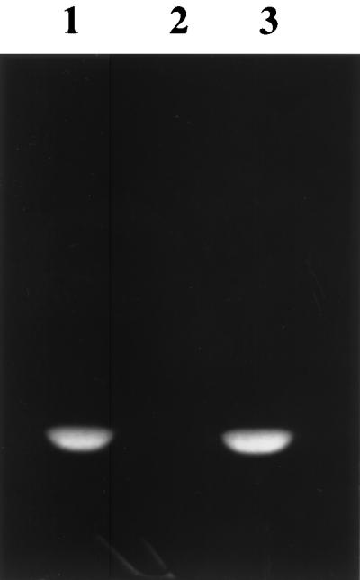
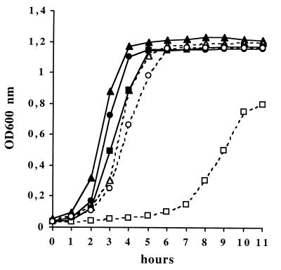
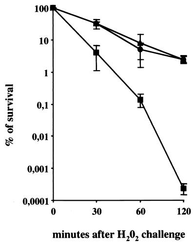
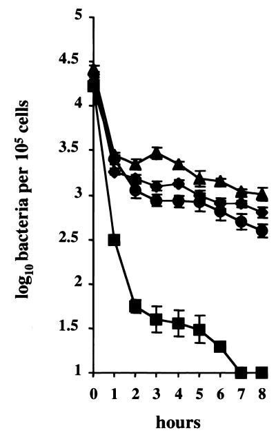
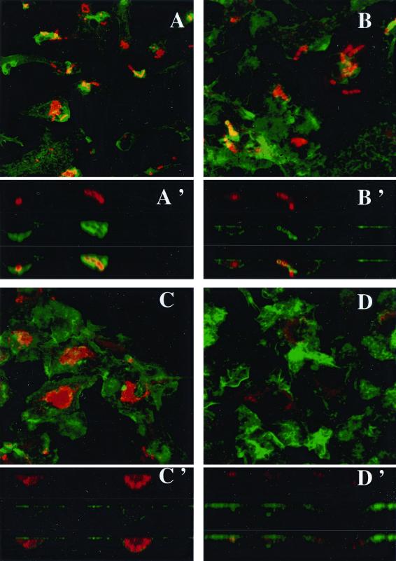
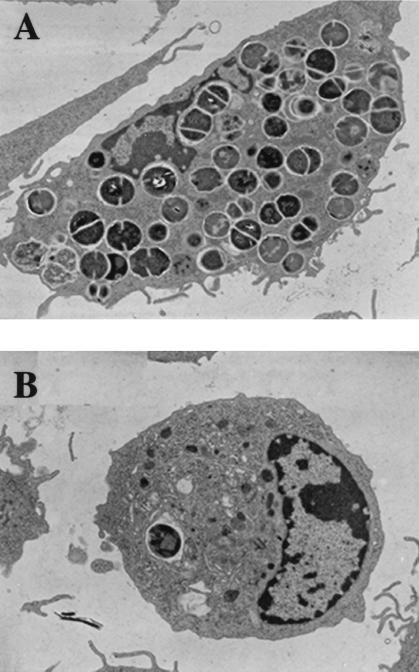
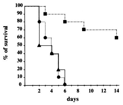
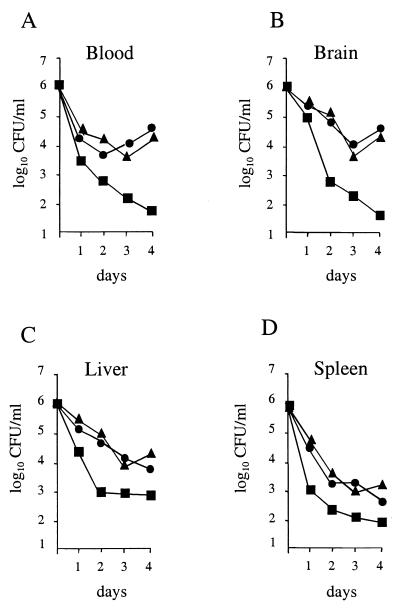
Superoxide dismutases convert superoxide anions to molecular oxygen and hydrogen peroxide, which, in turn, is metabolized by catalases and/or peroxidases. These enzymes constitute one of the major defense mechanisms of cells against oxidative stress and hence play a role in the pathogenesis of certain bacteria. We previously demonstrated that group B streptococci (GBS) possess a single Mn-cofactored superoxide dismutase (SodA). To analyze the role of this enzyme in the pathogenicity of GBS, we constructed a sodA-disrupted mutant of Streptococcus agalactiae NEM316 by allelic exchange. This mutant was subsequently cis complemented by integration into the chromosome of pAT113/Sp harboring the wild-type sodA gene. The SOD specific activity detected by gel analysis in cell extracts confirmed that active SODs were present in the parental and complemented strains but absent in the sodA mutant. The growth rates of these strains in standing cultures were comparable, but the sodA mutant was extremely susceptible to the oxidative stress generated by addition of paraquat or hydrogen peroxide to the culture medium and exhibited a higher mutation frequency in the presence of rifampin. In mouse bone marrow-derived macrophages, the sodA mutant showed an increased susceptibility to bacterial killing by macrophages. In a mouse infection model, after intravenous injection the survival of the sodA mutant in the blood and the brain was markedly reduced in comparison to that of the parental and complemented strains whereas only minor effects on survival in the liver and the spleen were observed. These results suggest that SodA plays a role in GBS pathogenesis.


Figures

FIG. 1
FIG. 1
SOD activity gel. Crude cell extracts (50 μg) of S. agalactiae were loaded onto a nondenaturing 10% polyacrylamide gel stained for SOD activity. Lanes 1, S. agalactiae NEM316; 2, sodA mutant NEM1640; 3, sodA-complemented mutant NEM1641.
FIG. 2
FIG. 2
Growth curves of S. agalactiae NEM316, the sodA mutant, NEM1640, and the complemented strain, NEM1641, in BHI broth at 37°C under aerobic conditions with and without paraquat (10 mM). Strains and growth conditions are represented as follows: wild-type strain without (▴) or with 10 mM paraquat (▵); sodA mutant without (■) or with 10 mM paraquat (□); sodA-complemented mutant without (●) or with 10 mM paraquat (○). The results shown are representative of at least three independent experiments showing less than 10% variation.
FIG. 3
FIG. 3
Sensitivity of S. agalactiae to H2O2. The wild-type strain, NEM316 (▴), the sodA mutant, NEM1640 (■), and the complemented strain, NEM1641 (●) were grown and treated with H2O2 as described in Materials and Methods. Exponential-phase cells were exposed to 20 mM H2O2 for 30, 60, or 120 min at 37°C. Viability was determined by plating on BHI agar. Error bars represent the standard deviations of three independent experiments.
FIG. 4
FIG. 4
Growth of S. agalactiae in macrophages. BMMs were cultured in vitro and exposed to S. agalactiae NEM316 (▴), the sodA mutant, NEM1640 (■), and the complemented strain, NEM1641 (●), or the cpsD mutant, NEM1871 (⧫). Error bars represent the standard deviation of three independent experiments done in triplicate for each strain studied.
FIG. 5
FIG. 5
Fluorescent confocal microscopy of BMMs infected (100 bacteria per cell) with S. agalactiae NEM316 (A and C) or the sodA mutant, NEM1640 (B and D). F-actin was stained with β-phalloidin (green). Bacteria were labeled with anti-S. agalactiae antibodies (red). F-actin sheets associated with bacteria are indicated by the overlapping of green and red light (orange-yellow). After 30 min of infection, characteristic chains are observed and the bacterial uptake is similar for both strains (A and B). Images reconstructed from confocal xz sections show that bacterial phagocytosis for both strains is associated with actin polymerization (A' and B'). After 3 h of infection, bacterial clusters are observed with the wild-type strain (C) whereas only bacterial degradation products are labeled in the mutant (D). Images reconstructed from confocal xz sections demonstrate the intracellular localization of bacteria (C'). Magnification, ×130.
FIG. 6
FIG. 6
Transmission electron micrographs of BMMs infected (100 bacteria per cell) with S. agalactiae NEM316 (A) or the sodA mutant, NEM1640 (B). Samples were taken 3 h postinfection. Magnification, ×4,558.
FIG. 7
FIG. 7
Mortality curves in mice infected with S. agalactiae NEM316 (▴), the sodA mutant, NEM1640 (■); or the complemented strain, NEM1641 (●). Mice (10 per group) were inoculated i.v. with 6 × 10⁶ bacteria.
FIG. 8
FIG. 8
Mouse virulence assays. Growth of S. agalactiae NEM316 (▴), the sodA mutant, NEM1640 (■), or the complemented strain, NEM1641 (●) was monitored in the blood (A), brain (B), liver (C), and spleen (D) of mice inoculated i.v. with 106 bacteria. Means of bacterial counts in four organs per time point are shown (standard deviation, ≤0.25).

Similar articles

Cited by

References

    1. Antal J M, Cunningham J V, Goodrum K J. Opsonin-independent phagocytosis of group B streptococcus: role of complement receptor type three. Infect Immun. 1992;60:1114–1121. - PMC - PubMed
    1. Baker C J, Edwards M S. Group B streptococcal infections. In: Remington J S, Klein J O, editors. Infectious diseases of the fetus and newborn infant. Philadelphia, Pa: The W. B. Saunders Co; 1990. pp. 743–811.
    1. Bannister J V, Bannister W H, Rotilio G. Aspects of the structure, function and applications of superoxide dismutase. Crit Rev Biochem. 1987;22:111–180. - PubMed
    1. Beaman L, Beaman B L. Monoclonal antibodies demonstrate that superoxide dismutase contributes to protection of Nocardia asteroides within the intact host. Infect Immun. 1992;58:3122–3128. - PMC - PubMed
    1. Beauchamp C, Fridovitch I. Superoxide dismutase: improved assays and an assay applicable to acrylamide gels. Anal Biochem. 1971;44:276–287. - PubMed

Publication types

MeSH terms