. 2001 Jul 17;98(15):8885-90.
doi: 10.1073/pnas.151217998. Epub 2001 Jul 10.

Prostaglandins are required for CREB activation and cellular proliferation during liver regeneration

Affiliations

Prostaglandins are required for CREB activation and cellular proliferation during liver regeneration

D A Rudnick et al. Proc Natl Acad Sci U S A. .

Abstract

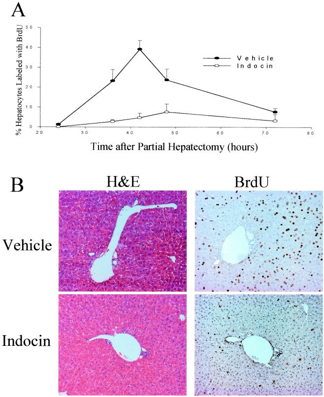
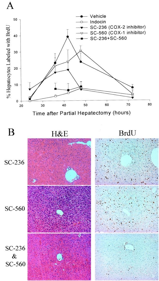
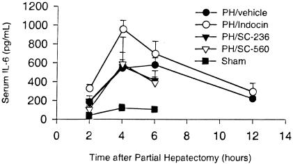
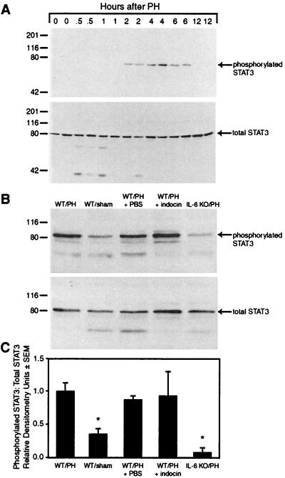
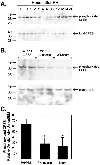
The liver responds to multiple types of injury with an extraordinarily well orchestrated and tightly regulated form of regeneration. The response to partial hepatectomy has been used as a model system to elucidate the molecular basis of this regenerative response. In this study, we used cyclooxygenase (COX)-selective antagonists and -null mice to determine the role of prostaglandin signaling in the response of liver to partial hepatectomy. The results show that liver regeneration is markedly impaired when both COX-1 and COX-2 are inhibited by indocin or by a combination of the COX-1 selective antagonist, SC-560, and the COX-2 selective antagonist, SC-236. Inhibition of COX-2 alone partially inhibits regeneration whereas inhibition of COX-1 alone tends to delay regeneration. Neither the rise in IL-6 nor the activation of signal transducer and activator of transcription-3 (STAT3) that is seen during liver regeneration is inhibited by indocin or the selective COX antagonists. In contrast, indocin treatment prevents the activation of CREB by phosphorylation that occurs during hepatic regeneration. These data indicate that prostaglandin signaling is required during liver regeneration, that COX-2 plays a particularly important role but COX-1 is also involved, and implicate the activation of CREB rather than STAT3 as the mediator of prostaglandin signaling during liver regeneration.


Figures

Figure 1
Figure 1
Hepatocyte BrdUrd labeling after PH. Percentage of hepatocytes that label with BrdUrd by the injection method is plotted in line form and by the osmotic pump method in bar form. Three to six animals were studied for each time point and treatment group.
Figure 2
Figure 2
Effect of indocin on hepatocyte BrdUrd labeling and liver histology. (A) Percentage of hepatocytes that label with BrdUrd by the injection method after vehicle or indocin treatment. Three to eight animals were studied for each time point and treatment group. Significant differences were found at 42 h (P < 0.002). (B) Liver sections from vehicle- and indocin-treated animals 42 h after PH stained with hematoxylin/eosin or BrdUrd.
Figure 3
Figure 3
Effect of COX-1- and COX-2-specific inhibitors on hepatocyte BrdUrd labeling and liver histology. (A) Percentage of hepatocytes that label with BrdUrd by the injection method is plotted for vehicle-, indocin-, SC-236-, SC-560-, and SC-236 + SC-560-treated animals. Three to eight animals were studied for each time point and treatment group. Significant differences between the vehicle control and the indocin, SC-236, and SC-236 + SC-560 treatment groups were found at 42 h (P < 0.002). (B) Liver sections from SC-236-, SC-560-, and SC-236 + SC-560-treated animals 42 h after PH stained with hematoxylin/eosin or BrdUrd.
Figure 4
Figure 4
Effect of COX inhibitors on serum IL-6 after PH. Serum IL-6 determination is plotted for vehicle-, indocin-, SC-236-, and SC-560-treated animals, and for sham-operated animals. Three to four animals were studied for each time point and treatment group.
Figure 5
Figure 5
Hepatic STAT3 activation after PH in the presence or absence of COX inhibition. (A) Western blot analysis of phosphorylated and total STAT3 in hepatic lysates at serial time points after PH. (B) Western blot analysis of phosphorylated and total STAT3 from hepatic lysates of wt mice subjected to PH, sham surgery (sham), subjected to PH while treated with PBS, subjected to PH while treated with indocin, or IL-6 null mice subjected to PH (IL-6 knockout) is shown. Lysates were prepared from liver harvested 2 h after surgery. (C) Densitometric analysis of the ratio of phosphorylated STAT3 to total STAT3 from three separate experiments.
Figure 6
Figure 6
Hepatic CREB activation after PH in the presence or absence of COX inhibition. (A) Western blot analysis of phosphorylated and total CREB in hepatic lysates at serial time points after PH. (B) Western blot analysis of phosphorylated and total CREB from hepatic lysates of mice subjected to PH in the presence of vehicle (PBS) or indocin or mice subjected to sham surgery is shown. Lysates were prepared from liver harvested 1 h after surgery. (C) Densitometric analysis of the relative amounts of p-CREB from three separate experiments.

References

    1. Michalopoulos G K, DeFrances M C. Science. 1997;276:60–66. - PubMed
    1. Diehl A M E, Rai R. In: Schiff's Diseases of the Liver. 8th Ed. Schiff R, Sorrell M C, Maddrey W C, editors. Philadelphia: Lippincott; 1999. pp. 39–54.
    1. Fausto N. J Hepatol. 2000;32,Supp. 1:19–31. - PubMed
    1. Higgins G M, Anderson R M. Arch Pathol. 1931;12:186–202.
    1. Boros P, Miller C M. Lancet. 1995;345:293–295. - PubMed

Publication types

MeSH terms

Substances