. 2001 Jul 17;98(15):8880-4.
doi: 10.1073/pnas.151244398. Epub 2001 Jul 10.

4-Hydroxytamoxifen binds to and deactivates the estrogen-related receptor gamma

Affiliations

4-Hydroxytamoxifen binds to and deactivates the estrogen-related receptor gamma

P Coward et al. Proc Natl Acad Sci U S A. .

Abstract

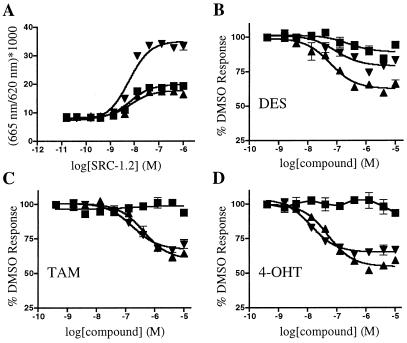
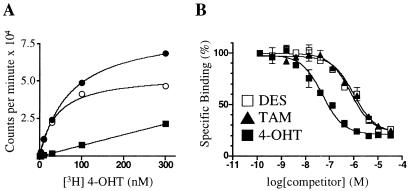
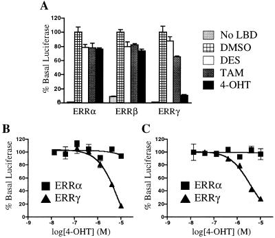
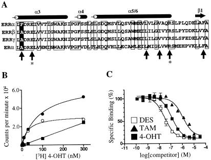
The estrogen-related receptors (ERR alpha, ERR beta, and ERR gamma) form a family of orphan nuclear receptors that share significant amino acid identity with the estrogen receptors, but for which physiologic roles remain largely unknown. By using a peptide sensor assay, we have identified the stilbenes diethylstilbestrol (DES), tamoxifen (TAM), and 4-hydroxytamoxifen (4-OHT) as high-affinity ligands for ERR gamma. In direct binding assays, 4-OHT had a K(d) value of 35 nM, and both DES and TAM displaced radiolabeled 4-OHT with K(i) values of 870 nM. In cell-based assays, 4-OHT binding caused a dissociation of the complex between ERR gamma and the steroid receptor coactivator-1, and led to an inhibition of the constitutive transcriptional activity of ERR gamma. ERR alpha did not bind 4-OHT, but replacing a single amino acid predicted to be in the ERR alpha ligand-binding pocket with the corresponding ERR gamma residue allowed high-affinity 4-OHT binding. These results demonstrate the existence of high-affinity ligands for the ERR family of orphan receptors, and identify 4-OHT as a molecule that can regulate the transcriptional activity of ERR gamma.


Figures

Figure 1
Figure 1
Development of a FRET assay for ERR family members. (A) Coactivator peptide interacts with ERR LBDs in the FRET assay. SRC-1.2 peptide shows a dose-dependent, constitutive interaction with ERRα (■), ERRβ (▴), and ERRγ (▾) LBDs. (B–D) DES, TAM, and 4-OHT disrupt the interaction between SRC-1.2 and ERR LBDs in the FRET assay. Various concentrations of DES (B), TAM (C), or 4-OHT (D) were added to 2 nM of ERRα (■), ERRβ (▴), and ERRγ (▾) and 300 nM SRC-1.2. EC50 values for each compound are listed in the text. For each panel, the data are from a single experiment preformed in duplicate. At least two additional experiments gave similar results.
Figure 2
Figure 2
A radioligand binding assay for ERRγ. (A) Saturation binding curve of [3H]4-OHT and GST-ERRγ. The graph shows total (●), specific (○), and nonspecific (■) binding. Excess unlabeled 4-OHT (30 μM) was used to determine nonspecific binding. The Kd of 4-OHT was 35 nM. (B) Nonradioactive DES, TAM, and 4-OHT compete with [3H]4-OHT for binding to ERRγ. The Ki value for DES and TAM was 870 nM; for 4-OHT the Ki was 75 nM. These data are from a single experiment preformed in duplicate. Two additional experiments gave similar results.
Figure 3
Figure 3
Cell-based activity of DES, TAM, and 4-OHT on ERR family members. CV1 cells were transfected as described in Materials and Methods. (A) Mammalian two-hybrid assay. Compounds were added at 10 μM. (B) Dose-response curves of 4-OHT on ERRα and ERRγ in the mammalian two-hybrid assay. (C) Dose-response curve of 4-OHT on full-length ERRα and ERRγ when a 3× ERE promoter-driven reporter gene was used. In all cases, luciferase activity was normalized to β-galactosidase activity. These data are from a single experiment preformed in triplicate. Two additional experiments gave similar results.
Figure 4
Figure 4
ERRαF232A has binding characteristics similar to those of ERRγ. (A) Alignment of the ERR and ER LBDs. The sequence begins with amino acid 231 for ERRα, 246 for ERRβ, 248 for ERRγ, and 349 for ERα. The positions of the α-helices and β-sheet are noted above the alignment. Conserved residues are boxed. Upright arrows indicate ER residues involved in recognition of DES and 4-OHT (26). The positions of glutamic acid-353 and arginine-394 in ERα, responsible for hydrogen bonding to the phenolic hydroxyl of the A ring of 4-OHT, are indicated by asterisks. The position of phenylalanine-232 in ERRα is indicated by a white letter on a black background. (B) Saturation binding curve of [3H]4-OHT and GST-ERRαF232A. The graph shows total (●), specific (○), and nonspecific (■) binding. Excess unlabeled 4-OHT (30 μM) was used to determine nonspecific binding. The Kd of 4-OHT was 40 nM. (C) Nonradioactive DES, TAM, and 4-OHT compete with [3H]4-OHT for binding to ERRαF232A. Ki values were 30 nM, 750 nM, and 110 nM, respectively. These data are from a single experiment preformed in duplicate. Two additional experiments gave similar results.

Similar articles

Cited by

References

    1. Freedman L P. Cell. 1999;97:5–8. - PubMed
    1. Heery D M, Kalkhoven E, Hoare S, Parker M G. Nature (London) 1997;387:733–736. - PubMed
    1. Makishima M, Okamoto A Y, Repa J J, Tu H, Learned R M, Luk A, Hull M V, Lustig K D, Mangelsdorf D J, Shan B. Science. 1999;284:1362–1365. - PubMed
    1. Xu L, Glass C K, Rosenfeld M G. Curr Opin Genet Dev. 1999;9:140–147. - PubMed
    1. Willson T M, Brown P J, Sternbach D D, Henke B R. J Med Chem. 2000;43:527–550. - PubMed

MeSH terms

LinkOut - more resources