. 2001 Jul 17;98(15):8632-7.
doi: 10.1073/pnas.151247698. Epub 2001 Jul 10.

Contact interactions between epitheliocytes and fibroblasts: formation of heterotypic cadherin-containing adhesion sites is accompanied by local cytoskeletal reorganization

Affiliations

Contact interactions between epitheliocytes and fibroblasts: formation of heterotypic cadherin-containing adhesion sites is accompanied by local cytoskeletal reorganization

T Omelchenko et al. Proc Natl Acad Sci U S A. .

Abstract

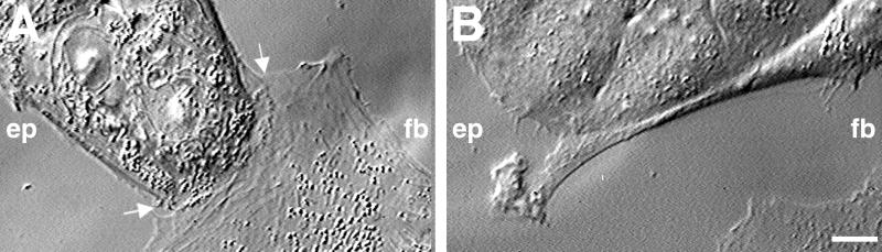
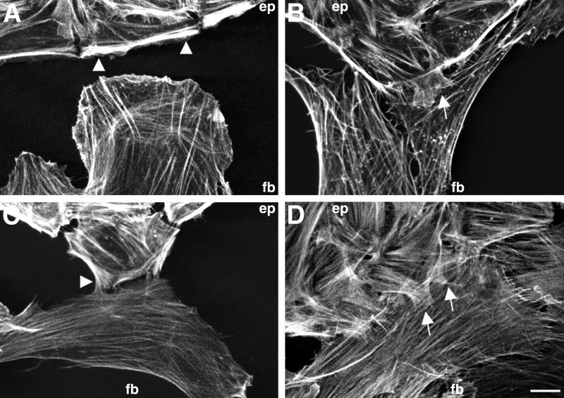
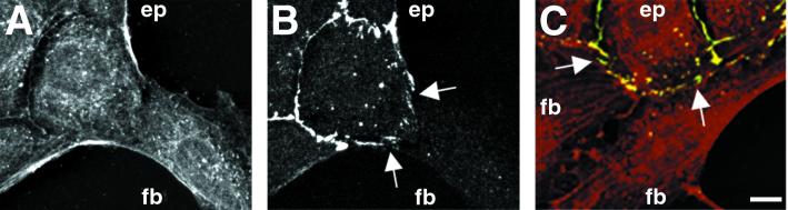
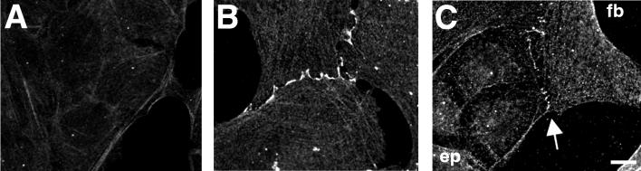
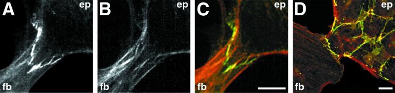
Contact interactions between different cell types play a number of important roles in development, for example in cell sorting, tissue organization, and ordered migration of cells. The nature of such heterocellular interactions, in contrast to interactions between cells of the same type, remains largely unknown. In this report, we present experimental data examining the dynamics of heterocellular interactions between epitheliocytes and fibroblasts, which express different cadherin cell adhesion molecules and possess different actin cytoskeletal organizations. Our analysis revealed two striking features of heterocellular contact. First, the active free edge of an epitheliocyte reorganizes its actin cytoskeleton after making contact with a fibroblast. Upon contact with the leading edge of a fibroblast, epitheliocytes disassemble their marginal bundle of actin filaments and reassemble actin filaments into a geometric organization more typical of a fibroblast lamella. Second, epitheliocytes and fibroblasts form cell--cell adhesion structures that have an irregular organization and are associated with components of cell adhesion complexes. The structural organization of these adhesions is more closely related to the type of contacts formed between fibroblasts rather than to those between epitheliocytes. Heterotypic epithelio-fibroblastic contacts, like homotypic contacts between fibroblasts, are transient and do not lead to formation of stable contact interactions. We suggest that heterocellular contact interactions in culture may be regarded as models of how tissue systems consisting of epithelia and mesenchyme interact and become organized in vivo.


Figures

Figure 1
Figure 1
Heterocellular collisions between IAR-2 epithelial cells and RAT-1 fibroblasts. (A) During the early stage of collision, the lamella of the fibroblast (fb) burrows under the free edge (see arrows) of an epithelial cell within a small island. In this example, the epithelial cell forms a wide lamellar extension at the site of contact. (B) At later stages of collision, fibroblasts tended to turn laterally and migrate along the edge of the epithelial island. Often short lamellar extensions reached out and would overlap the side of the fibroblast (also see Fig. 2 C and D). (Bar = 10 μm.)
Figure 2
Figure 2
Localization of actin filaments at different stages of heterocellular collisions between IAR-2 epitheliocytes and RAT-1 fibroblasts. (A) Before contact, the epithelial cells (ep) within an island exhibit typical marginal bundles (arrowheads) at their free edge, whereas fibroblasts have numerous bundles of filaments extending radially into a broad lamella. (B) Early stage of heterocellular collision, where the fibroblast has migrated beneath the edge of an epithelial island. Notice the localized disassembly of the marginal bundle in the area of collision and coincident formation of radially oriented small bundles of actin filaments within a small lamellipodial extension at the edge of the epithelial cell (see arrow). (C) At later stages of collision fibroblasts retracted the underlapped lamella and turned away from the epithelial edge. The marginal bundles within epithelial cells remain disassembled, and small, actin-rich lamellipodia (arrow) continue to retain contact with the surface of the turned fibroblast. (D) When grown in dense cultures numerous fibroblasts align themselves so that their side edge runs along the edge of epithelial islands. Epithelial cells form numerous lamellipodial extensions out onto the surface of the fibroblast (arrows), and the characteristic marginal bundles are completely disassembled. (Bar = 10 μm.)
Figure 3
Figure 3
Localization of E-cadherin in heterotypic contacts. (A and B) Cells were fixed and double-stained with anti-actin (red, A) and anti-E-cadherin (green, B) antibodies. After a head-on collision between a single fibroblast (fb) and an island of epithelial cells (ep), the marginal bundle of the epithelial cell is disassembled at the site of contact, and there is no distinguishing difference between the staining patterns of the two cells at the site of contact. Staining with E-cadherin revealed the presence of fragmented clusters of E-cadherin along the zone of overlap between the epitheliocyte and the fibroblast. Note the well-defined continuous staining for E-cadherin along the edges of neighboring epithelial cells. (C) Cells were double labeled with anti-actin (red) and anti-E-cadherin (green) antibodies. Punctate yellow spots along the epithelial cell–fibroblast border (arrows) indicate the colocalization of E-cadherin and actin. Note the absence of E-cadherin (green) staining along the edge between two IAR-2 fibroblasts. (Bar = 10 μm.)
Figure 4
Figure 4
Distribution of N-cadherin in IAR-2 epitheliocytes and RAT-1 fibroblasts. Cells were fixed and immunolabeled with anti-N-cadherin antibodies. (A) Anti-N-cadherin antibodies did not stain homotypic cell–cell contacts between adjacent cells in epithelial islands. (B) Homotypic contacts between fibroblasts exhibited N-cadherin staining along the edge between adjacent cells. (C) Anti-N-cadherin staining of heterotypic contacts between IAR-2 (ep) and RAT-1 (fb) cells identified the presence of discontinuous clusters of N-cadherin along the boundary between cells. (Bar = 10 μm.)
Figure 5
Figure 5
Immunofluorescence labeling for β-catenin in heterotypic contact between fibroblasts and epitheliocytes. (AC) MDCK (ep) and M19 (fb) cells were double-stained with anti-β-catenin (green) and anti-actin (red) antibodies while undergoing heterocellular contact. In this example, the MDCK cell formed a triangular extension over the M19 fibroblast. Note the presence of strong positive staining for β-catenin (A) and, in some locations, coincident positioning of actin filament bundles (B). (C) The merged image of A and B, with yellow designating areas of colocalization of β-catenin and actin. (D) Heterocellular contact between IAR-2 epithelial cells and RAT-1 fibroblasts stained with anti-β-catenin antibodies. (Bar = 10 μm.)

References

    1. Yap A S, Brieher W M, Gumbiner B M. Annu Rev Cell Dev Biol. 1997;13:119–146. - PubMed
    1. Troyanovsky S M. Curr Opin Cell Biol. 1999;11:561–566. - PubMed
    1. Adams C L, Nelson W J, Smith S J. J Cell Biol. 1996;135:1899–1911. - PMC - PubMed
    1. Gumbiner B M. Cell. 1996;84:345–357. - PubMed
    1. Adams C L, Nelson W J. Curr Opin Cell Biol. 1998;10:572–577. - PubMed

Publication types