. 2001 Jul 17;98(15):8578-83.
doi: 10.1073/pnas.161259898. Epub 2001 Jul 10.

Critical role for the docking-protein FRS2 alpha in FGF receptor-mediated signal transduction pathways

Affiliations

Critical role for the docking-protein FRS2 alpha in FGF receptor-mediated signal transduction pathways

Y R Hadari et al. Proc Natl Acad Sci U S A. .

Abstract

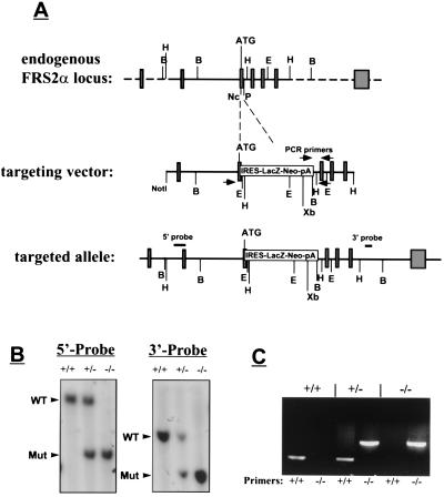
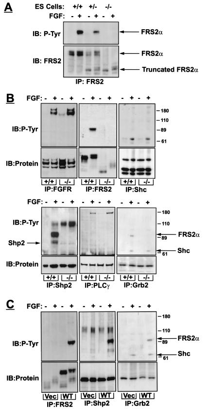
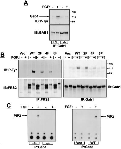
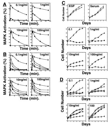
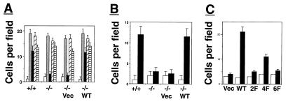
The docking protein FRS2 alpha has been implicated as a mediator of signaling via fibroblast growth factor receptors (FGFRs). We have demonstrated that targeted disruption of FRS2 alpha gene causes severe impairment in mouse development resulting in embryonal lethality at E7.0--E7.5. Experiments with FRS2 alpha-deficient fibroblasts demonstrate that FRS2 alpha plays a critical role in FGF-induced mitogen-activated protein (MAP) kinase stimulation, phosphatidylinositol-3 (PI-3) kinase activation, chemotactic response, and cell proliferation. Following FGF stimulation, tyrosine phosphorylated FRS2 alpha functions as a site for coordinated assembly of a multiprotein complex that includes Gab1 and the effector proteins that are recruited by this docking protein. Furthermore, we demonstrate that different tyrosine phosphorylation sites on FRS2 alpha are responsible for mediating different FGF-induced biological responses. These experiments establish the central role of FRS2 alpha in signaling via FGFRs and demonstrate that FRS2 alpha mediates multiple FGFR-dependent signaling pathways critical for embryonic development.


Figures

Figure 1
Figure 1
Targeted disruption of the Frs2α locus. (A) Strategy for generation of Frs2α mutation. The targeting vector contains a β-galactosidase neomycin resistance cassette in place of a 0.5-kb region of Frs2α gene that includes the first coding exon. Open boxes mark noncoding exons, and shaded boxes mark coding regions. Arrowheads mark the primers for genotyping by PCR, and the locations of the internal 5′ and external 3′ probes that were used for Southern blotting are also indicated. Restriction sites for several restriction enzymes are indicated. B, BamHI; E, EcoRI; H, HindIII; Nc, NcoI; P, PstI; Xb, XbaI. (B) Southern blot analysis of genomic DNA isolated from ES cell lines. For hybridization with the 5′ internal probe, DNA was digested with XbaI; for hybridization with the 3′ external probe, DNA was digested with BamHI. Arrows mark the positions of wild-type or mutant fragments. The sizes of these fragments are: 5′ probe: wild type 12 kb, mutant 7 kb; 3′ probe: wild type 8 kb, mutant 5 kb. (C) PCR analysis of Frs2α mutation. DNA was isolated from ES cell lines and PCR was performed as described in Methods. Two sets of primers were used, one for the wild-type allele (+/+) and the other for the Frs2α mutant allele (−/−).
Figure 2
Figure 2
Biochemical characterization of FRS2α-deficient fibroblasts. (A) Expression and tyrosine phosphorylation of FRS2α in wild-type, heterozygous, and homozygous FRS2α mutant cells. ES cells grown on gelatin in the presence of 1 μM retinoic acid for 24 h, starved overnight, treated with FGF1 (100 ng/ml) and heparin (5 μg/ml) for 5 min, lysed, and subjected to immunoprecipitation with anti-FRS2 antibodies followed by immunoblotting with anti-FRS2 or anti-pTyr antibodies. (B) Mouse embryo fibroblasts were serum-starved overnight, treated with FGF1 (100 ng/ml) and heparin (5 μg/ml) for 5 min, lysed, and immunoprecipitated with antibodies as indicated. (C) FRS2α mutant fibroblasts were infected with retroviral expression vector for wild-type FRS2α or with vector alone. Fibroblasts were treated with FGF1, and cell lysates were immunoprecipitated and immunoblotted with different antibodies as indicated.
Figure 3
Figure 3
FRS2α is essential for Gab1 recruitment and activation of PI-3 kinase. (A) Fibroblasts were stimulated with FGF1 and heparin. Cell lysates were immunoprecipitated with anti-Gab1 antibodies followed by immunoblotting with the indicated antibodies. (B) FRS2α-deficient fibroblasts were infected with expression vector for wild-type or FRS2α mutants or with vector alone. Following FGF1 stimulation, cell lysates were subjected to immunoprecipitation with anti-FRS2 or anti-Gab1 antibodies, followed by immunoblotting with antibodies as indicated. (C) PI3-kinase assay was performed on Gab1 immunoprecipitates derived from wild-type or mutant fibroblasts (Left) or from mutant fibroblasts transfected with expression vector encoding for FRS2α or vector alone (Right).
Figure 4
Figure 4
Impaired FGF-induced MAP kinase (ERK) stimulation and cell proliferation of FRS2α-deficient fibroblasts. (A) Comparison of MAP kinase activation in wild-type or FRS2α-deficient fibroblasts in response to FGF1 stimulation. Fibroblasts were starved, treated with different concentrations of FGF1, lysed, and immunoblotted with phospho-specific MAP kinase antibodies or with anti-Erk1 antibodies, and the intensity of the protein bands was quantitated by densitometry. ●, Wild-type fibroblasts; ■, FRS2α-deficient fibroblasts. (B) FRS2α mutant fibroblasts were infected with expression vector encoding for wild-type FRS2α or mutant FRS2α, or with vector alone. MAP kinase stimulation was revealed by immunoblotting with anti-phospho-MAP-kinase antibodies followed by quantitation by densitometry. ●, Wild-type FRS2α; ▴, ♦, or □, the 2F, 4F, or 6F FRS2α mutants; ■, vector alone. Similar results were obtained in three different experiments. (C) Growth curves of wild-type and FRS2α-deficient fibroblasts in response to growth factor stimulation. Wild-type or FRS2α mutant fibroblasts were grown in medium containing 10 ng/ml EGF, 10% serum, or different concentrations of FGF. The cells were trypsinized at the indicated time points and counted. ● Wild-type fibroblasts; ■ FRS2α-deficient fibroblasts. (D) FRS2α-deficient fibroblasts were infected with expression vector for wild-type FRS2α, FRS2α mutants, or vector alone. The cell lines were stimulated with different concentrations of FGF and growth curves were determined. ●, Wild-type FRS2α; ▴, ♦, or □, the 2F, 4F, or 6F FRS2α mutants; ■, vector alone.
Figure 5
Figure 5
FRS2α is essential for FGF-induced cell migration. (A) Migration assay using Boyden chamber was performed with wild-type fibroblasts, FRS2α-deficient fibroblasts, or FRS2α-deficient fibroblasts infected with expression vectors for FRS2α or vector alone. The fibroblasts were stimulated with 10% serum ░⃞, FGF1 (100 ng/ml) and heparin (5 μg/ml) ■, PDGF (50 ng/ml) ▨, or EGF (100 ng/ml) ▤ or medium alone □. (B) Fibroblasts were stimulated with FGF1 (100 ng/ml) and heparin (5 mg/ml) ■ or medium alone □. (C) Migration analysis of unstimulated □ or FGF-stimulated ■ FRS2α-deficient fibroblasts that were transfected with expression vectors for FRS2α (WT), for the 2F, 4F, or 6F mutants, or with vector alone.

References

    1. Hunter T. Cell. 2000;100:113–127. - PubMed
    1. Pawson T, Schlessinger J. Curr Biol. 1993;3:434–442. - PubMed
    1. Baker J M, Myers M G, Shoelson S E, Chin D J, Sun X J, Miralpeix M, Hu P, Margolis B, Skolnik E, Schlessinger J, White M F. EMBO J. 1992;11:3469–3479. - PMC - PubMed
    1. Kouhara H, Hadari Y R, Spivak-Kroizman T, Schilling J, Lax I, Schlessinger J. Cell. 1997;89:693–702. - PubMed
    1. Hadari Y R, Kouhara H, Lax I, Schlessinger J. Mol Cell Biol. 1998;18:3966–3973. - PMC - PubMed

Publication types

MeSH terms

Substances