. 2001 Jul 9;154(1):109-22.
doi: 10.1083/jcb.200102142.

Human Vam6p promotes lysosome clustering and fusion in vivo

Affiliations

Human Vam6p promotes lysosome clustering and fusion in vivo

S Caplan et al. J Cell Biol. .

Abstract

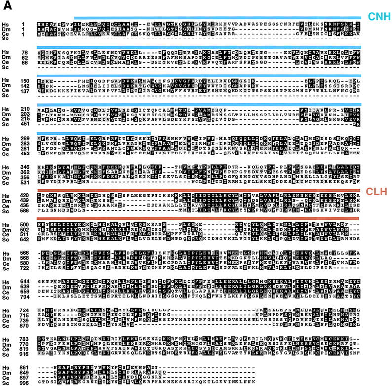
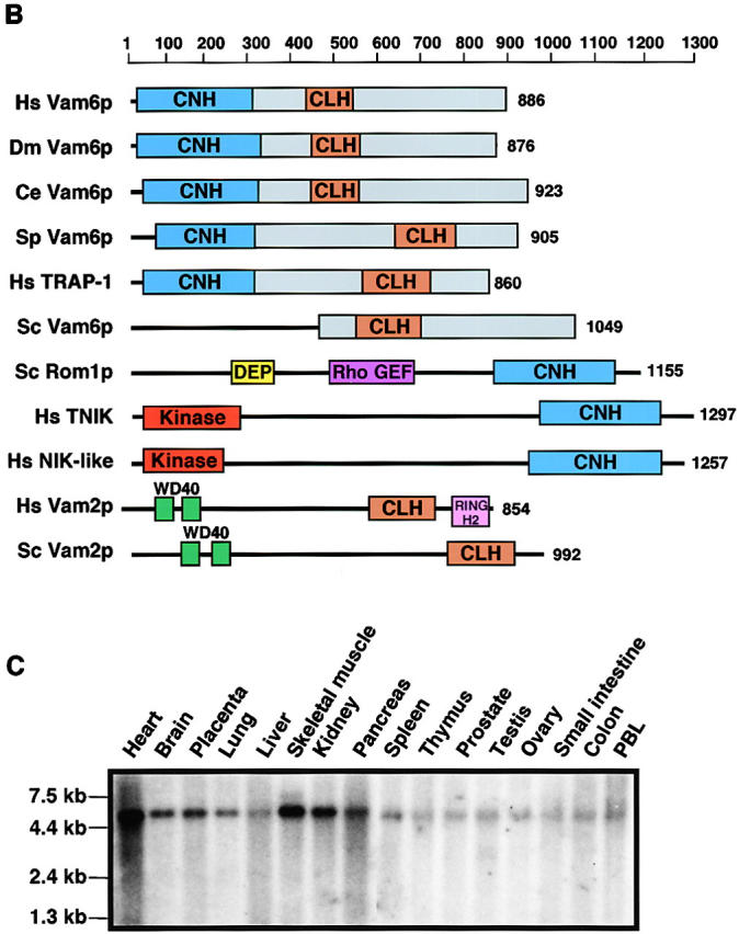
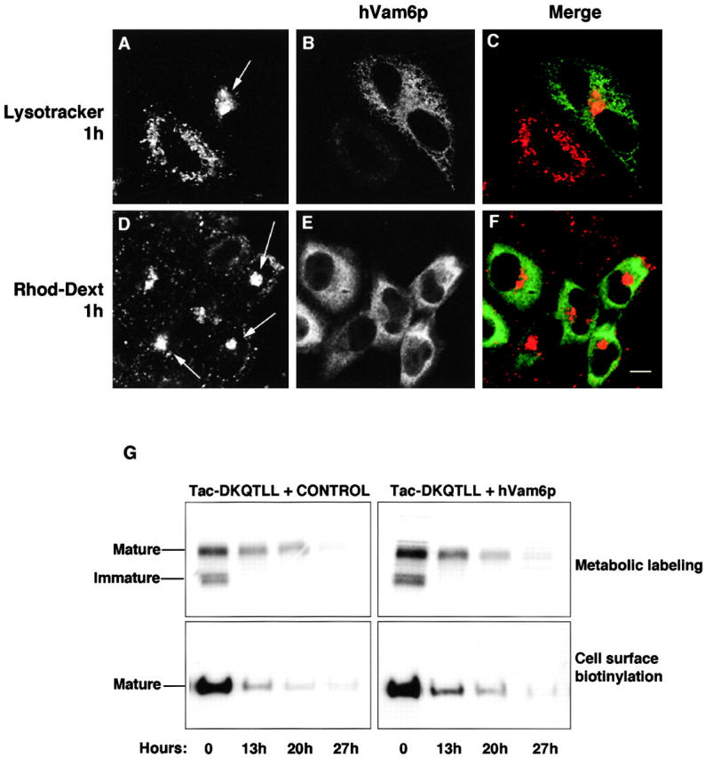
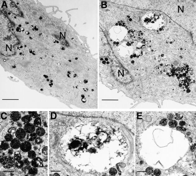
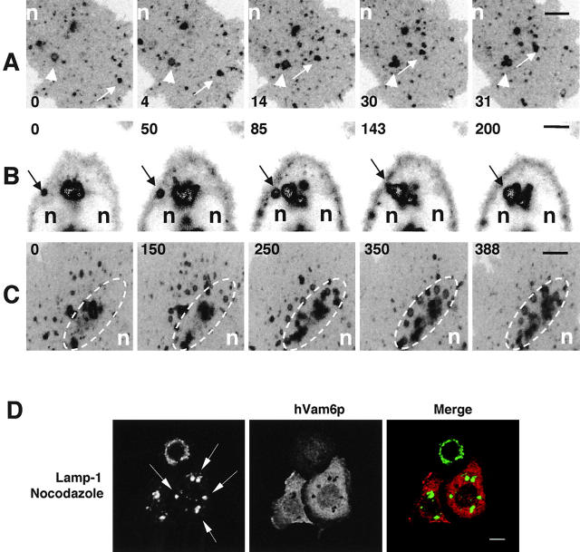
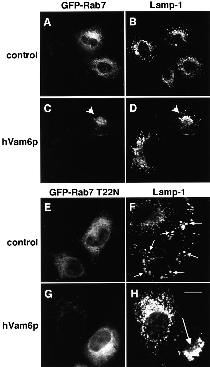
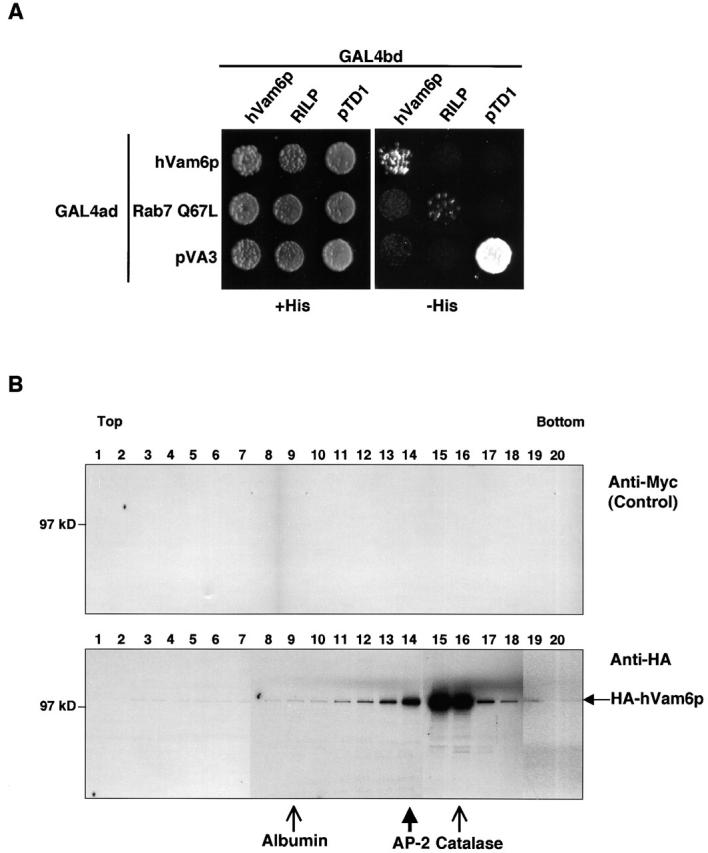
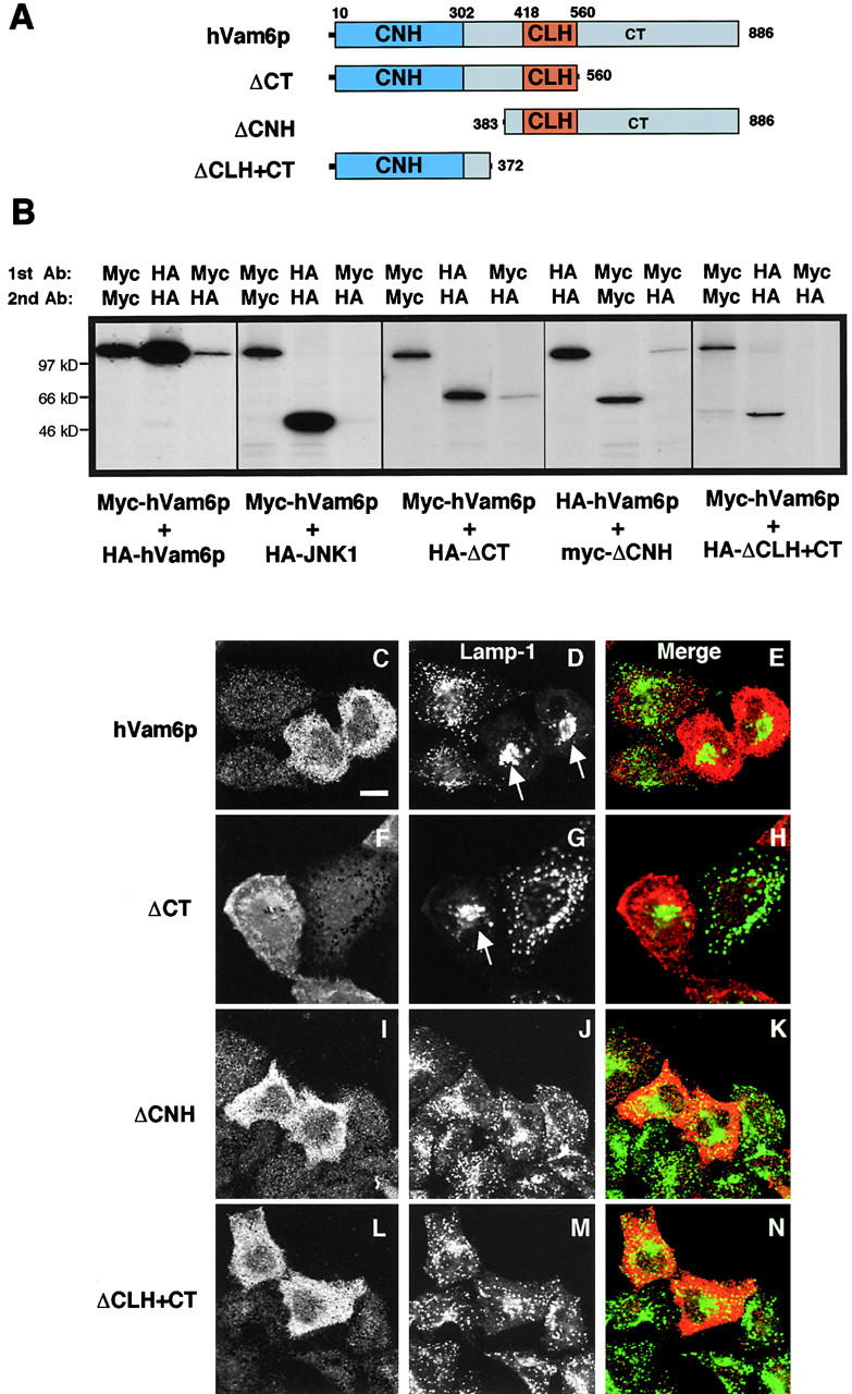
Regulated fusion of mammalian lysosomes is critical to their ability to acquire both internalized and biosynthetic materials. Here, we report the identification of a novel human protein, hVam6p, that promotes lysosome clustering and fusion in vivo. Although hVam6p exhibits homology to the Saccharomyces cerevisiae vacuolar protein sorting gene product Vam6p/Vps39p, the presence of a citron homology (CNH) domain at the NH(2) terminus is unique to the human protein. Overexpression of hVam6p results in massive clustering and fusion of lysosomes and late endosomes into large (2-3 microm) juxtanuclear structures. This effect is reminiscent of that caused by expression of a constitutively activated Rab7. However, hVam6p exerts its effect even in the presence of a dominant-negative Rab7, suggesting that it functions either downstream of, or in parallel to, Rab7. Data from gradient fractionation, two-hybrid, and coimmunoprecipitation analyses suggest that hVam6p is a homooligomer, and that its self-assembly is mediated by a clathrin heavy chain repeat domain in the middle of the protein. Both the CNH and clathrin heavy chain repeat domains are required for induction of lysosome clustering and fusion. This study implicates hVam6p as a mammalian tethering/docking factor characterized with intrinsic ability to promote lysosome fusion in vivo.


Figures

Figure 1.
Figure 1.
Sequence homology, domain organization, and expression of hVam6p. (A) Full-length amino acid sequences of human (Homo sapiens; Hs) Vam6p (hVam6p), a D. melanogaster (Dm) homologue, and a C. elegans (Ce) homologue were aligned together with residues 451–1049 of S. cerevisiae (Sc) Vam6p/Vps39p, using the ClustalW Multiple Sequences Alignment software (available at the European Bioinformatics Institute website http://www2.ebi.ac.uk/clustalw/) and shaded using the BOXSHADE program. Identical and similar residues are indicated by black and gray shading, respectively. The blue line denotes the hypothetical hVam6p CNH domain, which is conserved in Dm and Ce but not Sc. The brown line indicates the position of the hypothetical hVam6p CLH domain, which is conserved in all four orthologues. Domains were identified by Pfam HMM database searches (available from Washington University at http://pfam.wustl.edu/hmmsearch.shtml) (B) Domain organization of Vam6p/Vps39p family members. Specific domains are color-coded and regions of significant homology to hVam6p are shown in blue (CNH), brown (CLH), and green. GenBank/EMBL/DDBJ accession numbers are as follows: Hs Vam6p/Vps39p (AF280814), Ds Vam6p (AAF55525), Ce Vam6p (T24712), Sp Vam6p (T38314), Hs TRAP-1 (XP 002298), Sc Vam6p/Vps39p (BAA11758), Sc Rom1p (S64365), Hs Traf2 and NCK-interacting kinase (TNIK) (AF172270), Hs NCK-interacting kinase–like (AAC83079), Hs Vam2p/Vps41p (P49754), Sc Vam2p/Vps41p (BAA19071). Note that whereas the S. cerevisiae CLH domain has previously been situated between residues 716 and 900 (Wurmser et al., 2000); our domain analysis, using the Pfam program, positions it between residues 512 and 676. DEP is a domain found in Dishevelled, Egl-10, and Pleckstrin. (C) Analysis of hVam6 mRNA expression in different human tissues. Northern blots with mRNA from various human tissues were analyzed with a 32P-labeled probe specific for the complete hVam6 mRNA. The positions of RNA size markers are indicated.
Figure 1.
Figure 1.
Sequence homology, domain organization, and expression of hVam6p. (A) Full-length amino acid sequences of human (Homo sapiens; Hs) Vam6p (hVam6p), a D. melanogaster (Dm) homologue, and a C. elegans (Ce) homologue were aligned together with residues 451–1049 of S. cerevisiae (Sc) Vam6p/Vps39p, using the ClustalW Multiple Sequences Alignment software (available at the European Bioinformatics Institute website http://www2.ebi.ac.uk/clustalw/) and shaded using the BOXSHADE program. Identical and similar residues are indicated by black and gray shading, respectively. The blue line denotes the hypothetical hVam6p CNH domain, which is conserved in Dm and Ce but not Sc. The brown line indicates the position of the hypothetical hVam6p CLH domain, which is conserved in all four orthologues. Domains were identified by Pfam HMM database searches (available from Washington University at http://pfam.wustl.edu/hmmsearch.shtml) (B) Domain organization of Vam6p/Vps39p family members. Specific domains are color-coded and regions of significant homology to hVam6p are shown in blue (CNH), brown (CLH), and green. GenBank/EMBL/DDBJ accession numbers are as follows: Hs Vam6p/Vps39p (AF280814), Ds Vam6p (AAF55525), Ce Vam6p (T24712), Sp Vam6p (T38314), Hs TRAP-1 (XP 002298), Sc Vam6p/Vps39p (BAA11758), Sc Rom1p (S64365), Hs Traf2 and NCK-interacting kinase (TNIK) (AF172270), Hs NCK-interacting kinase–like (AAC83079), Hs Vam2p/Vps41p (P49754), Sc Vam2p/Vps41p (BAA19071). Note that whereas the S. cerevisiae CLH domain has previously been situated between residues 716 and 900 (Wurmser et al., 2000); our domain analysis, using the Pfam program, positions it between residues 512 and 676. DEP is a domain found in Dishevelled, Egl-10, and Pleckstrin. (C) Analysis of hVam6 mRNA expression in different human tissues. Northern blots with mRNA from various human tissues were analyzed with a 32P-labeled probe specific for the complete hVam6 mRNA. The positions of RNA size markers are indicated.
Figure 2.
Figure 2.
Overexpression of hVam6p induces coalescence of lysosomal vesicles. (A–O) HeLa cells were transiently transfected with a plasmid encoding Myc–hVam6p. Cells were fixed, permeabilized, and incubated with rabbit polyclonal antibodies to the Myc epitope together with mouse monoclonal antibodies to lamp-1 (A–C), lamp-2 (D–F), CD63 (G–I), or transferrin receptor (M–O), or rabbit polyclonal antibody to cathepsin D (J–L). Arrows (A, D, G, and J) indicate the coalescence of lysosomes into large juxtanuclear structures. Arrowhead (M) indicates a transfected cell. (P–R) HA–hVam6p-transfected HeLa cells were extracted for 1 min with 0.05% (wt/vol) saponin before fixation. Cells were then fixed and incubated with rabbit polyclonal antibodies to the HA epitope, together with mouse monoclonal antibodies to lamp-1. Bound antibodies were revealed by Cy3-conjugated donkey anti–rabbit IgG (red channel) and Alexa-488–conjugated donkey anti–mouse antibody (green channel). Bar, 10 μm.
Figure 3.
Figure 3.
Association of hVam6p with clusters of lysosomes and late endosomes. HeLa cells were transiently transfected with HA–hVam6p and processed for immunoelectron microscopy. Ultra-thin frozen sections were labeled with antibodies to HA (to detect hVam6p) and either endogenous lamp-2 (A) or endogenous CI-MPR (B). Bound antibodies were detected using conjugated protein A–gold. Arrows indicate the localization of hVam6p to a halo around the membranes of lysosomes and late endosomes (A and B, 10-nm gold particles), whereas arrowheads indicate the localization of lamp-2 (A, 15-nm gold particles) or CI-MPR (B, 15-nm gold particles), and the asterisk marks a structure labeled only for hVam6p. G, Golgi. Bars, 0.2 μm.
Figure 4.
Figure 4.
Functional characterization of hVam6p-induced lysosomal clusters. (A–F) Live HeLa cells transiently transfected with Myc–hVam6p were incubated with LysoTracker red (A–C) or rhodamine–dextran (D–F) for 1 h, and then fixed-permeabilized. Cells were incubated with a mouse monoclonal antibody to Myc, followed by Alexa-488–conjugated donkey antibody to mouse IgG (B and E, green channel). The LysoTracker red and rhodamine–dextran are visible in the red channel (A and D). Arrows denote the accumulation of acidotropic (A) and fluid phase (D) markers within clustered lysosomes in hVam6p-transfected cells. Bar, 10 μm. (G) HeLa cells were cotransfected with either Tac-DKQTLL and Myc–hVam6p, or Tac-DKQTLL and a nonmyristylated Arf6 control. After 24 h, cotransfection efficiency was monitored by indirect immunofluorescence (as described in the legend to Fig. 2), and cells were pulsed for 30 min either by metabolic labeling (top) or cell surface biotinylation (bottom). The cells were then chased for the time points indicated, harvested, lysed, subjected to immunoprecipitation analysis using antibodies directed against Tac, and resolved on 4–20% SDS-PAGE. Samples from metabolically labeled and biotinylated cells were visualized by autoradiography and blotting with streptavidin–HRP, respectively.
Figure 5.
Figure 5.
Ultrastructural analysis of hVam6p-induced lysosomal structures labeled with internalized HRP. Untransfected (A) or hVam6p-transfected (B–E) HeLa cells were subjected to a continuous 4 h fluid phase uptake of HRP 24 h after transfection. (A) In control cells, HRP-containing lysosomes and endosomes were small (0.2–0.6 μm) and distributed throughout cell. (B) In hVam6p-transfected cells, clusters of multivesicular HRP-positive vesicles (enlarged in C) and large (2–3 μm) vacuoles situated next to the nucleus were visible. Some of these large vacuoles contained HRP (enlarged in D), whereas others appeared to have smaller HRP-positive vesicles docking onto their membrane (enlarged in E). N, Nucleus. Bars: (A and B) 2 μm; (C–E) 0.4 μm.
Figure 6.
Figure 6.
Dynamics of hVam6p-induced lysosome clustering and fusion. COS-7 (A and C) or HeLa (B) cells were transfected with plasmids encoding GFP–lamp-1 (GFP–lgp120) and HA–hVam6p. 10 h after transfection, live cells were incubated at 37°C and scanned for GFP-expressing cells by confocal microscopy. Live images were acquired at 30-s time intervals and are displayed as inverted images with the time in minutes relative to the start of imaging indicated in the lower left corner (A) or upper left corner (B and C). The dotted oval region of interest (C) outlines juxtanuclear regions that accumulate lysosome clusters and giant lysosomes. Arrows (A and B) point at moving vesicles. Arrowheads (A) point at a lysosome clustering event. n, nucleus. (D) Microtubule depolymerization impairs formation of a unified, giant juxtanuclear lysosomal structure in hVam6p-transfected cells. HeLa cells were transfected with Myc-hVam6p and treated with 0.5 μM nocodazole for 16 h. Cells were then fixed-permeabilized, incubated with a rabbit polyclonal antibody to Myc and a mouse monoclonal antibody to lamp-1, followed by Alexa-488–conjugated donkey antibody to mouse IgG (left, green channel) and Cy3-conjugated donkey anti–rabbit IgG (middle, red channel). Bars, (A and C) 5 μm; (B) 10 μm. Quicktime movie sequence versions of this figure are available at http://www.jcb.org/cgi/content/full/200102142/DC1.
Figure 7.
Figure 7.
hVam6p affects lysosomal morphology independently of Rab7 nucleotide cycling. HeLa cells were transfected with either GFP–Rab7 (A and B, control) or dominant-negative GFP–Rab7 T22N (E and F, control), or were cotransfected with Myc–hVam6p together with either GFP–Rab7 (C and D) or GFP–Rab7 T22N (G and H). After 24 h, the cells were fixed-permeabilized and incubated with mouse monoclonal antibodies to lamp-1. Bound antibodies were revealed by Cy3-conjugated donkey anti–mouse IgG (B, D, F, and H), and expression of GFP–Rab7 proteins visualized by their intrinsic fluorescence (A, C, E, and G). Arrowheads depict the coalescence of both GFP–Rab7 (C) and lamp-1 (D) to a large juxtanuclear conglomerate. Small arrows (F) mark the localization of dispersed lysosomes in cells transfected with Rab7 T22N. Large arrow (H) denotes a giant juxtanuclear lysosome conglomerate in a cell overexpressing GFP–Rab7 T22N and Myc–hVam6p. Bar, 10 μm.
Figure 8.
Figure 8.
Homooligomerization of hVam6p. (A) The S. cerevisiae yeast strain AH109 was cotransformed with the following GAL4ad fusion constructs: GAL4ad–hVam6p, GAL4ad–Rab7 Q67L, and GAL4ad–pVA3 (murine p53 control), together with the GAL4bd fusion constructs GAL4bd–hVam6p, GAL4bd–RILP, and GAL4bd–pTD1 (SV40 large T-antigen control). Cotransformants were assayed for growth on nonselective (+His) and selective (−His) media. (B) Sedimentation velocity analysis of hVam6p from [35S]methionine–labeled HeLa cells. The cell extract was run on a 4–20% sucrose gradient, and fractions were analyzed by sequential immunoprecipitations. The fractions were first cleared with an irrelevant antibody and then subjected to immunoprecipitation with a second irrelevant antibody (mouse monoclonal anti-Myc). The fractions were then immunoprecipitated with a mouse monoclonal antibody to the HA epitope. Fractions from the anti-Myc (control, top) and anti-HA (bottom) immunoprecipitations were then resolved by 4–20% gradient SDS-PAGE. Thin arrows denote markers visualized by Coomassie blue staining of separated fractions subjected to the same gradient conditions (albumin and catalase). The thick arrow denotes the position of AP-2, as visualized by immunoblotting of 10% of the labeled fractions with anti–AP-2 antibody (100/2) upon completion of the immunoprecipitations.
Figure 9.
Figure 9.
Delineation of functional domains in hVam6p. (A) Schematic representation of full-length hVam6p and various deletion constructs used in these experiments. (B) HeLa cells were cotranfected with plasmids encoding Myc– and HA–hVam6p, or hVam6p deletion mutants, or a control epitope-tagged protein (HA–JNK1), as indicated. After 18 h, cells were labeled for 8 h with [35S]methionine, detergent extracted, and subjected to immunoprecipitation–recapture with the antibodies indicated. (C-N) HeLa cells were transfected with plasmids encoding Myc– or HA–Vam6p full-length or deletion constructs. After 24 h, fixed-permeabilized cells were coincubated with rabbit polyclonal antibodies to either the Myc or HA epitopes together with mouse monoclonal antibody to lamp-1. Bound antibodies were revealed by Alexa-488–conjugated donkey anti–mouse antibody (green channel) and Cy3-conjugated donkey anti–rabbit IgG (red channel). The third panel in each row was generated by merging of the images in the red and green channels. Arrows mark the coalescence of lysosomes into juxtanuclear regions. Bar, 10 μm.

References

    1. Aguilar, R.C., H. Ohno, K.W. Roche, and J.S. Bonifacino. 1997. Functional mapping of the clathrin-associated adaptor medium chains μ1 and μ2. J. Biol. Chem. 272:27160–27166. - PubMed
    1. Bakker, A.C., P. Webster, W.A. Jacob, and N.W. Andrews. 1997. Homotypic fusion between aggregated lysosomes triggered by elevated [Ca2+]i in fibroblasts. J. Cell Sci. 110:2227–2238. - PubMed
    1. Bucci, C., P. Thomsen, P. Nicoziani, J. McCarthy, and B. van Deurs. 2000. Rab7: a key to lysosome biogenesis. Mol. Biol. Cell. 11:467–480. - PMC - PubMed
    1. Cantalupo, G., P. Alifano, V. Roberti, C.B. Bruni, and C. Bucci. 2001. Rab-interacting lysosomal protein (RILP): the Rab7 effector required for transport to lysosomes. EMBO J. 20:683–693. - PMC - PubMed
    1. Caplan, S., E.C. Dell'Angelica, W.A. Gahl, and J.S. Bonifacino. 2000. Trafficking of major histocompatibility complex class II molecules in human B-lymphoblasts deficient in the AP-3 adaptor complex. Immunol. Lett. 72:113–117. - PubMed

Publication types

MeSH terms

Substances