. 2001 Jul 9;154(1):123-36.
doi: 10.1083/jcb.200102110.

Obscurin, a giant sarcomeric Rho guanine nucleotide exchange factor protein involved in sarcomere assembly

Affiliations

Obscurin, a giant sarcomeric Rho guanine nucleotide exchange factor protein involved in sarcomere assembly

P Young et al. J Cell Biol. .

Abstract

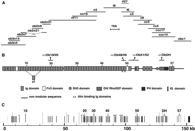
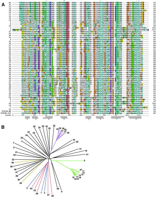
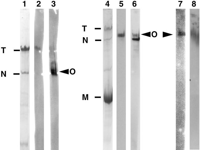
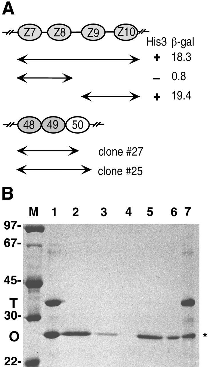
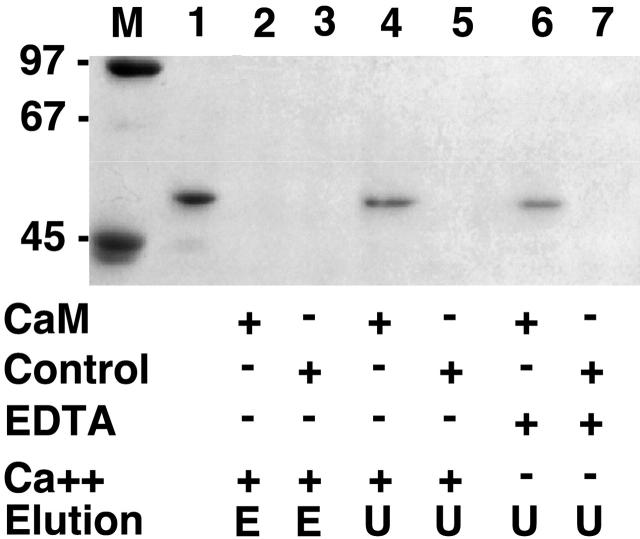
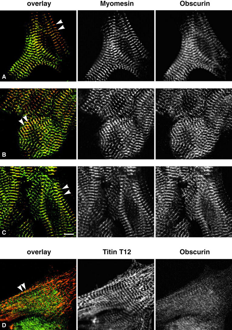
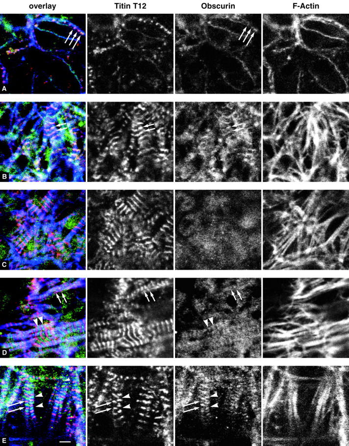
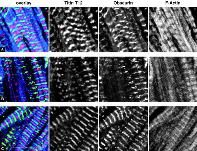
Vertebrate-striated muscle is assumed to owe its remarkable order to the molecular ruler functions of the giant modular signaling proteins, titin and nebulin. It was believed that these two proteins represented unique results of protein evolution in vertebrate muscle. In this paper we report the identification of a third giant protein from vertebrate muscle, obscurin, encoded on chromosome 1q42. Obscurin is approximately 800 kD and is expressed specifically in skeletal and cardiac muscle. The complete cDNA sequence of obscurin reveals a modular architecture, consisting of >67 intracellular immunoglobulin (Ig)- or fibronectin-3-like domains with multiple splice variants. A large region of obscurin shows a modular architecture of tandem Ig domains reminiscent of the elastic region of titin. The COOH-terminal region of obscurin interacts via two specific Ig-like domains with the NH(2)-terminal Z-disk region of titin. Both proteins coassemble during myofibrillogenesis. During the progression of myofibrillogenesis, all obscurin epitopes become detectable at the M band. The presence of a calmodulin-binding IQ motif, and a Rho guanine nucleotide exchange factor domain in the COOH-terminal region suggest that obscurin is involved in Ca(2+)/calmodulin, as well as G protein-coupled signal transduction in the sarcomere.


Figures

Figure 1.
Figure 1.
The cDNA and genomic sequence of obscurin. (A) Positions of isolated obscurin cDNAs. A total of 20,435 bp of continuous cDNA sequence can be assembled from the depicted overlapping cDNAs. This cDNA sequence contains an ORF encoding 6,620 amino acids. (B) Domain pattern of obscurin. The deduced amino acid sequence predicts a protein of 721,685 kD. Within this sequence, known protein domains were identified by database searches and visual inspection. Obscurin is a polar molecule with signal transduction domains in the COOH-terminal region and repetitive tandem Ig domains NH2-terminal to the signaling region. The Ig and Fn3 modules are numbered consecutively from the NH2 terminus. Ig, immunoglobulin-like; IQ, IQ calmodulin binding motif. One shorter alternatively spliced variant was sequenced and the extra Ig domain from this splice variant is denoted a1. The positions of the epitopes for antibodies used in this study are indicated above the domain structure. (C) Partial structure of the obscurin gene. A >150-kb obscurin genomic sequence was compiled by analyzing the draft human genome sequence. Exons are indicated as vertical black lines with the position of the exons encoding selected domains indicated above. Putative exons encoding 10 Ig domains which are not present in the cDNA sequence are marked by an asterisk. Long vertical grey lines indicate gaps of undefined length in the sequence.
Figure 2.
Figure 2.
Comparison of obscurin Ig domains. (A) Multiple sequence alignment of all obscurin Ig domains using ClustalW (Higgins et al., 1996). Note the extremely high homology of some groups of adjacent domains. Also included are titin M5 and I27 domains whose structures have been solved. The seven β-strands of M5 (top) and I27 (bottom) are indicated as bars under the alignment (Pfuhl and Pastore, 1995; Improta et al., 1996). This alignment was used to generate the phylogenetic tree in B. Residue coloring: all prolines, yellow; all glycines, brown; conserved basic residues, pink; conserved acidic residues, purple; conserved hydrophobic residues, blue. (B) A phylogenetic tree of the obscurin Ig domains does not reveal any patterns of super repeats as seen in titin. In some regions, consecutive domains cluster together and are highly homologous, for example domains Ob9–15 and Ob36–42 (green and purple, respectively). This suggests a rapid expansion of the domain pattern during the evolution of the protein. The three NH2-terminal domains (blue) cluster together with the five most COOH-terminal ones (red). The titin binding domains Ob48 and Ob49 are colored yellow.
Figure 3.
Figure 3.
Detection of obscurin by Western blotting. (A) The polyclonal antibody α-Ob48–49 against obscurin detects a large protein of approximately the same size as nebulin in striated muscle. Lanes 1, 2, and 3: human Vastus lateralis muscle sample run on 3% polyacrylamide gel. Lane 1, Coomassie stain of a lane from the gel; lane 2, titin detected with S53 monoclonal antibody on a Western blot of one lane from the same gel; lane 3, obscurin detected with α-Ob48–49 polyclonal antibody on a Western blot of an adjacent lane. Note, titin and nebulin were well blotted and their positions are marked on the blot. Titin is resolved as a single band. Obscurin is detected slightly above the position of nebulin as marked on the blot. Lanes 4–7, human Vastus lateralis muscle sample run on 4% polyacrylamide gel. Lane 4, Coomassie stain of a lane from the gel; lane 5; obscurin detected with α-Ob48–49 on a lane cut from of a Western blot of the same gel; lane 6, the same blot as lane 5 stripped and reprobed with antinebulin NSH3-ra. Note, blots have been accurately aligned and obscurin can be distinguished from nebulin. The α-Ob48–49 antibody has not been completely stripped from the blot, so, in lane 6 there is some carry over of the obscurin signal appearing just above the darker nebulin band. Lane 7, obscurin detected with α-ObDH; lane 8, human cardiac muscle sample run on a 4% polyacrylamide gel. Obscurin detected with α-Ob48–49 on a Western blot. Note, a protein of a similar size is detected in cardiac and skeletal muscle. M, myosin; T, titin; N, nebulin; O, obscurin.
Figure 4.
Figure 4.
Binding of titin and obscurin in yeast two-hybrid system and in vitro. (A) Yeast two-hybrid analysis of the interaction between obscurin and titin. Titin domains Z7–Z10 (depicted as ovals) which were used as the primary two-hybrid bait is shown. One of the interacting clones obtained (clone no. 27) was used to further map the binding site on titin. The region Z9–Z10 interacted as strongly as the original bait whereas Z7–Z8 did not interact in this assay. Interactions were assayed by cotransformation of bait and prey plasmids into L40 yeast cells and monitoring reporter gene activation. His3 gene activation is marked as +/− and β-galactosidase activity is given as arbitrary units from liquid assays. Further separation of either titin Ig domains Z9 and Z10 or obscurin Ig 48 and 49 abolishes the interaction, demonstrating that the tandem domains of both proteins are required to form a functional binding site. (B) In vitro binding of titin to obscurin. The histidine-tagged titin fragment Z9–Z10 was assayed for binding to an untagged obscurin fragment Ig48–49 on mini Ni-NTA agarose columns. Lane 1 shows a mixture of both proteins as used in the assay. Either such a mixture of both proteins (lanes 5–7), or the obscurin fragment alone (lanes 2–4), were loaded on the column. The obscurin fragment is retained on the column when mixed with the titin Z9–Z10 (lane 7, asterisk), whereas there is no unspecific binding (lane 4). Lanes 2 and 5, flow through fractions; lanes 3 and 6, wash fraction; lanes 4 and 7, eluate fraction; M, marker lane (sizes given in kD). (C) Ob48–51 is targeted to the Z-disk in neonatal rat cardiomyocytes. The T7-tagged fragment was transfected into neonatal rat cardiomyocytes and detected by a tag-specific monoclonal antibody at the sarcomeric Z-disk (green), as demonstrated by the counterstain with myomesin at the M-band (red) in the overlay. Occasional weak M-band localization can also be observed (arrowhead). Overexpressed protein is also accumulating in the nuclei. Note that expression of Ob48–51 does not disrupt myofibrils. Bar, 8 μm.
Figure 4.
Figure 4.
Binding of titin and obscurin in yeast two-hybrid system and in vitro. (A) Yeast two-hybrid analysis of the interaction between obscurin and titin. Titin domains Z7–Z10 (depicted as ovals) which were used as the primary two-hybrid bait is shown. One of the interacting clones obtained (clone no. 27) was used to further map the binding site on titin. The region Z9–Z10 interacted as strongly as the original bait whereas Z7–Z8 did not interact in this assay. Interactions were assayed by cotransformation of bait and prey plasmids into L40 yeast cells and monitoring reporter gene activation. His3 gene activation is marked as +/− and β-galactosidase activity is given as arbitrary units from liquid assays. Further separation of either titin Ig domains Z9 and Z10 or obscurin Ig 48 and 49 abolishes the interaction, demonstrating that the tandem domains of both proteins are required to form a functional binding site. (B) In vitro binding of titin to obscurin. The histidine-tagged titin fragment Z9–Z10 was assayed for binding to an untagged obscurin fragment Ig48–49 on mini Ni-NTA agarose columns. Lane 1 shows a mixture of both proteins as used in the assay. Either such a mixture of both proteins (lanes 5–7), or the obscurin fragment alone (lanes 2–4), were loaded on the column. The obscurin fragment is retained on the column when mixed with the titin Z9–Z10 (lane 7, asterisk), whereas there is no unspecific binding (lane 4). Lanes 2 and 5, flow through fractions; lanes 3 and 6, wash fraction; lanes 4 and 7, eluate fraction; M, marker lane (sizes given in kD). (C) Ob48–51 is targeted to the Z-disk in neonatal rat cardiomyocytes. The T7-tagged fragment was transfected into neonatal rat cardiomyocytes and detected by a tag-specific monoclonal antibody at the sarcomeric Z-disk (green), as demonstrated by the counterstain with myomesin at the M-band (red) in the overlay. Occasional weak M-band localization can also be observed (arrowhead). Overexpressed protein is also accumulating in the nuclei. Note that expression of Ob48–51 does not disrupt myofibrils. Bar, 8 μm.
Figure 5.
Figure 5.
The obscurin IQ domain interacts with calmodulin in a Ca ++ -independent manner. The expressed and purified obscurin fragment Ob51–52, which contains the IQ motif, is shown in lane 1. The binding of this fragment to either calmodulin-coupled Sepharose beads (lanes 2, 4, and 6), or to control beads coupled with an unrelated protein (lanes 3, 5, and 7), was analyzed in a pulldown assay. The assay was carried out in the presence of 1 mM CaCl2 (lanes 2–5) or 1 mM EDTA (lanes 6 and 7). Bound protein was eluted from the beads with either 5 mM EDTA (lanes 2 and 3) or 6 M urea (lanes 4 to 7) and analyzed by SDS-PAGE and Coomassie blue staining. The obscurin fragment binds specifically to calmodulin both in the presence and absence of Ca++ and can be eluted under denaturing conditions (6 M urea), but not by chelating agents (EDTA). Elution by EDTA or urea is indicated by E or U, respectively.
Figure 6.
Figure 6.
Localization of endogenous obscurin. Localization of endogenous obscurin (green with all antibodies) in neonatal rat cardiomyocytes demonstrates all four obscurin epitopes in association with the sarcomeric M-band (arrowheads). Obscurin visualized with α-Ob48–49 colocalizes with myomesin (red in A–C) at the M-band (A) similar to the staining observed with α-Ob51–52 (B) and α–Ob-DH (C). The epitope of α-Ob19–20 is first detected after birth and is initially only weakly expressed (D), counterstain with the titin Z-disk antibody T12 in red). Bar, 5 μm.
Figure 7.
Figure 7.
Localization of endogenous obscurin in developing hearts demonstrates a relocalization of epitopes during development. Hearts of embryonic chicken (A–D) or mouse (E) are stained with antibodies raised against various obscurin epitopes (green). The titin Z-disk epitope T-12 is stained in red and the actin cytoskeleton in blue. The resulting color of titin and obscurin colocalization is yellow to bluish white here and in Fig. 8, depending on the background of blue actin staining. (A) Chick, 8-somite stage: the α-Ob48–49 epitope colocalizes with titin T12 in dots or cross-striated patterns on actin-filaments at the cell periphery (arrows). (B) Chick, 10-somite stage: α-Ob48–49 is predominantly colocalized with titin T12 at the Z-disk (arrows). (C) and (D) Chick, 10-somite stage: the α-Ob51–52 and α–Ob-DH epitopes, respectively, are diffusely localized or partly detected at the M-band (arrowheads in D). M-band localization is observed in myofibrils with parallel arrangements but unaligned myofibrils show diffuse localization (arrows in D). (E) Mouse (E9.5) relocalization of the α-Ob48–49 epitope is observed which shows prominent M-band and weaker Z-disk staining with α-Ob48–49. Note that the myofibrils in these hearts begin to align in parallel compared with the criss-cross patterns observed in early chicken hearts. Bar, 4 μm.
Figure 8.
Figure 8.
Localization of endogenous obscurin (green for all antibodies) in E14.5 rat hearts. At this developmental stage, myofibrils are arranged in mature, parallel bundles and all obscurin epitopes detectable at this stage are found at the M-bands, as shown by the alternating staining pattern with Z-disk titin (T12, red; F-actin, blue). (A) α-Ob48–49; (B) α-Ob51–52; (C) α–Ob-DH. Bar, 10 μm.

Comment in

Similar articles

Cited by

References

    1. Aghazadeh, B., W.E. Lowry, X.-Y. Huang, and M.K. Rosen. 2000. Structural basis for relief of autoinhibition of the Dbl homology domain of proto-oncogene Vav by tyrosine phosphorylation. Cell. 102:625–633. - PubMed
    1. Altschul, S.F., T.L. Madden, A.A. Schaffer, J. Zhang, Z. Zhang, W. Miller, and D.J. Lipman. 1997. Gapped BLAST and PSI-BLAST: a new generation of protein database search programs. Nucleic Acids Res. 25:3389–3402. - PMC - PubMed
    1. Andersson, S., D.N. Davis, H. Dahlbäck, H. Jörnvall, and D.W. Russell. 1989. Cloning, structure, and expression of the mitochondrial cytochrome P-450 sterol 26-hydroxylase, a bile acid biosynthetic enzyme. J. Biol. Chem. 264:8222–8229. - PubMed
    1. Ausubel, F.M., R. Brent, R.E. Kingston, D.D. Moore, J.G. Seidman, J.A. Smith, and K. Struhl. 1995. Short protocols in molecular biology. Wiley and Sons, Inc., New York.
    1. Benian, G.M., J.E. Kiff, N. Neckelmann, D.G. Moerman, and R.H. Waterston. 1989. Sequence of an unusually large protein implicated in regulation of myosin activity in C. elegans. Nature. 342:45–50. - PubMed

Publication types

MeSH terms