. 2001 Jul;13(7):1639-52.
doi: 10.1105/tpc.010008.

Overexpression of the heterotrimeric G-protein alpha-subunit enhances phytochrome-mediated inhibition of hypocotyl elongation in Arabidopsis

Affiliations

Overexpression of the heterotrimeric G-protein alpha-subunit enhances phytochrome-mediated inhibition of hypocotyl elongation in Arabidopsis

H Okamoto et al. Plant Cell. 2001 Jul.

Abstract

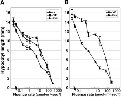
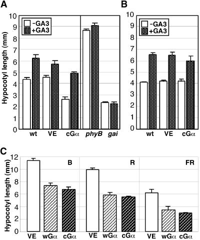
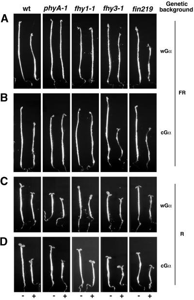
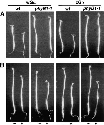
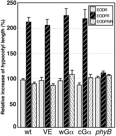
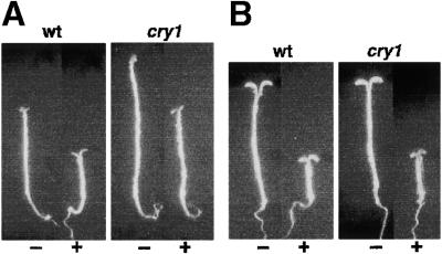
Plant heterotrimeric G-proteins have been implicated in a number of signaling processes. However, most of these studies are based on biochemical or pharmacological approaches. To examine the role of heterotrimeric G-proteins in plant development, we generated transgenic Arabidopsis expressing the Galpha subunit of the heterotrimeric G-protein under the control of a glucocorticoid-inducible promoter. With the conditional overexpression of either the wild type or a constitutively active version of Arabidopsis Galpha, transgenic seedlings exhibited a hypersensitive response to light. This enhanced light sensitivity was more exaggerated in a relatively lower intensity of light and was observed in white light as well as far-red, red, and blue light conditions. The enhanced responses in far-red and red light required functional phytochrome A and phytochrome B, respectively. Furthermore, the response to far-red light depended on functional FHY1 but not on FIN219 and FHY3. This dependence on FHY1 indicates that the Arabidopsis Galpha protein may act only on a discrete branch of the phytochrome A signaling pathway. Thus, our results support the involvement of a heterotrimeric G-protein in the light regulation of Arabidopsis seedling development.


Figures

Figure 1.
Figure 1.
Protein Blot Analysis of Gα Overexpression in Transgenic Plants. (A) Total protein extracts (35 μg/lane) of wild-type, VE, wGα, and cGα seedlings grown for 5 days in darkness on DEX-containing plates were separated on a 10% acrylamide gel and probed with anti-Gα and anti–α-tubulin polyclonal antibodies. (B) Total protein extracts (35 μg/lane) of wild-type, VE, wGα, and cGα seedlings grown for 5 days in the light (160 μmol·m−2·sec−1) on DEX-containing plates were separated on a 10% acrylamide gel and probed with anti-Gα and anti–α-tubulin polyclonal antibodies. (C) Gel filtration chromatography of dark-grown seedling extracts. (D) Gel filtration chromatography of light-grown seedling extracts. wt, wild type.
Figure 2.
Figure 2.
Gα Overexpression Results in Inhibition of Hypocotyl Elongation. (A) Photographs of representative 5-day-old VE, wGα, and cGα seedlings (left to right) grown under continuous white light irradiation (10 μmol·m−2·sec−1) in the presence of DEX. Bar = 2 mm. (B) Representative cotyledons of VE, wGα, and cGα (left to right) seedlings grown under continuous white light irradiation (160 μmol·m−2·sec−1) for 5 days in the presence of DEX. Bar = 2 mm. (C) CAB and RBCS expression of VE, wGα, and cGα (left to right) seedlings grown under continuous white light irradiation (12 μmol·m−2·sec−1) for 5 days in the presence of DEX. (D) Mean hypocotyl length of 5-day-old seedlings grown in darkness (left) or continuous white light (right) irradiation (10 μmol·m−2·sec−1) in the presence of DEX. (E) Total chlorophyll content of VE, wGα, and cGα seedlings grown under the same conditions as in (A). Error bars represent the standard deviation.
Figure 3.
Figure 3.
Fluence Responses of Gα Overexpressor Seedlings. (A) Fluence response curves of wGα, VE, and wild-type (wt) seedlings grown for 5 days in continuous white light on a plate containing 30 nM DEX. (B) Fluence response curves of cGα and VE control seedlings grown for 5 days in continuous white light on a plate containing 70 nM DEX. Error bars represent the standard deviation.
Figure 4.
Figure 4.
Stomata Cell Differentiation in the Hypocotyl Epidermis. (A) Scheme of an Arabidopsis seedling and a transverse section of a hypocotyl. The double-headed arrow shows the region in which hypocotyl cells were counted. The solid line shows the region in which stomata cells were observed and counted, and cells from this region are shown in (B) to (D). b, burrowed cells; p, protruding cells. (B) Scanning electron microscopy image of the upper hypocotyl cells of a 5-day-old VE seedling. (C) Scanning electron microscopy image of the upper hypocotyl cells of a 5-day-old wGα overexpressor seedling. (D) Scanning electron microscopy image of the upper hypocotyl cells of a 5-day-old cGα overexpressor seedling. The seedlings shown in (B) to (D) were grown under continuous white light (10 μmol·m−2·sec−1). The seedlings of the VE line (B) and the cGα line (D) were grown in the presence of 70 nM DEX, and the wGα line (C) was grown in the presence of 30 nM DEX. White arrowheads indicate stomatal structures.
Figure 5.
Figure 5.
Responsiveness of the Gα Overexpressors to Exogenous Application of GA3. (A) The wild type, VE, and cGα, phyB, and gai mutants were grown in the absence or presence of 50 μM GA3 on plates containing DEX. The plants were grown under continuous white light (46 μmol·m−2· sec−1) for 6 days, and hypocotyl lengths were measured. (B) The wild type, VE, and cGα were grown in the absence or presence of 50 μM GA3 without DEX in the medium. (C) Mean hypocotyl lengths of seedlings grown under continuous B (1.8 μmol·m−2·sec−1), R (8.9 μmol·m−2·sec−1), or FR (13.2 μmol· m−2·sec−1) light for 5 days. wt, wild type. Error bars represent the standard deviation.
Figure 6.
Figure 6.
Dependence of the Gα Overexpression Phenotype on the phyA Signaling Pathway. (A) Representative seedlings of the wGα line with the genetic backgrounds indicated at the top, grown in the absence (−) or presence (+) of 30 nM DEX, were exposed to 3-min pulses of FR light (16 μmol·m−2·sec−1) given hourly for 5 days. (B) Representative seedlings of the cGα line with the genetic backgrounds indicated at the top, grown in the absence (−) or presence (+) of 70 nM DEX, were exposed to 3-min pulses of FR light (16 μmol·m−2·sec−1) given hourly for 5 days. (C) Representative seedlings of the wGα line with the genetic backgrounds indicated at the top, grown in the absence (−) or presence (+) of 30 nM DEX, were exposed to 3-min pulses of R light (16 μmol·m−2·sec−1) given hourly for 5 days. (D) Representative seedlings of the cGα line with the genetic backgrounds indicated at the top, grown in the absence (−) or presence (+) of 70 nM DEX, were exposed to 3-min pulses of R light (16 μmol· m−2·sec−1) given hourly for 5 days. The seedlings were grown on plates containing half-strength Murashige and Skoog medium (in Mes/KOH buffer, pH 5.8) without sucrose. wt, wild type.
Figure 7.
Figure 7.
The Effect of Gα Overexpression on R Light Inhibition of Hypocotyl Elongation Requires Functional phyB. (A) Representative seedlings of the Gα overexpressor lines in the phyB mutant background were grown in the absence (−) or presence (+) of DEX and exposed to 3-min pulses of R light (16 μmol·m−2·sec−1) given hourly for 5 days. The DEX concentrations used for wGα and cGα seedlings were 30 and 70 nM, respectively. (B) Representative seedlings of the Gα overexpressor lines in the phyB mutant background were grown in the absence (−) or presence (+) of DEX and exposed to 3-min pulses of FR light (16 μmol·m−2·sec−1) given hourly for 5 days. The DEX concentrations used for wGα and cGα seedlings were 30 and 70 nM, respectively. The seedlings were grown on plates containing half-strength Murashige and Skoog medium (in Mes/KOH buffer, pH 5.8) without sucrose. wt, wild type.
Figure 8.
Figure 8.
The EODFR Response in Gα-Overexpressing Lines. Seed were sown on plates containing DEX and were grown under 8-hr-white light (160 μmol·m−2·sec−1)/16-hr-dark cycles for 2 days. Each plate was then treated with R light (5 min; 50 μmol·m−2·sec−1) and/or FR light (5 min; 50 μmol·m−2·sec−1) at the end of the 8-hr-light cycle for 3 additional days. The hypocotyl lengths were measured, and the relative increases in hypocotyl length were calculated by comparison with control plates that had received no EODFR treatment. wt, wild type.
Figure 9.
Figure 9.
The Effect of Gα Overexpression on B Light Inhibition of Hypocotyl Elongation Does Not Require Functional CRY1. (A) Representative seedlings of the wGα overexpressor line in the wild-type (left) and the cry1-304 mutant (right) background were grown in the absence (−) or presence (+) of DEX and exposed to continuous B light irradiation (5 μmol·m−2·sec−1) for 5 days. The DEX concentration used for wGα was 30 nM. (B) Representative seedlings of the wGα overexpressor line in the wild-type (left) and the cry1-304 mutant (right) background were grown in the absence (−) or presence (+) of DEX and exposed to continuous FR light irradiation (16 μmol·m−2·sec−1) for 5 days. The DEX concentration used for wGα was 30 nM. The seedlings were grown on plates containing half-strength Murashige and Skoog medium (in Mes/KOH buffer, pH 5.8) without sucrose. wt, wild type.
Figure 10.
Figure 10.
Schemes of Gα Interaction with phyA and phyB Signal Transduction. (A) The FR light signaling pathway mediated by phyA branches after photoperception. The heterotrimeric G-protein is involved in the pathway defined by FHY1 but not in the pathway that requires FHY3 and FIN219. (B) phyB mediates R light–dependent hypocotyl regulation and the EODFR response. The heterotrimeric Gα protein is involved in the R light inhibition of hypocotyl elongation but not in the EODFR response.

Similar articles

Cited by

References

    1. Aharon, G., Gelli, A., Snedden, W.A., and Blumwald, E. (1998). Activation of a plant plasma membrane Ca2+ channel by TGα1, a heterotrimeric G protein α-subunit homologue. FEBS Lett. 424, 17–21. - PubMed
    1. Aoyama, T., and Chua, N.-H. (1997). A glucocorticoid-mediated transcriptional induction system in transgenic plants. Plant J. 11, 605–612. - PubMed
    1. Arabidopsis Genome Initiative. (2000). Analysis of the genome sequence of the flowering plant Arabidopsis thaliana. Nature 408, 796–815. - PubMed
    1. Ashikari, M., Wu, J., Yano, M., Sasaki, T., and Yoshimura, A. (1999). Rice gibberellin-insensitive dwarf mutant gene Dwarf1 encoded the α-subunit of GTP-binding protein. Proc. Natl. Acad. Sci. USA 96, 9207–9211. - PMC - PubMed
    1. Assmann, S.M. (1996). Guard cell G proteins. Trends Plant Sci. 1, 73–74.

Publication types

MeSH terms

LinkOut - more resources