Comparative Study
. 2001 Aug;45(8):2198-203.
doi: 10.1128/AAC.45.8.2198-2203.2001.

Growth and development of tetracycline-resistant Chlamydia suis

Affiliations
Comparative Study

Growth and development of tetracycline-resistant Chlamydia suis

J Lenart et al. Antimicrob Agents Chemother. 2001 Aug.

Abstract

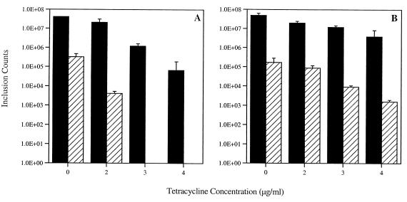
Tetracycline (TET) is a front-line antibiotic for the treatment of chlamydial infections in both humans and animals, and the emergence of TET-resistant (Tet(r)) Chlamydia is of significant clinical importance. Recently, several Tet(r) chlamydial strains have been isolated from swine (Sus scrofa) raised in production facilities in Nebraska. Here, the intracellular development of two Tet(r) strains, R19 and R27, is characterized through the use of tissue culture and immunofluorescence. The strains grow in concentrations of up to 4 microg of TET/ml, while a TET-sensitive (Tet(s)) swine strain (S45) and a strain of the human serovar L2 (LGV-434) grow in up to 0.1 microg of TET/ml. Although inclusions form in the presence of TET, many contain large aberrant reticulate bodies (RBs) that do not differentiate into infectious elementary bodies. The percentage of inclusions containing typical developmental forms decreases with increasing TET concentrations, and at 3 microg of TET/ml 100% of inclusions contain aberrant RBs. However, upon removal of TET the aberrant RBs revert to typical RBs, and a productive developmental cycle ensues. In addition, inclusions were found that contained both C. suis R19 and Chlamydia trachomatis L2 after sequential infection, demonstrating that two biologically distinct chlamydial strains could both develop within a single inclusion.


Figures

FIG. 1
FIG. 1
TET resistance patterns of several chlamydial strains. Infected cells were fixed at 50 hpi, and inclusion-forming units (IFUs) were enumerated using immunofluorescent microscopy and a monoclonal antibody against HSP60. The human C. trachomatis serovar, L2, was sensitive to a TET concentration of 0.25 μg/ml. Similar results were observed with the sensitive C. suis strain, S45. The two resistant C. suis strains, R19 and R27, formed inclusions at a TET concentration of 4 μg/ml, and no growth of R19 or R27 was observed at 5 μg of TET/ml. Values are averages of results from three wells.
FIG. 2
FIG. 2
Temporal analysis of EB production by C. suis strain R19. HeLa cells were infected with C. suis R19, and EBs were harvested at the time points indicated. Cells were lysed, and the lysate was used to infect new monolayers of HeLa cells. At 50 hpi, cells were fixed and inclusion-forming units (IFUs) were enumerated using immunofluorescense with antibodies directed against HSP60. Values are averages of results from three wells, and standard deviations are represented with positive error bars.
FIG. 3
FIG. 3
Decreased growth of R19 and the effects of increasing TET concentrations. Infected cells were fixed at 50 hpi, and total numbers of inclusions were enumerated using immunofluorescence with antibodies against HSP60. These numbers, indicated with black bars, include both typical and aberrant inclusions. The hatched bars represent the number of inclusions formed after cells were lysed at 50 hpi, used to reinfect new cells, and cultured in the absence of TET. At 3 and 4 μg/ml, 100% of the RBs were aberrant and no EBs were present. Values are averages of results from three wells, and standard deviations are represented with positive error bars. IFUs, inclusion-forming units.
FIG. 4
FIG. 4
Inclusion morphology. (A and B) C. suis strain S45 formed typical inclusions when grown in the absence of TET. (C and D) C. suis strain R19 predominately formed typical inclusions when grown in the absence of TET. These inclusions are heavily laden with developmental forms. When grown in 2 μg of TET/ml, however, the majority of the inclusions contained only a few large aberrant RBs (E and F). Panels A, C, and E represent DIC micrographs; panels B, D, and F are fluorescent images labeled with antibodies against HSP60. Bar, 5 μm.
FIG. 5
FIG. 5
Production of EBs from aberrant RBs upon removal of tetracycline. Infected cells were fixed at 50 hpi, and total numbers of inclusions were enumerated using immunofluorescence with antibodies against HSP60. These numbers, indicated with black bars, include both typical and aberrant inclusions. The hatched bars represent the number of inclusions formed after cells were lysed at 50 hpi, used to reinfect new cells, and cultured in the absence of TET. (A) EB production when R19 is cultured for 50 h in the indicated concentrations of TET. (B) EB production when R19 is cultured for the first 24 h in the indicated concentrations of TET and then cultured in no TET for the final 26 h. Values are averages of results from three wells, and standard deviations are represented with positive error bars.
FIG. 6
FIG. 6
Overall protein production in response to TET. Overall protein production was examined through the use of radioactive methionine and cysteine. Lane 1, uninfected HeLa cell control; lane 2, L2-infected HeLa cells grown in the absence of TET; lane 3, L2 grown in the presence of 0.5 μg of TET/ml; lane 4, L2 grown in the presence of 1 μg of TET/ml; lane 5, S45-infected HeLa cells grown in the absence of TET; lane 6, S45 grown in the presence of 0.5 μg of TET/ml; lane 7, S45 grown in the presence of 1 μg of TET/ml; lane 8, R19-infected HeLa cells grown in the absence of TET; lanes 9 (0.5 μg/ml), 10 (1 μg/ml), 11 (2 μg/ml), 12 (3 μg/ml), 13 (4 μg/ml), and 14 (5 μg/ml) all represent R19 protein synthesis in the presence of TET. Molecular size markers are shown on the left in kilodaltons.
FIG. 7
FIG. 7
C. trachomatis L2 and C. suis R19 growth within a single inclusion. HeLa cells sequentially infected with R19 (time, 0 h) and L2 (time, 16 h) were fixed with methanol and labeled with antibodies directed against L2 and R19. (A) DIC micrograph of an infected cell containing a single inclusion 50 h after infection with R19. (B) Fluorescent image of the cells shown in panel A labeled with antisera against L2 MOMP (red) and R19 MOMP (green). (C) DIC micrograph of infected cells fixed 32 h after infection with R19. (D) Fluorescent image of the cells shown in panel C labeled with antisera against L2 IncA (red) and R19 MOMP (green). The nuclei of cells shown in panel D are colored blue with 4′,6-diamino-2-phenylindole (Sigma). Bar, 5 μm.

Similar articles

Cited by

References

    1. Andersen A A, Rogers D G. Resistance to tetracycline and sulfadiazine in swine C. trachomatis isolates. In: Stephens R S, et al., editors. Chlamydial Infections. Proceedings of the Ninth International Symposium on Human Chlamydial Infection. 1998. pp. 313–316.
    1. Anonymous. Government advisory committee calls for a reduction in antibiotic use. Vet Rec. 1999;145:266–267. - PubMed
    1. Bannantine J P, Griffiths R S, Viratyosin W, Brown W J, Rockey D D. A secondary structure motif predictive of protein localization to the chlamydial inclusion membrane. Cell Microbiol. 2000;2:35–47. - PubMed
    1. Bannantine J P, Stamm W E, Suchland R J, Rockey D D. Chlamydia trachomatis IncA is localized to the inclusion membrane and is recognized by antisera from infected humans and primates. Infect Immun. 1998;66:6017–6021. - PMC - PubMed
    1. Barth W F, Segal K. Reactive arthritis (Reiter's syndrome) Am Fam Physician. 1999;60:499–503. - PubMed

Publication types

LinkOut - more resources