. 2001 Aug;45(8):2229-37.
doi: 10.1128/AAC.45.8.2229-2237.2001.

Sodium lauryl sulfate abrogates human immunodeficiency virus infectivity by affecting viral attachment

Affiliations

Sodium lauryl sulfate abrogates human immunodeficiency virus infectivity by affecting viral attachment

J Bestman-Smith et al. Antimicrob Agents Chemother. 2001 Aug.

Abstract

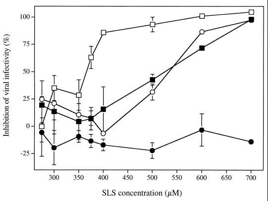
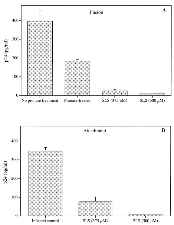
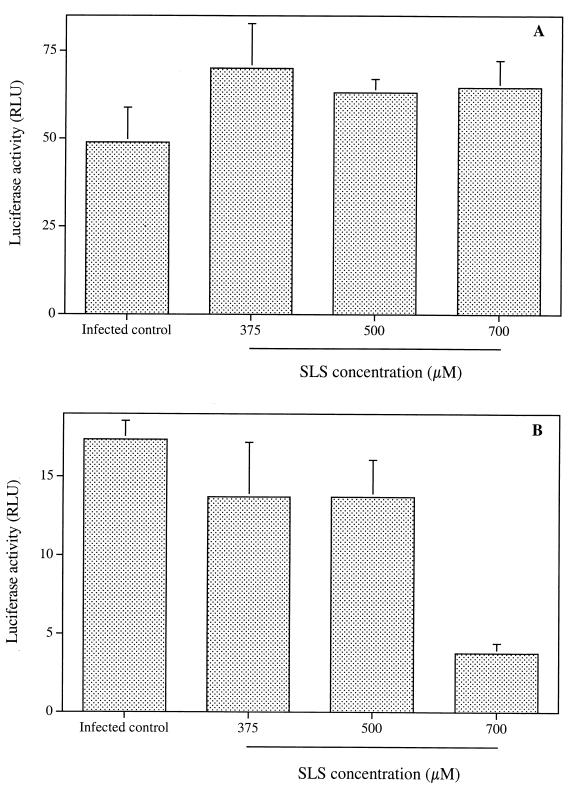
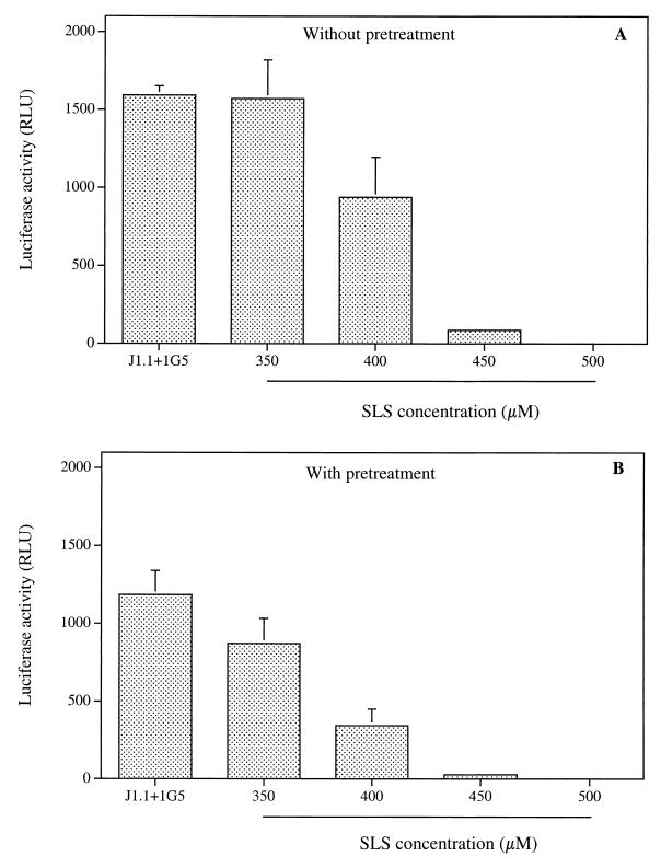
The microbicidal activity of sodium lauryl sulfate (SLS) against human immunodeficiency virus type 1 (HIV-1) was studied in cultured cells. Pretreatment of HIV-1(NL4-3) with SLS decreased, in a concentration-dependent manner, its infectivity when using 1G5 as target cells. In the absence of a viral pretreatment period or when 1G5 cells were pretreated with SLS, the surfactant-induced inactivation of viral infectivity was less pronounced, especially at concentrations between 375 and 550 microM. SLS had no effect on HIV-1 when the virus was adsorbed to 1G5 cells by a 2-h incubation period. SLS almost completely inhibited the fusion process by decreasing the attachment of HIV-1 to target cells. SLS also inhibited the infectivity of HIV-1-based luciferase reporter viruses pseudotyped with the amphotropic murine leukemia virus envelope (which enters cells in a CD4-, CCR5-, and CXCR4-independent manner), indicating that SLS may inactivate other envelope viruses. In contrast, no effect was seen with vesicular stomatitis virus envelope glycoprotein G (which enters cells through receptor-mediated endocytosis) pretreated with up to 700 microM SLS. SLS also decreased, in a dose-dependent manner, the HIV-1-dependent syncytium formation between 1G5 and J1.1 cells after a 24-h incubation. The reduction of luciferase activity was more pronounced when J1.1 cells (which express HIV-1 proteins on their surface) were pretreated with SLS rather than 1G5 cells. Taken together, our results suggest that SLS could represent a candidate of choice for use in vaginal microbicides to prevent the sexual transmission of HIV and possibly other pathogens causing sexually transmitted diseases.


Figures

FIG. 1
FIG. 1
Effect of SLS on the infectivity of HIV-1NL4-3 to 1G5 cells under different experimental (or infection) conditions. (i) Cell-free virus (□) was pretreated with increasing SLS concentrations for 1 h at 37°C and washed with PBS. 1G5 cells were then infected with HIV-1NL4-3 (10 ng of p24/105 cells) for 2 h at 37°C, washed, and maintained in fresh culture medium in the absence of SLS (control luciferase activity was 33.1 ± 6.5 relative light units [RLU]). (ii) 1G5 cells (■) were pretreated with increasing SLS concentrations for 1 h at 37°C and washed with PBS. 1G5 cells were then infected with HIV-1NL4-3 (10 ng of p24/105 cells) for 2 h at 37°C, washed, and maintained in fresh culture medium in the absence of SLS (control luciferase activity was 23.6 ± 2.9 RLU). (iii) SLS and the virus were added simultaneously to cells, incubated for 2 h, washed, and maintained in fresh culture medium in the absence of SLS (○) (control luciferase activity was 36.8 ± 3.3 RLU). (iv) HIV-1NL4-3 was adsorbed to cells by a 2-h incubation period prior to the addition of SLS, which was maintained in contact with cells for 72 h (●) (control luciferase activity was 96.3 ± 10.5 RLU). Luciferase activity was measured 72 h after infection. Results are means (± standard deviations) of triplicates and are representative of three independent experiments.
FIG. 2
FIG. 2
Effect of SLS on the fusion (A) and attachment (B) of HIV-1NL4-3 to Jurkat E6.1 cells. Panel A shows untreated and SLS-treated HIV-1NL4-3 viruses (30 ng of p24) incubated with Jurkat E6.1 cells (3 × 10⁵) for 2 h at 37°C and then treated with pronase (0.1 mg/ml) for 5 min. Cells not treated with pronase correspond to bound and fused virus whereas cells treated with pronase correspond to fused virus only. Standard p24 quantification was subsequently performed after cell lysis. Panel B shows untreated and SLS-treated viruses (30 ng of p24) incubated with Jurkat E6.1 cells (3 × 10⁵) for 90 min at 4°C. After extensive washing, cells were lysed and p24 quantification was performed. p24 levels measured in uninfected cells were subtracted from data as background values. Results are means (± standard deviations) of triplicates and are representative of three independent experiments.
FIG. 3
FIG. 3
Effect of SLS on the infectivity to Jurkat E6.1 cells. of HIV-1-based luciferase reporter viruses pseudotyped with VSV-G (A) and A-MLV (B) envelopes. Single-round infections were made with viruses pretreated with increasing SLS concentrations for 1 h at 37°C and then washed with PBS. Jurkat E6.1 cells were then infected with pretreated viruses (10 ng of p24) for 2 h at 37°C, washed, and maintained in fresh culture medium in the absence of SLS. Luciferase activity was measured at 72 h after infection. Luciferase activities for uninfected control cells were subtracted from data as background values. Results are means (± standard deviations) of triplicates and are representative of two independent experiments.
FIG. 4
FIG. 4
Effect of SLS on the syncytium formation between J1.1 and 1G5 cells. Panel A shows 1G5 and J1.1 cells (105 cells) mixed in the absence (positive control) or in the presence of increasing SLS concentrations and incubated for 24 h at 37°C. Panel B shows J1.1 cells pretreated with increasing SLS concentrations, washed, and then incubated with 1G5 cells for 24 h at 37°C. Cells were lysed and assayed for luciferase activity. Luciferase activities for 1G5 cells alone were subtracted from data as background values. Results are means (± standard deviations) of triplicates and are representative of two independent experiments.

Similar articles

Cited by

References

    1. Adachi A, Koenig S, Gendelman H E, Daugherty D, Gattoni-Celli S, Fauci A S, Martin M A. Productive, persistent infection of human colorectal cell lines with human immunodeficiency virus. J Virol. 1986;61:209–213. - PMC - PubMed
    1. Aguilar-Cordova E, Chinen J, Donehower L, Lewis D E, Belmont J W. A sensitive reporter cell line for HIV-1 tat activity, HIV-1 inhibitors, and T cell activation effects. AIDS Res Hum Retrovir. 1994;10:295–301. - PubMed
    1. Alexander N J. Future contraceptives. Sci Am. 1995;273:136–141. - PubMed
    1. Barbeau B, Fortin J F, Genois N, Tremblay M J. Modulation of human immunodeficiency virus type 1-induced syncytium formation by the conformational state of LFA-1 determined by a new luciferase-based syncytium quantitative assay. J Virol. 1998;72:7125–7136. - PMC - PubMed
    1. Bourinbaiar A S, Lee-Huang S. Comparative in vitro study of contraceptive agents with anti-HIV activity: gramicidin, nonoxynol-9 and gossypol. Contraception. 1994;49:131–137. - PubMed

Publication types

MeSH terms