. 2001 Jul;12(7):1925-35.
doi: 10.1091/mbc.12.7.1925.

Gamma subunit of the AP-1 adaptor complex binds clathrin: implications for cooperative binding in coated vesicle assembly

Affiliations
Free PMC article

Gamma subunit of the AP-1 adaptor complex binds clathrin: implications for cooperative binding in coated vesicle assembly

B Doray et al. Mol Biol Cell. 2001 Jul.
Free PMC article

Abstract

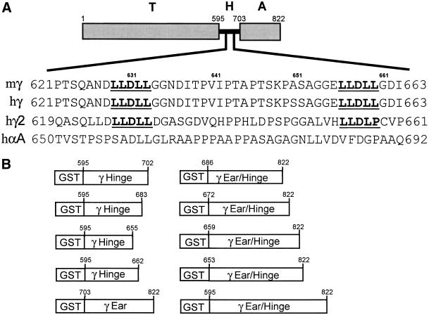
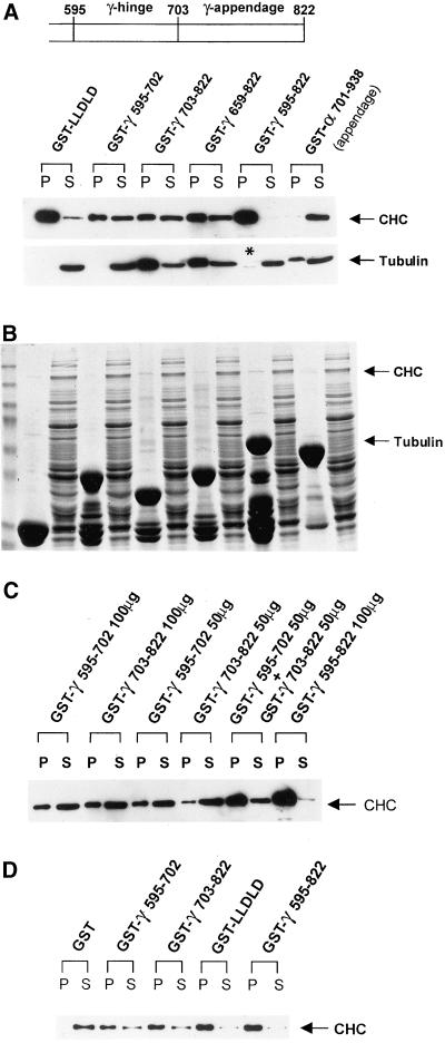
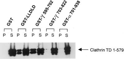
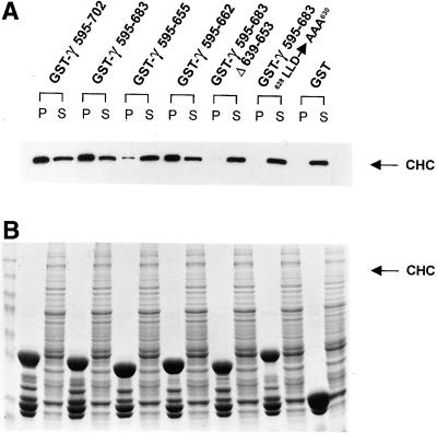
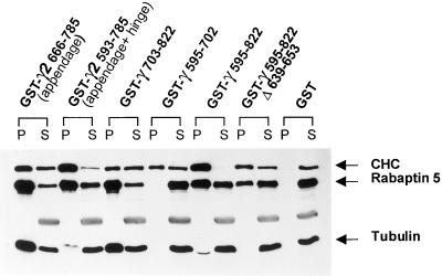
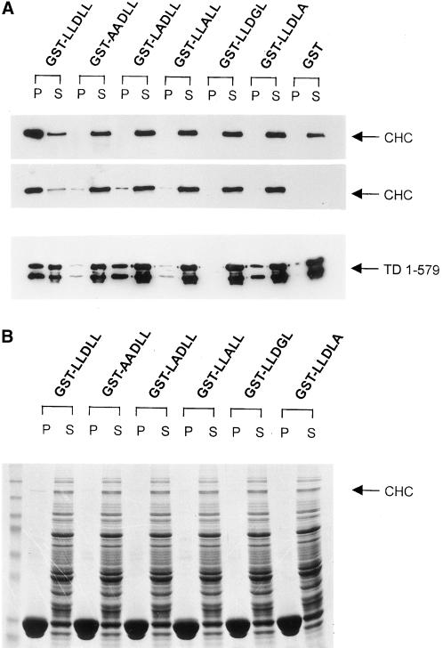
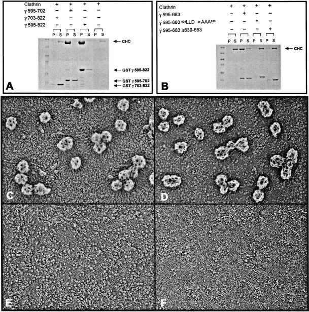
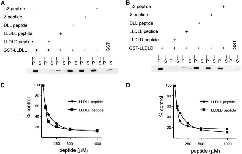
The heterotetrameric AP-1 adaptor complex is involved in the assembly of clathrin-coated vesicles originating from the trans-Golgi network (TGN). The beta 1 subunit of AP-1 is known to contain a consensus clathrin binding sequence, LLNLD (the so-called clathrin box motif), in its hinge segment through which the beta chain interacts with the N-terminal domains of clathrin trimers. Here, we report that the hinge region of the gamma subunit of human and mouse AP-1 contains two copies of a new variant, LLDLL, of the clathrin box motif that also bind to the terminal domain of the clathrin heavy chain. High-affinity binding of the gamma hinge to clathrin trimers requires both LLDLL sequences to be present and the spacing between them to be maintained. We also identify an independent clathrin-binding site within the appendage domain of the gamma subunit that interacts with a region of clathrin other than the N-terminal domain. Clathrin polymerization is promoted by glutathione S-transferase (GST)-gamma hinge, but not by GST-gamma appendage. However, the hinge and appendage domains of gamma function in a cooperative manner to recruit and polymerize clathrin, suggesting that clathrin lattice assembly at the TGN involves multivalent binding of clathrin by the gamma and beta1 subunits of AP-1.


Figures

Figure 1
Figure 1
Sequence comparison of the hinge of mouse γ (mγ), human γ (hγ), human γ2 (hγ2), and human α (hα) adaptins, and GST-γ adaptin fusion constructs. (A) Schematic of γ adaptin showing the trunk (T), the hinge (H), and the appendage (A) also called the ear. An alignment of part of the hinge region shows that only γ and γ2 adaptin but not α adaptin possess a variant of the consensus clathrin binding sequence (underlined). (B) Construction of the various GST-γ adaptin fusion proteins is described under MATERIALS AND METHODS.
Figure 2
Figure 2
GST-γ hinge and GST-γ appendage bind clathrin independently and cooperatively. (A) Immunoblot of GST pull-down assay with 200 μg of immobilized fusion protein, which had been incubated with rat liver cytosol at a final concentration of 7.5 mg/ml. Portions of the blot were probed with the anti-clathrin HC mAb TD.1 or an anti-tubulin mAb. (B) Coomassie blue-stained gel of the same samples as in A indicates the approximately equivalent loading of the different fusion proteins. In this case, each pellet lane corresponds to 1/20th of the total pellet fraction, whereas 1/60th of each supernatant was loaded. (C) Immunoblot of GST pull-down assay with the use of 50 or 100 μg of immobilized fusion proteins, individually or in combination, with rat liver cytosol. (D) Immunoblot of GST pull-down assay with the use of purified soluble clathrin triskelia isolated from bovine brain cytosol.
Figure 3
Figure 3
GST-γ hinge but not GST-γ appendage binds clathrin terminal domain. Clathrin TD 1–579 was expressed as described under MATERIALS AND METHODS and separated from GST after cleavage with the protease thrombin. Purified TD (50 μg) was incubated with 200 μg of the immobilized GST fusion proteins as indicated. Blots were probed with the anti-clathrin HC mAB TD.1. Only the GST-γ 595–702 (hinge) contains the LLDLL sequence and binds terminal domain.
Figure 4
Figure 4
Both γ hinge LLDLL sequences with the correct spacing are required for clathrin binding. GST-γ 595–655 lacks the second 656LLDLL660 sequence, whereas GST-γ 595–683 628LLD → AAA630 has the first LLDLL sequence mutated. (A) Immunoblot of the various GST-γ hinge fusion proteins incubated with rat liver cytosol, probed with the TD.1 mAb. (B) Coomassie blue-stained gel of the same samples indicated in A.
Figure 5
Figure 5
γ 2 Adaptin binds the same subset of proteins as does γ adaptin. Immunoblots of the pull-downs of GST-γ2 and GST-γ fusion proteins. Portions of the blot were probed with the anti-clathrin TD.1 mAB, an anti-tubulin mAb, or an anti-rabaptin 5 mAb.
Figure 6
Figure 6
Each residue within the γ hinge LLDLL sequence is important for binding clathrin trimers. (A) Binding assays performed with rat liver cytosol in the presence of 0.1% Triton X-100 (top) and in the absence of detergent (middle), or with purified clathrin TD 1–579 (bottom). (B) Coomassie blue-stained gel of the same samples indicated in A incubated with rat liver cytosol in the presence of 0.1% Triton X-100.
Figure 7
Figure 7
γ Hinge/appendage facilitates the polymerization and assembly of clathrin lattices. The polymerization assays were carried out as described under MATERIALS AND METHODS. (A) Coomassie blue-stained gel of the polymerization of cytosolic clathrin in the presence of GST γ appendage, GST γ hinge, and GST γ appendage + hinge. By densitometric analysis, 3% of the clathrin was in the high-speed pellet for GST γ appendage, 50% for GST γ hinge, 90% for GST γ appendage + hinge, and 9% for clathrin alone. The values represent the average of two independent experiments with the use of two different preparations of clathrin. (B) Coomassie blue-stained gel of the polymerization of cytosolic clathrin in the presence of GST γ 595–683, GST γ 595–683Δ639–653, and GST γ 595–683 628LLD → AAA630. (C–F) Electron microscopy images obtained from high-speed pellets of assembled clathrin of the samples indicated in A. (C) GST γ appendage + hinge and clathrin; (D) GST γ hinge and clathrin; (E) GST γ appendage and clathrin; (F) clathrin alone.
Figure 8
Figure 8
Both LLDLL and LLDLD peptides inhibit GST-LLDLL and GST-LLDLD. Inhibition assays were performed as described under MATERIALS AND METHODS. (A–B) Concentration of each free peptide was 1 mM. (A) GST-LLDLL immobilized on glutathione-Sepharose 4B. (B) Immobilized GST-LLDLD. (C and D) Free peptide concentrations varied from 50 μM to 1 mM. Curves were generated from densitometric analysis of the pellet fractions of the pull-down assays at different peptide concentrations. (C) GST-LLDLL immobilized on glutathione-Sepharose 4B. (D) Immobilized GST-LLDLD.

References

    1. Ahle S, Ungewickell E. Identification of a clathrin binding subunit in the HA2 adaptor protein complex. J Biol Chem. 1989;264:20089–20093. - PubMed
    1. Ahle S, Ungewickell E. Auxilin, a newly identified clathrin-associated protein in coated vesicles from bovine brain. J Cell Biol. 1990;111:2047–2054. - PMC - PubMed
    1. Dell'Angelica EC, Klumperman J, Stoorvogel W, Bonifacino JS. Association of the AP-3 adaptor complex with clathrin. Science. 1998;280:431–434. - PubMed
    1. Drake MT, Downs MA, Traub LM. Epsin binds to clathrin by associating directly with the clathrin terminal domain. J Biol Chem. 2000;275:6479–6489. - PubMed
    1. Gallusser A, Kirchhausen T. The β1and β2 subunits of the AP complexes are the clathrin coat assembly components. EMBO J. 1993;13:5237–5244. - PMC - PubMed

Publication types

MeSH terms